. 2022 Dec 14;15(12):1558.
doi: 10.3390/ph15121558.

Protective Effects of Naringin-Dextrin Nanoformula against Chemically Induced Hepatocellular Carcinoma in Wistar Rats: Roles of Oxidative Stress, Inflammation, Cell Apoptosis, and Proliferation

Affiliations

Protective Effects of Naringin-Dextrin Nanoformula against Chemically Induced Hepatocellular Carcinoma in Wistar Rats: Roles of Oxidative Stress, Inflammation, Cell Apoptosis, and Proliferation

Eman E Mohamed et al. Pharmaceuticals (Basel). .

Abstract

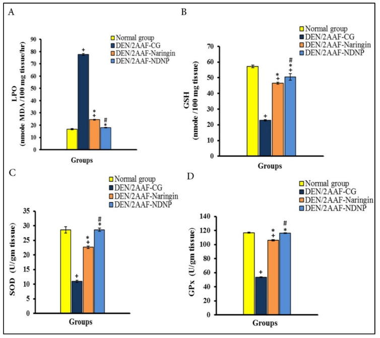
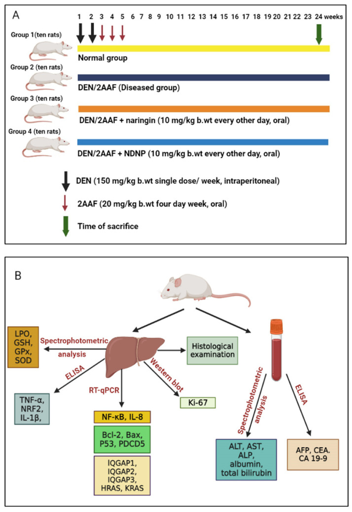
Nanotechnology holds great promise for the development of treatments for deadly human diseases, such as hepatocellular carcinoma (HCC). In the current study, we compared the hepatoprotective effects of naringin-dextrin nanoparticles (NDNPs) against HCC in male Wistar rats with those of pure naringin and investigated the underlying cellular and molecular mechanisms. HCC was induced by intraperitoneal injection of diethylnitrosamine (DEN, 150 mg/kg body weight (b.w.) per week) for two weeks, followed by oral administration of 2-acetylaminofluorene (2AAF, 20 mg/kg b.w.) four times per week for three weeks. DEN/2AAF-administered rats were divided into three groups that respectively received 1% carboxymethyl cellulose (as vehicle), 10 mg/kg b.w. naringin, or 10 mg/kg b.w. NDNP every other day by oral gavage for 24 weeks. Both naringin and NDNP significantly attenuated the harmful effects of DEN on liver function. Both compounds also suppressed tumorigenesis as indicated by the reduced serum concentrations of liver tumor markers, and this antitumor effect was confirmed by histopathological evaluation. Additionally, naringin and NDNP prevented DEN-induced changes in hepatic oxidative stress and antioxidant activities. In addition, naringin and NDNP suppressed inflammation induced by DEN. Moreover, naringin and NDNP significantly reduced the hepatic expression of Bcl-2 and increased Bax, p53, and PDCD5 expressions. Naringin and NDNP also reduced expression of IQGAP1, IQGAP3, Ras signaling, and Ki-67 while increasing expression of IQGAP2. Notably, NDNP more effectively mitigated oxidative stress and inflammatory signaling than free naringin and demonstrated improved antitumor efficacy, suggesting that this nanoformulation improves bioavailability within nascent tumor sites.

Keywords: acetylaminofluorene; anti-inflammatory effects; antiapoptotic effects; diethylnitrosamine; naringin; naringin–dextrin nanoparticles.


Conflict of interest statement

The authors declare no conflict of interest.

Figures

Figure 1
Figure 1
Naringin and naringin–dextrin nanoparticles (NDNPs) reduced serum tumor biomarker concentrations in DENA/2AAF-treated rats. (A) Serum AFP. (B) Serum CEA. (C) Serum CA19.9. Data are expressed as means ± standard error. Number of detected samples in each group was six. + Significant compared to normal group; * significant compared to DEN/2AAF-CG; and # significant compared to DEN/2AAF + naringin group at p < 0.05. AFP: alpha fetoprotein; CEA: carcinoembryonic antigen; CA19.9: carbohydrate antigen 19.9.
Figure 2
Figure 2
Naringin and NDNP suppressed hepatic oxidative stress in HCC model rats. (A) LPO. (B) GSH concentration. (C) SOD activity. (D) GPx activity. Data are expressed as means ± standard error. Number of detected samples in each group was six. + Significant compared to normal group; * significant compared to DEN/2AAF-CG; and # significant compared to DEN/2AAF + naringin group at p < 0.05. LPO: lipid peroxidation; GSH: reduced glutathione; SOD: superoxide dismutase; GPx: glutathione peroxidase.
Figure 3
Figure 3
Effects of naringin and NDNP on (A) NF-κB and IL-8 mRNA expression; (B) antiapoptotic Bcl-2 expression and proapoptotic Bax, p53, and PDCD5 expression; (C) IQGAP1, IQGAP2, and IQGAP3 mRNA expression; and (D) HRAS and KRAS mRNA expression in DENA/2AAF-treated rats. Data are expressed as means ± standard error. Number of detected samples in each group was six. + Significant compared to normal group; * significant compared to DEN/2AAF-CG; and # significant compared to DEN/2AAF + naringin group at p < 0.05. NF-κB: nuclear factor-kappa B; IL-8: interleukin-8; Bcl-2: B-cell lymphoma-2; Bax: Bcl-2-associated X protein; p53: tumor suppressor protein 53; PDCD5: programmed cell death 5; IQGAP1: isoleucine–glutamine motif-containing GTPase-activating protein 1; IQGAP2: isoleucine–glutamine motif-containing GTPase-activating protein 2; IQGAP3: Isoleucine–glutamine motif-containing GTPase-activating protein 3; HRAS: Harvey rat sarcoma viral oncogene homolog; KRAS: Kirsten rat sarcoma viral oncogene homolog.
Figure 4
Figure 4
Effect of naringin and NDNP on liver Ki-67 expression in DENA/2AAF-treated rats. Data are expressed as means ± standard error. Number of detected samples in each group was 3. + Significant compared to normal group; * significant compared to DEN/2AAF-CG; and # significant compared to DEN/2AAF + naringin group at p < 0.05. Ki-67: Proliferator protein.
Figure 5
Figure 5
Histological features of liver sections from untreated control rats. (A,B) Sections stained with H&E showing intact lobules with portal triads among them. Each lobule consisted of cords formed from regularly arranged hepatocytes (black arrow) enclosing sinusoids (red arrow) lined with Kupffer cells (green arrow) and a central vein (CV) located in the center (H&E stain, scale bar: 50 µm).
Figure 6
Figure 6
Liver sections from DEN/2AAF-adminstered rats showing extensive histopathology, including disruption of the lobular structure, degeneration, and tumorigenesis. (A) Disruption of hepatic lobule structure. Tumor cells appeared well-differentiated or resembled hepatocytes and formed trabeculae (arrow), cords (green arrow), and nests (blue arrow). Note the growing septa between hepatic lobules (red arrow) (H&E stain, scale bar: 50 µm). (B) Disruption of the normal structure of the hepatic lobule (arrowhead) and tumor cells arranged in glandular or lamellar form (arrow) (H&E stain, scale bar: 50 µm). (C) Clear hepatocyte foci, deeply eosinophilic hepatocyte foci, vacuolated hepatocytes, and dysplastic hepatocytes with mitotic figures (H&E stain, scale bar: 50 µm). (D) Dilated central veins (arrow), some with signet ring appearance (green arrow), binucleated hepatocytes (yellow arrow), and hepatocytic steatosis (blue arrow) (H&E stain, scale bar: 50 µm). (E) Fibrotic tissue with inflammatory cells (yellow arrow), hepatocytes with dark shrunken nuclei (blue arrow), activated Kupffer cells, binucleated hepatocytes (green arrow), and hepatocytes with large hyperchromatic nuclei and prominent enlarged nucleoli (arrowhead) (H&E stain, scale bar: 50 µm). (F) Large hyperchromatic nuclei with prominent multiple nucleoli (arrow) (H&E stain, scale bar: 100 µm).
Figure 7
Figure 7
(A,B) Liver sections from DEN/2AAF-administered rats treated with naringin showing marked amelioration of histopathology and fewer degenerated and vacuolated hepatocytes, together with less extensive dysplasia, milder hepatic sinusoid dilation, fewer proliferating Kupffer cells, and maintenance of normal lobular architecture. Some sporadic tumor cells were found but rarely in a glandular form (H&E stain, scale bar: 50 µm). (C,D) NDNP treatment showing similar hepatoprotection as free naringin but with an absence of tumor cells (H&E stain, scale bar: 50 µm).
Figure 8
Figure 8
Schematics of the experimental design and animal grouping: Wistar adult male rats were divided into four groups and the number of animals in each group was ten (A); blood, tissue sampling, and techniques (B).
Figure 9
Figure 9
Schematic diagram showing the hepatoprotective mechanisms of naringin–dextrin nanoparticles against DEN/2AAF: suppression of oxidative stress, inflammation, and proliferation, as well as induction of transformed cell apoptosis. DEN: diethylnitrosamine; 2AAF: 2-acetylaminofluorene; ROS: reactive oxygen species; IGs: inflammatory genes; IQGAP1: isoleucine–glutamine motif-containing GTPase-activating protein 1; IQGAP2: isoleucine–glutamine motif-containing GTPase-activating protein 2; IQGAP3: Isoleucine–glutamine motif-containing GTPase-activating protein 3; NF-κB: nuclear factor-kappa B; IL-8: interleukin-8; IL-1β: interleukin-1β; Bcl-2: B-cell lymphoma-2; Bax: Bcl-2-associated X protein; p53: tumor suppressor protein 53; PDCD5: programmed cell death 5. RAS: rat sarcoma viral oncogene homolog; ALT: alanine transaminase; AST: aspartate transaminase; ALP: alkaline phosphatase; NDNP: naringin-dextrin nanoparticle; TNF-α: tumor necrosis factor alpha; TNFR: tumor necrosis factor alpha receptor; NRF2: nuclear factor E2-related factor 2; LPO: lipid peroxidation; GSH: Glutathione; SOD: Superoxide dismutase; GPx: glutathione peroxidase.

References

    1. Saber S., Mahmoud A., Goda R., Helal N.S., El-Ahwany E., Abdelghany R.H. Perindopril, fosinopril and losartan inhibited the progression of diethylnitrosamine-induced hepatocellular carcinoma in mice via the inactivation of nuclear transcription factor kappa-B. Toxicol. Lett. 2018;295:32–40. doi: 10.1016/j.toxlet.2018.05.036. - DOI - PubMed
    1. Saber S., Khodir A.E., Soliman W.E., Salama M.M., Abdo W.S., Elsaeed B., Nader K., Abdelnasser A., Megahed N., Basuony M., et al. Telmisartan attenuates N-nitrosodiethylamine-induced hepatocellular carcinoma in mice by modulating the NF-κB-TAK1-ERK1/2 axis in the context of PPARγ agonistic activity. Naunyn-Schmiedeberg’s Arch. Pharmacol. 2019;392:1591–1604. doi: 10.1007/s00210-019-01706-2. - DOI - PubMed
    1. Younis N.S., Ghanim A.M., Saber S. Mebendazole augments sensitivity to sorafenib by targeting MAPK and BCL-2 signalling in n-nitrosodiethylamine-induced murine hepatocellular carcinoma. Sci. Rep. 2019;9:19095. doi: 10.1038/s41598-019-55666-x. - DOI - PMC - PubMed
    1. Seydi E., Motallebi A., Dastbaz M., Dehghan S.G., Salimi A., Nazemi M., Pourahmad J. Selective toxicity of Persian Gulf sea cucumber (Holothuria parva) and sponge (Haliclona oculata) methanolic extracts on liver mitochondria isolated from an animal model of hepatocellular carcinoma. Hepat Mon. 2015;15:e33073. doi: 10.5812/hepatmon.33073. - DOI - PMC - PubMed
    1. Asafo-Agyei K.O., Samant H. Hepatocellular Carcinoma. StatPearls Publishing LLC, National Center for Biotechnology Information, U.S. National Library of Medicine; Bethesda, MD, USA: 2021.