. 2022 Nov 30;14(12):2662.
doi: 10.3390/pharmaceutics14122662.

Effect of Ciprofloxacin-Loaded Niosomes on Escherichia coli and Staphylococcus aureus Biofilm Formation

Affiliations

Effect of Ciprofloxacin-Loaded Niosomes on Escherichia coli and Staphylococcus aureus Biofilm Formation

Linda Maurizi et al. Pharmaceutics. .

Abstract

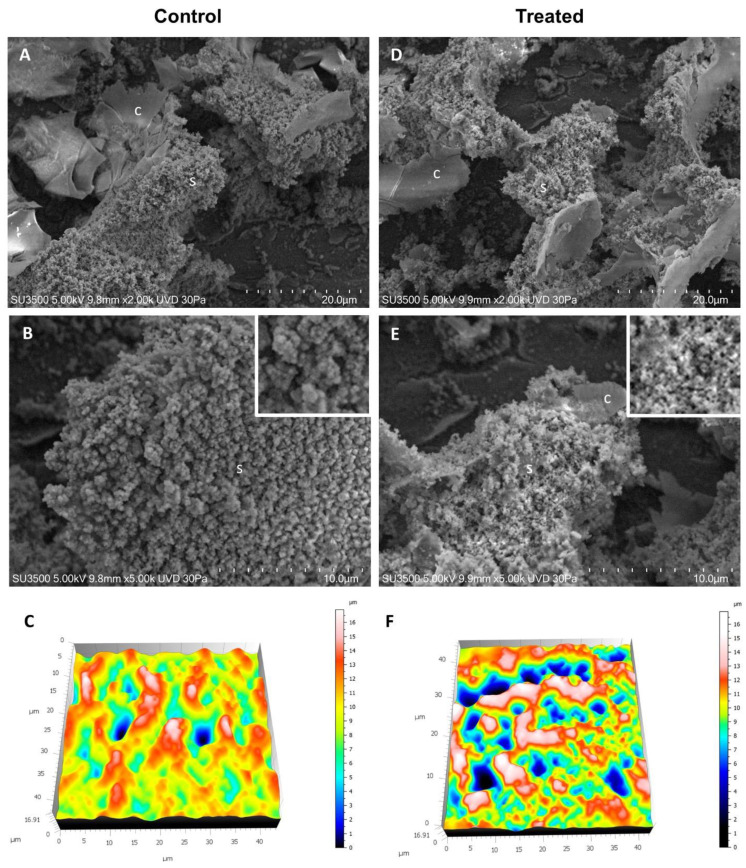
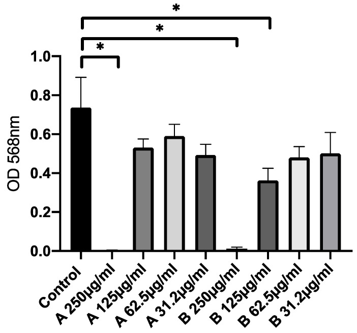
Infections caused by bacterial biofilms represent a global health problem, causing considerable patient morbidity and mortality in addition to an economic burden. Escherichia coli, Staphylococcus aureus, and other medically relevant bacterial strains colonize clinical surfaces and medical devices via biofilm in which bacterial cells are protected from the action of the immune system, disinfectants, and antibiotics. Several approaches have been investigated to inhibit and disperse bacterial biofilms, and the use of drug delivery could represent a fascinating strategy. Ciprofloxacin (CIP), which belongs to the class of fluoroquinolones, has been extensively used against various bacterial infections, and its loading in nanocarriers, such as niosomes, could support the CIP antibiofilm activity. Niosomes, composed of two surfactants (Tween 85 and Span 80) without the presence of cholesterol, are prepared and characterized considering the following features: hydrodynamic diameter, ζ-potential, morphology, vesicle bilayer characteristics, physical-chemical stability, and biological efficacy. The obtained results suggest that: (i) niosomes by surfactants in the absence of cholesterol are formed, can entrap CIP, and are stable over time and in artificial biological media; (ii) the CIP inclusion in nanocarriers increase its stability, with respect to free drug; (iii) niosomes preparations were able to induce a relevant inhibition of biofilm formation.

Keywords: anti biofilm activity; bladder cells; ciprofloxacin; drug delivery; niosomes.


Conflict of interest statement

The authors declare no conflict of interest.

Figures

Figure 1
Figure 1
SAXS spectrum of empty niosomes, sample A (22.5 mM, room temperature, open dots) reported in log-log scale. The green line is the best fit.
Figure 2
Figure 2
Transmission electron microscopy images of empty and CIP-loaded niosomes.
Figure 3
Figure 3
(a) Result of investigation on physicochemical stability of empty niosomes (A) and CIP-loaded niosomes (B) in terms of hydrodynamic diameter and ζ-potential until up 90 days at 4 °C and room temperature. (b) Stability studies in the presence of artificial body urine, following variation of hydrodynamic diameter and ζ-potential values of CIP-loaded niosomes; Pre-exp values refer to CIP-loaded niosomes before the presence of artificial body urine. (c) Stability studies over time of free CIP and CIP-loaded into niosomes at two different storage temperatures over a 90-day period.
Figure 4
Figure 4
CIP release profile until up 24 h. Data were obtained as the mean of three independent experiments.
Figure 5
Figure 5
Susceptibility test with empty niosomes (A) and CIP-loaded niosomes (B). Data were expressed as mean ± SD. All considered conditions were compared to untreated control. * p value ≤ 0.05.
Figure 6
Figure 6
VP-SEM images of E. coli (AC) and E. coli treated with CIP-loaded niosomes (DF). (A) Low magnification (2.00K), ECM shows compact and smooth ECM areas (c), as well as spongy and rough areas (c). (B) Higher magnification (5.00K) of the ECM spongy area, ECM trabeculae show a globular structure (inset). (C) 3D reconstruction of sample surface topography, ECM trabeculae are represented in white and red areas, and the channels that perforate the ECM are represented with color shades from green to blue. (D) Low magnification (2.00K), ECM shows both compact and smooth ECM areas (c), both spongy and rough areas. (E) Higher magnification (5.00K) of the ECM spongy area, ECM trabeculae show a fine filamentous network structure (inset). (F) 3D reconstruction of sample surface topography, ECM trabeculae are represented in white and red areas, and the channels that perforate the ECM are represented with color shades from green to blue. Note that ECM presents more channels than the control, and trabeculae are thinner than the control sample.
Figure 7
Figure 7
Analysis of globular structures in control (A) vs. treated samples (B). The particle analysis tool of Hitachi 3D map software revealed that the globular structures in the control sample are about twice those of the treated sample. This may be explained by thinking of a disassembling effect of the treatment on the filament that forms the ECM trabeculae.
Figure 8
Figure 8
Untreated T-24 cells (A), cells treated for 7h with Nile Red loaded niosomes (B) and Nile Red and CIP co-loaded niosomes (C).
Figure 9
Figure 9
Cytotoxic activity of CIP-loaded niosomes (OD values). MTT assay on human bladder cancer cells (T24). Data were expressed as mean ± SD. All considered conditions were compared to untreated control. * p value ≤ 0.05.

References

    1. Flemming H.C., Wingender J., Szewzyk U., Steinberg P., Rice S.A., Kjelleberg S. Biofilms: An emergent form of bacterial life. Nat. Rev. Microbiol. 2016;14:563–575. doi: 10.1038/nrmicro.2016.94. - DOI - PubMed
    1. Wi Y.M., Patel R. Understanding Biofilms and Novel Approaches to the Diagnosis, Prevention, and Treatment of Medical Device-Associated Infections. Infect. Dis. Clin. 2018;32:915–929. doi: 10.1016/j.idc.2018.06.009. - DOI - PMC - PubMed
    1. Flores-Mireles A.L., Walker J.N., Caparon M., Hultgren S.J. Urinary Tract Infections: Epidemiology, Mechanisms of Infection and Treatment Options. Nat. Rev. Microbiol. 2015;13:269–284. doi: 10.1038/nrmicro3432. - DOI - PMC - PubMed
    1. Walker J.N., Flores-Mireles A.L., Pinkner C.L., Schreiber H.L., Joens M.S., Park A.M., Potretzke A.M., Bauman T.M., Pinkner J.S., Fitzpatrick J.A.J., et al. Catheterization Alters Bladder Ecology to Potentiate Staphylococcus Aureus Infection of the Urinary Tract. Proc. Natl. Acad. Sci. USA. 2017;114:E8721–E8730. doi: 10.1073/pnas.1707572114. - DOI - PMC - PubMed
    1. Terlizzi M.E., Gribaudo G., Maffei M.E. UroPathogenic Escherichia Coli (UPEC) Infections: Virulence Factors, Bladder Responses, Antibiotic, and Non-Antibiotic Antimicrobial Strategies. Front. Microbiol. 2017;8:1566. doi: 10.3389/fmicb.2017.01566. - DOI - PMC - PubMed