. 2022 Dec 15;11(24):3527.
doi: 10.3390/plants11243527.

TPC1-Type Channels in Physcomitrium patens: Interaction between EF-Hands and Ca2

Affiliations

TPC1-Type Channels in Physcomitrium patens: Interaction between EF-Hands and Ca2

Franko Mérida-Quesada et al. Plants (Basel). .

Abstract

Two-pore channels (TPCs) are members of the superfamily of ligand-gated and voltage-sensitive ion channels in the membranes of intracellular organelles of eukaryotic cells. The evolution of ordinary plant TPC1 essentially followed a very conservative pattern, with no changes in the characteristic structural footprints of these channels, such as the cytosolic and luminal regions involved in Ca2+ sensing. In contrast, the genomes of mosses and liverworts encode also TPC1-like channels with larger variations at these sites (TPC1b channels). In the genome of the model plant Physcomitrium patens we identified nine non-redundant sequences belonging to the TPC1 channel family, two ordinary TPC1-type, and seven TPC1b-type channels. The latter show variations in critical amino acids in their EF-hands essential for Ca2+ sensing. To investigate the impact of these differences between TPC1 and TPC1b channels, we generated structural models of the EF-hands of PpTPC1 and PpTPC1b channels. These models were used in molecular dynamics simulations to determine the frequency with which calcium ions were present in a coordination site and also to estimate the average distance of the ions from the center of this site. Our analyses indicate that the EF-hand domains of PpTPC1b-type channels have a lower capacity to coordinate calcium ions compared with those of common TPC1-like channels.

Keywords: EF-domain; Physcomitrella; TPC1; coordination of Ca2+; vacuole.


Conflict of interest statement

The authors declare no conflict of interest.

Figures

Figure 1
Figure 1
Topology of a plant Two-Pore Cation Channel 1 (TPC1). A TPC1-subunit is built of two concatenated copies of a structural 6-TM motif, each with six transmembrane domains (1–6). The cytosolic linker between them contains two EF-hands, which sense the cytosolic Ca2+ concentration. Two subunits assemble into a functional channel and form a central permeation pathway.
Figure 2
Figure 2
Ancestry and homology analysis of TPC1-like sequences from P. patens. (a). Cladogram of the TPC1 sequences in A. thaliana and P. patens; (b) Percent identity matrix corresponding to the alignment of the TPC1 channel sequences of A. thaliana and P. patens. The colors indicate cluster 1 (yellow), cluster 2 (red), cluster 3 (blue), and cluster 4 (green).
Figure 3
Figure 3
EF-hand sequences of TPC1-type channels from A. thaliana and P. patens. (a) Sequence alignment, separating between TPC1 and TPC1b. The TPC1 group consisted of AtTPC1 and the PpTPC1-sequences from cluster 1. The TPC1b group consisted of the PpTPC1-sequences of clusters 2–4. For each sequence, the residue numbers of the beginning and end of the EF-hand domains are shown. Zappo staining was used to distinguish the characteristics of the residues: negatively charged (red), positively charged (blue), polar (green), aromatic (mustard), hydrophobic (pink), cysteine (yellow), and proline and glycine (purple). Colored bars on top of the alignment indicate the alpha-helix structure of the EF-hands of AtTPC1. Red dots indicate residues reported for calcium-binding in the AtTPC1 EF-hands. (b) Identity/similarity matrix of the EF-hand sequences of TPC1 channels from A. thaliana and P. patens. Numbers above the diagonal represent the percentage of similarity and those below the diagonal represent the percentage of identity. (c) Fragment of the AtTPC1 channel (PDBID:6E1N) corresponding to the EF-hands. The alpha helices E and F are color-coded as on the top of panel (a). Residues reported for Ca2+ binding that form hydrogen bonds in this crystal are highlighted.
Figure 4
Figure 4
Electrostatic surface of the EF-hands. The electrostatic surface images (a) of the AtTPC1 crystal and the models for (b) AtTPC1, (c) PpTPC1.1.1, (d) PpTPC1.2.1, and (e) PpTPC1.4.1 are shown in front (upper panel) and dorsal view (lower panel). The red regions refer to areas with negatively charged residues; the blue regions refer to areas with positively charged residues; the grey regions refer to hydrophobic areas. The green and yellow dotted circles refer to the Ca2+ binding sites of the EF1-hand and EF2-hand, respectively.
Figure 5
Figure 5
Analysis of the root-mean-square deviation (RMSD) and root-mean-square fluctuation (RMSF) of the MD simulations of the EF-hand domains from (a) AtTPC1, (b) PpTPC1.1.1, (c) TPC1.2.1, and (d) TPC1.4.1. In both RMSD and RMSF, the blue, green, and orange curves represented the three replicates of the molecular dynamics simulations performed for each model, with a duration of 1050 ns for each. The RMSD and RMSF analyses were performed using the first molecular dynamics frame as a reference.
Figure 6
Figure 6
Relative frequencies of the interaction of the residues with Ca2+. Calculating the interaction frequencies with Ca2+ in molecular dynamics simulations for each model was performed with a cutoff of 5 Å to calcium. The frequencies were estimated from a total of 1500 ns of molecular dynamics, corresponding to the last 500 ns, with three replicates per model. (a) Alignment of amino acid sequences per model; the blue color shows the frequency of interaction of each residue with the Ca2+ ion within at a distance of up to 5 Å. The higher the frequency, the higher the probability of being part of the Ca2+-binding site. Alpha helices are represented by black bars, and residues reported as part of the calcium-binding site in the AtTPC1 crystal structure are represented by red dots. The numbering corresponds to the consecutive numbering used in the models. Please note the gap at position 47 of the alignment of PpTPC1.2.1 and PpTPC1.4.1. To cope with this gap, for values ≥ 47, one position needs to be subtracted for these two channels (indicated by an asterisk *). (be) Relative interaction frequency of the residues with Ca2+: (b) AtTPC1 and (c) PpTPC1.1.1 correspond to the EF-hand models of the TPC1 type channels, and (d) PpTPC1.2.1 and (e) PpTPC1.4.1 correspond to the TPC1b-type channels.
Figure 7
Figure 7
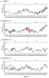
Distribution of distances between each residue and Ca2+. Boxplots show the distances between calcium and the center of mass of the residues in the vicinity of the EF-hand binding sites from (a) AtTPC1, (b) PpTPC1.1.1, (c) PpTPC1.2.1, and (d) PpTPC1.4.1. Zappo staining was used to distinguish the characteristics of the residues: red: negatively charged, blue: positively charged, green: polar, ochre: aromatic, pink: hydrophobic, yellow: cysteine, and magenta: proline and glycine. For both the EF1-hand and the EF2-hand, the box amplitude indicated the dispersion of the distances, where the greater dispersion indicated a more significant absence of Ca2+ binding and vice versa.

Similar articles

References

    1. Yu F.H., Catterall W.A. The VGL-chanome: A protein superfamily specialized for electrical signaling and ionic homeostasis. Sci. STKE. 2004;2004:re15. doi: 10.1126/stke.2532004re15. - DOI - PubMed
    1. Hedrich R., Marten I. TPC1—SV Channels Gain Shape. Mol. Plant. 2011;4:428–441. doi: 10.1093/mp/ssr017. - DOI - PubMed
    1. Peiter E., Maathuis F.J.M., Mills L.N., Knight H., Pelloux J., Hetherington A.M., Sanders D. The vacuolar Ca2+-activated channel TPC1 regulates germination and stomatal movement. Nature. 2005;434:404–408. doi: 10.1038/nature03381. - DOI - PubMed
    1. Hedrich R., Neher E. Cytoplasmic calcium regulates voltage-dependent ion channels in plant vacuoles. Nature. 1987;329:833–836. doi: 10.1038/329833a0. - DOI
    1. Gradogna A., Carpaneto A. Electrophysiology and fluorescence to investigate cation channels and transporters in isolated plant vacuoles. Stress Biol. 2022;2:42. doi: 10.1007/s44154-022-00064-z. - DOI - PMC - PubMed

LinkOut - more resources