. 2022 Dec 7;14(24):5343.
doi: 10.3390/polym14245343.

Regeneration of Osteochondral Defects by Combined Delivery of Synovium-Derived Mesenchymal Stem Cells, TGF-β1 and BMP-4 in Heparin-Conjugated Fibrin Hydrogel

Affiliations

Regeneration of Osteochondral Defects by Combined Delivery of Synovium-Derived Mesenchymal Stem Cells, TGF-β1 and BMP-4 in Heparin-Conjugated Fibrin Hydrogel

Madina Sarsenova et al. Polymers (Basel). .

Abstract

The regeneration of cartilage and osteochondral defects remains one of the most challenging clinical problems in orthopedic surgery. Currently, tissue-engineering techniques based on the delivery of appropriate growth factors and mesenchymal stem cells (MSCs) in hydrogel scaffolds are considered as the most promising therapeutic strategy for osteochondral defects regeneration. In this study, we fabricated a heparin-conjugated fibrin (HCF) hydrogel with synovium-derived mesenchymal stem cells (SDMSCs), transforming growth factor-β1 (TGF-β1) and bone morphogenetic protein-4 (BMP-4) to repair osteochondral defects in a rabbit model. An in vitro study showed that HCF hydrogel exhibited good biocompatibility, a slow degradation rate and sustained release of TGF-β1 and BMP-4 over 4 weeks. Macroscopic and histological evaluations revealed that implantation of HCF hydrogel with SDMSCs, TGF-β1 and BMP-4 significantly enhanced the regeneration of hyaline cartilage and the subchondral bone plate in osteochondral defects within 12 weeks compared to hydrogels with SDMSCs or growth factors alone. Thus, these data suggest that combined delivery of SDMSCs with TGF-β1 and BMP-4 in HCF hydrogel may synergistically enhance the therapeutic efficacy of osteochondral defect repair of the knee joints.

Keywords: BMP-4; TGF-β1; controlled release; heparin-conjugated fibrin hydrogel; osteochondral defect; regeneration; synovium-derived mesenchymal stem cells.


Conflict of interest statement

The authors declare no conflict of interest.

Figures

Figure 1
Figure 1
1H-NMR spectra of fibrinogen, heparin and heparin-conjugated fibrinogen.
Figure 2
Figure 2
Profiles of cumulative release of TGF-β1 (A) and BMP-4 (B) from fibrin and HCF hydrogels. The values represent mean ± standard deviation (n = 6). * Significant difference from fibrin group, p ≤ 0.05.
Figure 3
Figure 3
In vitro degradation kinetics of fibrin and HCF hydrogels. (A) Enzyme-mediated degradation of the hydrogels. (B) Cell-mediated degradation of the hydrogels.
Figure 4
Figure 4
Cell viability and proliferation of rabbit SDMSCs encapsulated in HCF hydrogel. (A) Representative image of SDMSCs encapsulated in HCF hydrogel. Scale bar 20 µm. (B) Proliferation of SDMSCs encapsulated in HCF hydrogel. (C) Cell viability percentage of SDMSCs from the total cell number (n = 3). (D) Fluorescent images of Live/Dead staining of SDMSCs encapsulated in HCF hydrogel. Live cells (Green) and dead cells (Red). Scale bar 100 µm. * p < 0.05, ** p < 0.01.
Figure 5
Figure 5
Macroscopic evaluation for osteochondral defect repair. (A) Macroscopic appearance of the defects at week 12 after implantation of hydrogels. Scale bar 4 mm. (B) ICRS scores of repaired osteochondral defects at week 12. Data are presented as mean ± SD (n = 4). * p < 0.05, ** p < 0.01.
Figure 6
Figure 6
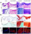
Histological analysis of the osteochondral defect repair at week 12 after hydrogel implantation. (A) H&E staining; (B) Safranin O staining; (C) Immunohistochemical staining of collagen II (red). Cell nuclei are stained with DAPI (blue). Scale bar, 100 µm.
Figure 7
Figure 7
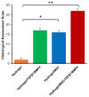
Histological assessment scale for repaired osteochondral defects. Data are presented as mean ± SD (n = 4). * p < 0.05, ** p < 0.01.

Similar articles

Cited by

References

    1. Sukarto A., Yu C., Flynn L.E., Amsden B.G. Co-delivery of adipose-derived stem cells and growth factor-loaded microspheres in RGD-grafted N-methacrylate glycol chitosan gels for focal chondral repair. Biomacromolecules. 2012;13:2490–2502. doi: 10.1021/bm300733n. - DOI - PubMed
    1. Huang Y.Z., Xie H.Q., Silini A., Parolini O., Zhang Y., Deng L., Huang Y.C. Mesenchymal stem/progenitor cells derived from articular cartilage, synovial membrane and synovial fluid for cartilage regeneration: Current status and future perspectives. Stem Cell Rev. Rep. 2017;13:575–586. doi: 10.1007/s12015-017-9753-1. - DOI - PubMed
    1. Makris E.A., Gomoll A.H., Malizos K.N., Hu J.C., Athanasiou K.A. Repair and tissue engineering techniques for articular cartilage. Nat. Rev. Rheumatol. 2015;11:21–34. doi: 10.1038/nrrheum.2014.157. - DOI - PMC - PubMed
    1. Lee H.P., Gu L., Mooney D.J., Levenston M.E., Chaudhuri O. Mechanical confinement regulates cartilage matrix formation by chondrocytes. Nat. Mater. 2017;16:1243–1251. doi: 10.1038/nmat4993. - DOI - PMC - PubMed
    1. Li X., Ding J., Wang J., Zhuang X., Chen X. Biomimetic biphasic scaffolds for osteochondral defect repair. Regen. Biomater. 2015;2:221–228. doi: 10.1093/rb/rbv015. - DOI - PMC - PubMed

LinkOut - more resources