. 2022 Dec 9;10(12):2106.
doi: 10.3390/vaccines10122106.

The Inhibition of SARS-CoV-2 and the Modulation of Inflammatory Responses by the Extract of Lactobacillus sakei Probio65

Affiliations

The Inhibition of SARS-CoV-2 and the Modulation of Inflammatory Responses by the Extract of Lactobacillus sakei Probio65

Irfan A Rather et al. Vaccines (Basel). .

Abstract

In the three years since the first outbreak of COVID-19 in 2019, the SARS-CoV-2 virus has continued to be prevalent in our community. It is believed that the virus will remain present, and be transmitted at a predictable rate, turning endemic. A major challenge that leads to this is the constant yet rapid mutation of the virus, which has rendered vaccination and current treatments less effective. In this study, the Lactobacillus sakei Probio65 extract (P65-CFS) was tested for its safety and efficacy in inhibiting SARS-CoV-2 replication. Viral load quantification by RT-PCR showed that the P65-CFS inhibited SARS-CoV-2 replication in human embryonic kidney (HEK) 293 cells in a dose-dependent manner, with 150 mg/mL being the most effective concentration (60.16% replication inhibition) (p < 0.05). No cytotoxicity was inflicted on the HEK 293 cells, human corneal epithelial (HCE) cells, or human cervical (HeLa) cells, as confirmed by the 3-(4,5-dimethylthiazol-2-yl)-2,5-diphenyl-2H-tetrazolium bromide (MTT) assay. The P65-CFS (150 mg/mL) also reduced 83.40% of reactive oxidizing species (ROS) and extracellular signal-regulated kinases (ERK) phosphorylation in virus-infected cells, both of which function as important biomarkers for the pathogenesis of SARS-CoV-2. Furthermore, inflammatory markers, including interferon-α (IFN-α), IFN-ß, and interleukin-6 (IL-6), were all downregulated by P65-CFS in virus-infected cells as compared to the untreated control (p < 0.05). It was conclusively found that L. sakei Probio65 showed notable therapeutic efficacy in vitro by controlling not only viral multiplication but also pathogenicity; this finding suggests its potential to prevent severe COVID-19 and shorten the duration of infectiousness, thus proving useful as an adjuvant along with the currently available treatments.

Keywords: COVID-19; ERK; Lactobacillus sakei; PROBIO65; ROS; SARS-CoV-2; inflammatory; viral replication.


Conflict of interest statement

The authors declare no conflict of interest.

Figures

Figure 1
Figure 1
Cytotoxicity of the cell-free supernatant of L. sakei Probio65 (P65-CFS) for human cell lines, assessed by the MTT assay. (A) Human embryonic kidney (HEK) 293, (B) human corneal epithelial (HCE), and (C) human cervical (HeLa) cells were used to determine the toxicity of P65-CFS in a range of increasing concentrations. Cells were seeded in 96-well plates at a density of 4 × 10⁴ cells/well, then treated with P65-CFS for 24 h; the percentage of viability was determined in relation to an untreated control. Analysis of each sample was performed in triplicate, and data are represented as the means ± SD of three independent experiments.
Figure 2
Figure 2
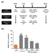
SARS-CoV-2 infection assay and the inhibitive effect of a cell-free supernatant of L. sakei Probio65 (P65-CFS) on viral replication. (A) Schematic of the SARS-CoV-2 infection assay and treatment groups. (B) Viral replication of SARS-CoV-2 in HEK293 quantified by RT-PCR. All wells were treated with an active virus, except for the positive controls, where the viruses were heat-inactivated at 95 °C for 15 min prior to their addition to cells. Cells were treated with P65-CFS (50–150 mg/mL) during the infection and after the infection; meanwhile, the positive control (+ve control) samples were treated in the same manner with the same volume of DMSO and the negative control (−ve control) samples were treated with PBS. Analysis of each sample were performed in triplicate, and data are represented as the means ± SD of three independent experiments. The results are presented as average values with the standard error of means (error bars). Independent t-tests were performed to compare the data with controls, where (###) represents p < 0.001, as compared to the positive control; (**) represents p < 0.01, and (*) represents p < 0.05, as compared to the virus (infected negative control).
Figure 3
Figure 3
Accumulation of cellular reactive oxygen species (ROS) in HEK 293 cells upon infection with SARS-CoV-2. (A) Representative fluorescence images of ROS staining with the fluorogenic dye dichlorofluorescein (H2DC FDA), and (B) intensity quantitation by image J. At the end of the treatment, SARS-CoV-2-infected cells were stained with H2DCFDA for 30 min, washed with PBS, and imaged under a confocal microscope (scale is 500 µm). Analysis of each sample was performed in triplicate, and data are represented as the means ± SD of three independent experiments. Independent T-tests were performed to compare the data with the controls, where (###) represents p < 0.001 as compared to the control; (***) represents p < 0.001 and (**) represents p < 0.01 as compared to the virus (infected, untreated negative control).
Figure 4
Figure 4
Expression of phosphorylated extracellular signal-regulated kinase (p-ERK) in HEK293 cells after infection with SARS-CoV-2 at MOI 0.1. During and after viral adsorption for 2 h, cells were treated with 50, 100, and 150 mg/mL of L. sakei Probio65 cell-free supernatant (P65-CFS). DMSO (0.1%) was used as positive control, while negative control cells were only treated with PBS. Cells were fixed 12 h after infection and double immunofluorescence staining was performed. The expression of p-ERK was visualized using confocal microscopy. Cell nuclei are stained blue, and p-ERK is stained green (scale bar = 5 μm).
Figure 5
Figure 5
Modulation of the expression of inflammatory markers in HEK 293 cells after infection in the presence and absence of L. sakei Probio65 cell-free supernatant (P65-CFS). The expression of (A) IFN-α, (B) IFN-β, and (C) IL-6 were measured using quantitative PCR and normalized to GAPDH. All the experiments were conducted in triplicate and statistical analysis were performed using the t-test. Analysis of each sample was performed in triplicate, and data are represented as the means ± SD of three independent experiments. * p < 0.05 compared with virus-infected cells; ## p < 0.05 compared with the control.

References

    1. Lew L.-C., Hor Y.-Y., Jaafar M.-H., Lau A.-S., Lee B.-K., Chuah L.-O., Yap K.-P., Azlan A., Azzam G., Choi S.-B., et al. Lactobacillus Strains Alleviated Hyperlipidemia and Liver Steatosis in Aging Rats via Activation of AMPK. Int. J. Mol. Sci. 2020;21:5872. doi: 10.3390/ijms21165872. - DOI - PMC - PubMed
    1. Xu Z., Shi L., Wang Y., Zhang J., Huang L., Zhang C., Liu S., Zhao P., Liu H., Zhu L., et al. Pathological findings of COVID-19 associated with acute respiratory distress syndrome. Lancet Respir. Med. 2020;8:420–422. doi: 10.1016/S2213-2600(20)30076-X. - DOI - PMC - PubMed
    1. Sencio V., Machado M.G., Trottein F. The lung-gut axis during viral respiratory infections: The impact of gut dysbiosis on secondary disease outcomes. Mucosal. Immunol. 2021;14:296–304. doi: 10.1038/s41385-020-00361-8. - DOI - PMC - PubMed
    1. Nobel Y.R., Phipps M., Zucker J., Lebwohl B., Wang T.C., Sobieszczyk M.E., Freedberg D.E. Gastrointestinal Symptoms and Coronavirus Disease 2019: A Case-Control Study From the United States. Gastroenterology. 2020;159:373–375.e2. doi: 10.1053/j.gastro.2020.04.017. - DOI - PMC - PubMed
    1. Pouya F., Imani Saber Z., Kerachian M.A. Molecular Aspects of Co-morbidities in COVID-19 Infection. Arch. Bone Jt. Surg. 2020;8:226–230. - PMC - PubMed

LinkOut - more resources