. 2022 Dec 9;14(12):2744.
doi: 10.3390/v14122744.

Cell-Type-Dependent Role for nsP3 Macrodomain ADP-Ribose Binding and Hydrolase Activity during Chikungunya Virus Infection

Affiliations

Cell-Type-Dependent Role for nsP3 Macrodomain ADP-Ribose Binding and Hydrolase Activity during Chikungunya Virus Infection

Taewoo Kim et al. Viruses. .

Abstract

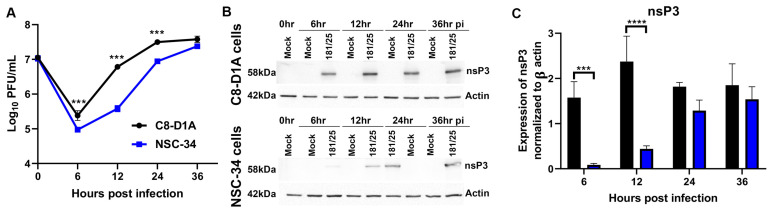
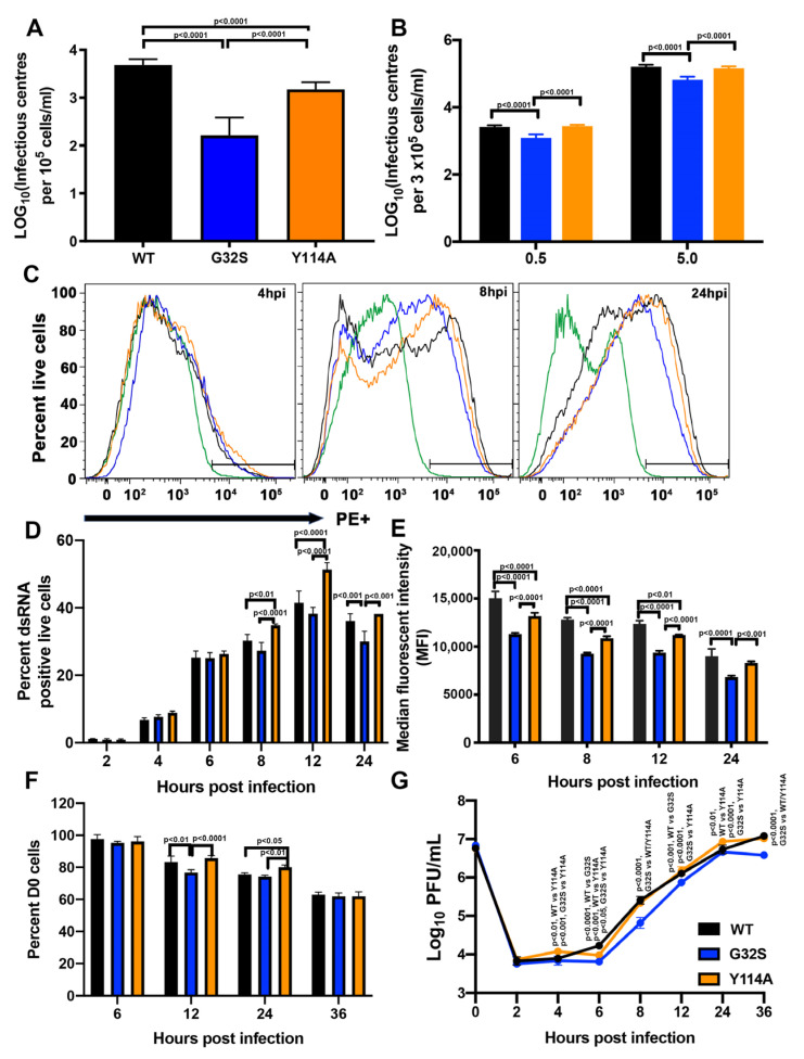
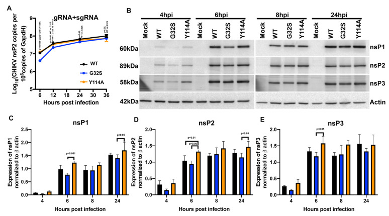
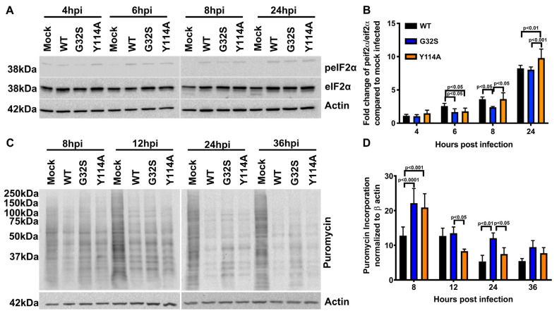
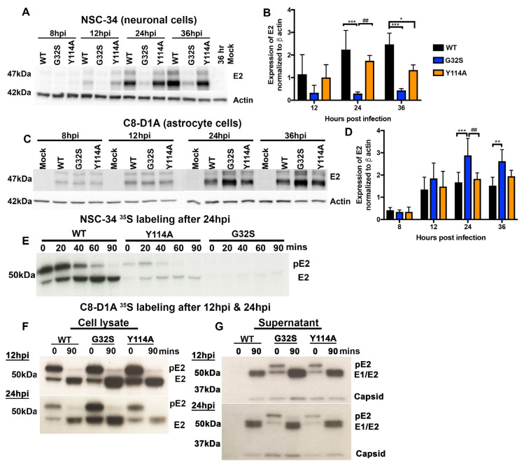
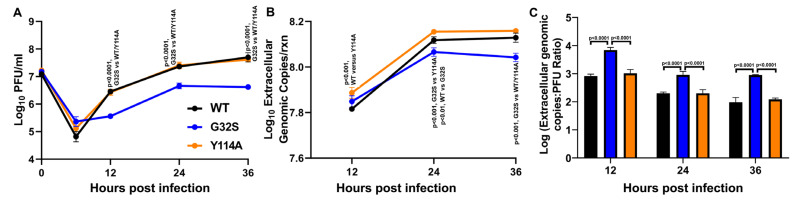
Chikungunya virus (CHIKV) causes outbreaks of rash, arthritis, and fever associated with neurologic complications, where astrocytes are preferentially infected. A determinant of virulence is the macrodomain (MD) of nonstructural protein 3 (nsP3), which binds and removes ADP-ribose (ADPr) from ADP-ribosylated substrates and regulates stress-granule disruption. We compared the replication of CHIKV 181/25 (WT) and MD mutants with decreased ADPr binding and hydrolase (G32S) or increased ADPr binding and decreased hydrolase (Y114A) activities in C8-D1A astrocytic cells and NSC-34 neuronal cells. WT CHIKV replication was initiated more rapidly with earlier nsP synthesis in C8-D1A than in NSC-34 cells. G32S established infection, amplified replication complexes, and induced host-protein synthesis shut-off less efficiently than WT and produced less infectious virus, while Y114A replication was close to WT. However, G32S mutation effects on structural protein synthesis were cell-type-dependent. In NSC-34 cells, E2 synthesis was decreased compared to WT, while in C8-D1A cells synthesis was increased. Excess E2 produced by G32S-infected C8-D1A cells was assembled into virus particles that were less infectious than those from WT or Y114A-infected cells. Because nsP3 recruits ADP-ribosylated RNA-binding proteins in stress granules away from translation-initiation factors into nsP3 granules where the MD hydrolase can remove ADPr, we postulate that suboptimal translation-factor release decreased structural protein synthesis in NSC-34 cells while failure to de-ADP-ribosylate regulatory RNA-binding proteins increased synthesis in C8-D1A cells.

Keywords: ADP ribosylation; alphavirus; astrocytes; chikungunya virus; host-virus interaction; macrodomain; neurons; neurovirulence; replication complexes; viral replication.


Conflict of interest statement

J.X.Y. is an employee of Takeda Pharmaceuticals. D.E.G. is a member of the GlaxoSmithKline Vaccines Research and Development Advisory Board, the Takeda Pharmaceuticals Zika virus Vaccine Data Monitoring Committee, Academia Sinica Genome Research Center Advisory Board, and GreenLight Biosciences Vaccine Development Scientific Advisory Board. T.K., R.A. and L.P. declare no competing interest.

Figures

Figure 1
Figure 1
CHIKV replicates more efficiently in mouse C8-D1A astrocytic cells than NSC-34 neuronal cells. Cells were infected with CHIKV 181/25 at an MOI of 5 as measured by plaque assay on Vero cells. (A) Production of infectious virus by C8-D1A cells and NSC-34 cells. Data are presented as the geometric mean +/− SD from two independent experiments each performed in triplicate. *** p < 0.001 (B) Representative immunoblots for expression of nsP3 6, 12, 24 and 36 h post infection (hpi) of C8-D1A (top) and NSC-34 (bottom) cells. (C) Amounts of nsP3 produced in C8-D1A compared to NSC-34 cells after infection. Blots from three separate experiments were scanned, quantified with Image J, band densities normalized to actin, and ratios compared with Student’s t test. *** p < 0.001; **** p < 0.0001.
Figure 2
Figure 2
Effect of nsP3 MD mutations affecting ADPr-binding and hydrolase activities on initiation of infection and amplification of replication complexes in C8-D1A cells. (A) Infectious center assays for C8-D1A cells (105 cells) electroporated with 10 µg of viral RNA of CHIKV 181/25 (WT) or nsp3 MD mutants G32S and Y114A. Electroporated cells were plated onto subconfluent BHK-21 cells and overlaid with bacto agar to identify virus-producing cells by plaque formation at 48 h. (B) Infectious center assays for C8-D1A cells infected with WT (black) or nsP3 MD mutants G32S (blue) and Y114A (orange) at MOIs of 0.5 and 5, incubated an hour at 4 °C and then 37 °C for 4 h. Cells were trypsinized, serially diluted, plated on Vero cells, and overlaid with bacto agar to assess plaque formation at 48 h. Data are presented as log10 infectious centers per 105 cells and represent the mean ± SD from three independent experiments. (C) Representative histograms showing the formation and amplification of replication complexes in C8-D1A cells infected with WT (black) and nsP3 MD mutants G32S (blue) and Y114A (orange). Cells were infected at an MOI of 5, live/dead stained, fixed, permeabilized, and stained for dsRNA with J2 mAb and analyzed by flow cytometry. Green line indicates uninfected cells. (D) The percentages of live cells positive for dsRNA were quantified and compared at each time point and data presented as a bar graph. (E) Median fluorescent intensities presented as the mean ± SD from two independent experiments. (F) Cells infected with CHIKV WT, G32S and Y114A mutants at an MOI 5 were counted and cell viability was determined by trypan blue exclusion at 6, 12, 24 and 36 h after infection. (G) Supernatant fluids were collected, and infectious virus produced was determined by plaque formation. The data are presented as the mean ± SD from three independent experiments each performed in triplicate. p values indicate the significance of differences between WT and G32S/Y114A or between G32S and Y114A at the indicated time points.
Figure 3
Figure 3
Effect of nsP3 MD function on astrocyte synthesis of viral RNAs and nsPs. C8-D1A cells were infected with CHIKV WT, G32S, and Y114A at an MOI of 5 and analyzed for synthesis of viral RNA and translation of nsPs. (A) Genomic and subgenomic (gRNA + sgRNA; E2) viral RNAs were quantified by qRT-PCR. The mean ± SD of log10 nsP2 or E2 copies/106 copies of Gapdh from three independent experiments are plotted. (B) Representative immunoblots of viral nsP proteins at 4, 6, 8, and 24 h post infection (hpi). Lysates from C8-D1A cells infected with WT or nsP3 MD mutants were immunoblotted with antibodies to nsP1, nsP2, nsP3, and β-actin followed by secondary HRP-labeled anti-IgG were developed using a chemiluminescent detection reagent. Expression levels of nsP1 (C), nsP2 (D), nsP3 (E) normalized to actin were measured by densitometry from three independent experiments and the values plotted as bar graphs of the mean ± SD. p values indicate the significance of differences between WT and G32S/Y114A or between G32S and Y114A at the indicated time points.
Figure 4
Figure 4
Effect of nsP3 MD function on host translational shut-off in CHIKV-infected astrocytes. C8-D1A cells were infected with CHIKV 181/25 (WT) or nsP3 MD mutants G32S and Y114A at an MOI of 5. (A) Representative immunoblots of lysates from cells 4, 6, 8, and 24 h post infection (hpi) probed for phospho-eIF2ɑ, total eIF2ɑ, and β actin. (B) Ratios of phospho-eIF2ɑ to total eIF2ɑ were determined by densitometry and normalized to β actin. Data from 3 experiments are plotted as the mean fold change relative to mock-infected C8-D1A cells. (C) Representative immunoblots of lysates from C8-D1A cells incubated at 8, 12, 24, and 36 h after infection with medium containing puromycin and probed with antibodies to puromycin and β actin. (D) Puromycin incorporation was normalized to β actin and data plotted as a graph. Change in puromycin incorporation is indicated as the average ± SD from three independent experiments. p values indicate the significance of differences between WT and G32S/Y114A or between G32S and Y114A at the indicated time points.
Figure 5
Figure 5
The G32S nsP3 MD mutation that affects both ADPr binding and hydrolase functions differentially affects structural protein synthesis by NSC-34 neuronal and C8-D1A astrocytic cells. NSC-34 and C8-D1A cells were infected with CHIKV WT, G32S, and Y114A at an MOI of 5. Representative immunoblots (A) and densitometry quantification (B) data on lysates from infected NSC-34 cells probed with antibodies to E2 glycoprotein and β-actin at 12, 24, and 36 h from Abraham et al., 2018 [53]. Representative immunoblots (C) and densitometry quantification (D) of lysates from infected C8-D1A cells probed with antibodies to the E2 glycoprotein and β-actin at 8, 12, 24, and 36 h. The averages from four independent experiments +/− SD are plotted as a bar graph. * p < 0.05; ** p < 0.01, *** p < 0.001 compared to WT; ## p < 0.01 G32S compared to Y114A. For analysis of pE2 processing, cells were pulsed with 35S-methionine at 12 hpi and/or 24 hpi and chased for 90 min. Cell lysates were immunoprecipitated with antibody to E2 then subjected to polyacrylamide gel electrophoresis, fixed, dried, exposed to X-ray film, and developed. Representative images from pulse-chase analyses of infected NSC-34 (E) and C8-D1A (F) cells. Pulse-chase analysis of NSC-34 cells was performed once. The image for C8-D1A cells is representative of three independent experiments. (G) PEG-precipitated virions from 12 and 24 h pulse-chase C8-D1A cell supernatant fluids (panel E) were subjected to polyacrylamide gel electrophoresis, fixed, dried, exposed to X-ray film, and developed. Image is representative of three independent experiments.
Figure 6
Figure 6
Infectivity of virions released from C8-D1A cells infected with CHIKV WT, G32S, and Y114A. C8-D1A cells were infected with CHIKV WT and nsP3 MD mutants G32S and Y114A at an MOI 5 measured by plaque assay on Vero cells. (A) Infectious extracellular virus released into the supernatant fluid as measured by plaque formation on Vero cells. (B) Genomic viral RNA (gRNA) released into supernatant fluid as quantified by qRT-PCR and displayed as log10 extracellular genome copies. (C) Ratios of genome copies: PFU for extracellular virions as calculated by converting qRT-PCR genomic copy per reaction data to genome copies per milliliter and dividing by PFU/mL at that time point. Values are an average of 3 biological replicates ± SD. p values indicate the significance of differences between G32S and WT or Y114A at each time point.

References

    1. Weaver S.C., Lecuit M. Chikungunya virus and the global spread of a mosquito-borne disease. N. Engl. J. Med. 2015;372:1231–1239. doi: 10.1056/NEJMra1406035. - DOI - PubMed
    1. Mehta R., Gerardin P., de Brito C.A.A., Soares C.N., Ferreira M.L.B., Solomon T. The neurological complications of chikungunya virus: A systematic review. Rev. Med. Virol. 2018;28:e1978. doi: 10.1002/rmv.1978. - DOI - PMC - PubMed
    1. Das T., Hoarau J.J., Jaffar Bandjee M.C., Maquart M., Gasque P. Multifaceted innate immune responses engaged by astrocytes, microglia and resident dendritic cells against Chikungunya neuroinfection. J. Gen. Virol. 2015;96:294–310. doi: 10.1099/vir.0.071175-0. - DOI - PubMed
    1. Inglis F.M., Lee K.M., Chiu K.B., Purcell O.M., Didier P.J., Russell-Lodrigue K., Weaver S.C., Roy C.J., MacLean A.G. Neuropathogenesis of chikungunya infection: Astrogliosis and innate immune activation. J. Neurovirol. 2016;22:140–148. doi: 10.1007/s13365-015-0378-3. - DOI - PMC - PubMed
    1. Hucke F.I.L., Bestehorn-Willmann M., Bassetto M., Brancale A., Zanetta P., Bugert J.J. CHIKV strains Brazil (wt) and Ross (lab-adapted) differ with regard to cell host range and antiviral sensitivity and show CPE in human glioblastoma cell lines U138 and U251. Virus Genes. 2022;58:188–202. doi: 10.1007/s11262-022-01892-x. - DOI - PMC - PubMed

Publication types

MeSH terms