. 2022 Dec 9;14(12):2753.
doi: 10.3390/v14122753.

Downregulation of the Protein C Signaling System Is Associated with COVID-19 Hypercoagulability-A Single-Cell Transcriptomics Analysis

Affiliations

Downregulation of the Protein C Signaling System Is Associated with COVID-19 Hypercoagulability-A Single-Cell Transcriptomics Analysis

Bruna Rafaela Santos Silva et al. Viruses. .

Abstract

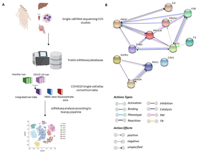
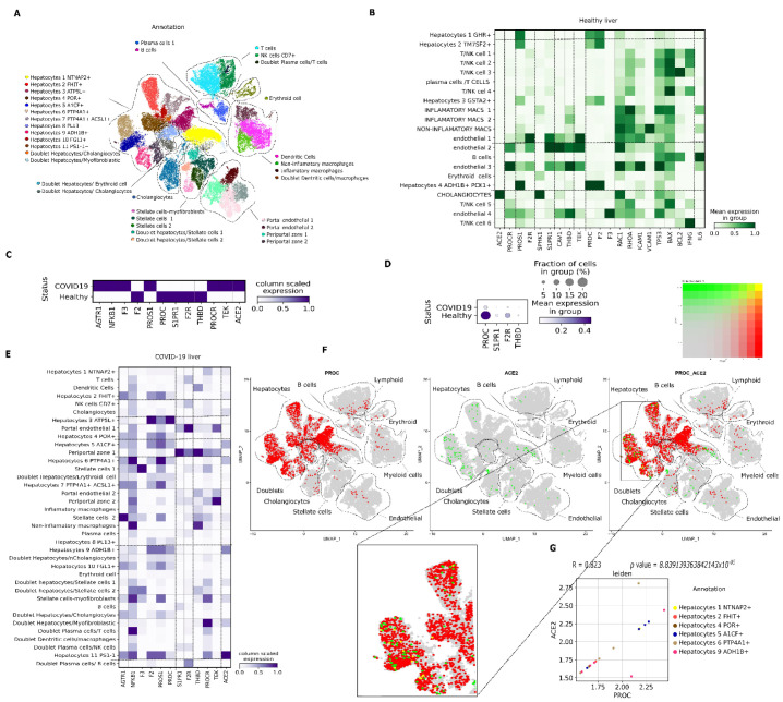
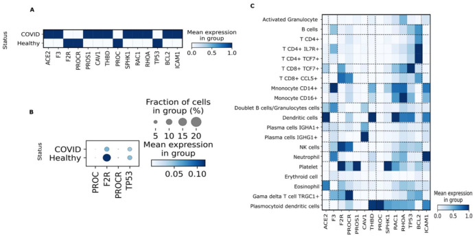
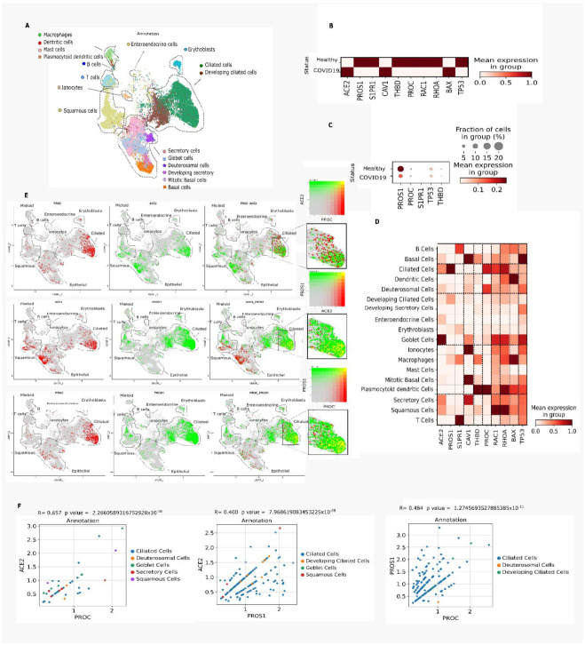
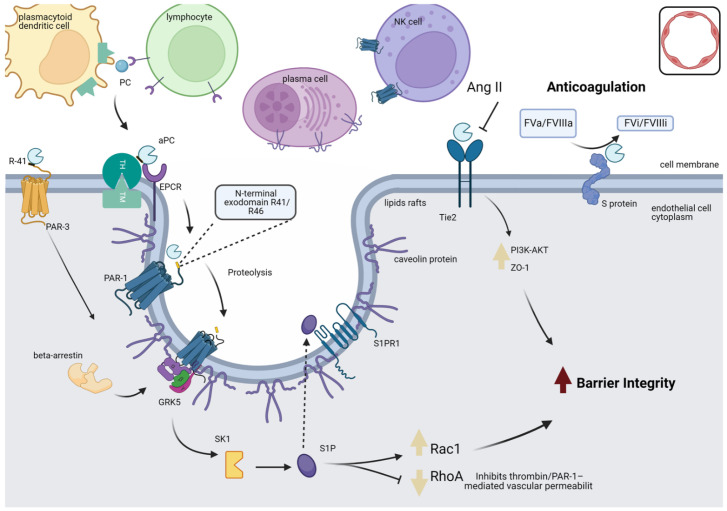
Because of the interface between coagulation and the immune response, it is expected that COVID-19-associated coagulopathy occurs via activated protein C signaling. The objective was to explore putative changes in the expression of the protein C signaling network in the liver, peripheral blood mononuclear cells, and nasal epithelium of patients with COVID-19. Single-cell RNA-sequencing data from patients with COVID-19 and healthy subjects were obtained from the COVID-19 Cell Atlas database. A functional protein-protein interaction network was constructed for the protein C gene. Patients with COVID-19 showed downregulation of protein C and components of the downstream protein C signaling cascade. The percentage of hepatocytes expressing protein C was lower. Part of the liver cell clusters expressing protein C presented increased expression of ACE2. In PBMC, there was increased ACE2, inflammatory, and pro-coagulation transcripts. In the nasal epithelium, PROC, ACE2, and PROS1 were expressed by the ciliated cell cluster, revealing co-expression of ACE-2 with transcripts encoding proteins belonging to the coagulation and immune system interface. Finally, there was upregulation of coagulation factor 3 transcript in the liver and PBMC. Protein C could play a mechanistic role in the hypercoagulability syndrome affecting patients with severe COVID-19.

Keywords: SARS-CoV-2; antigen-presenting cell; blood coagulation disorders; computational biology; endothelial cells.


Conflict of interest statement

The authors declare that they have no conflict of interest.

Figures

Figure 1
Figure 1
Experimental design and protein–protein interaction network. (A). Public data from single-cell RNA sequencing (scRNA-seq) studies on the human tissues (liver, peripheral blood mononuclear cells (PBMC), and nasal epithelia) were retrieved from the website portal www.COVID-19cellatlas.org accessed on 16 April 2022 and Gene Expression Omnibus (GEO) accession numbers GSE171668 and GSE115469. (B). The PROC interactome was retrieved with the string-db data mining toolkit (https://version-11-0.string-db.org/cgi/network.pl?taskId=lu4XDH7F7cOZ, accessed on 16 April 2022). The sources used for functional interactions were neighborhood, experiments, gene fusion, databases, co-occurrence and co-expression, and visualized by the ‘molecular actions’. The network nodes represent proteins and the edges represent protein–protein associations. The edges indicate the known molecular action of a protein node relative to another protein node. PM: post-translational modification; TR: transcriptional regulation.
Figure 2
Figure 2
Expression of genes involved in activated protein C (APC) signaling in the liver from healthy individuals and patients with coronavirus disease 2019 (COVID-19) based on single-cell transcriptomics. (A). Uniform Manifold Approximation and Projection (UMAP) embedding of liver cells via single-cell RNA sequencing (scRNA-Seq) droplet-based single-cell platform, showing single liver parenchymal cells grouped into 36 clusters. (B). Matrix plot showing expression of genes related to APC signaling according to the cell type in a healthy liver. (C). Matrix plot showing expression of genes related to APC signaling in patients with COVID-19 versus healthy individuals. (D). Dot plot representation of downregulated genes in patients with COVID-19. (E). Matrix plot showing expression of genes related to APC signaling according to the cell type in an autopsy liver sample from a patient with COVID-19. (F). Visualization of co-expression (yellow) by color overlap. PROC (red) is barely co-expressed with ACE2 (green) in hepatocyte clusters. (G). Spearman’s correlation of subset of cells that co-expressed PROC and ACE2.
Figure 3
Figure 3
Expression of genes involved in activated protein C (APC) signaling in human peripheral blood mononuclear cells (PBMC) from patients with coronavirus disease 2019 (COVID-19) based on single-cell transcriptomics. (A). Matrix plot showing expression of genes related to APC signaling in patients with COVID-19 versus healthy individuals. (B). Dot plot representation of downregulated genes in patients with COVID-19. (C). Matrix plot showing expression of the main genes related to APC signaling according to cell types.
Figure 4
Figure 4
Expression of genes involved in activated protein C (APC) signaling in nasal epithelial cells from patients with coronavirus disease 2019 (COVID-19) based on single-cell transcriptomics. (A). Uniform manifold approximation and projection (UMAP) embedding of nasal epithelial cells via single-cell RNA sequencing (scRNA-Seq) droplet-based single-cell platform grouped into 18 clusters. (B). Matrix plot showing expression of genes related to APC signaling in patients with COVID-19 versus uninfected individuals. (C). Dot plot representation of downregulated genes in patients with COVID-19. (D). Matrix plot showing expression of the main genes related to APC signaling according to cell types retrieved from Nasal epithelial samples. (E). Visualization of co-expression (yellow) by color overlap. PROC (red) is barely co-expressed with ACE2 (green) in ciliated cell clusters. ACE2 (red) is co-expressed with PROS1 (green) in ciliated cell clusters. PROC (red) is co-expressed with PROS1 (green) in ciliated cell clusters. (F). Spearman’s correlation of a subset of cells that expressed PROC, PROS1, and ACE2.
Figure 5
Figure 5
PC activation and signaling in the endothelial cell. When thrombin binds to TM on endothelial cell surfaces, PC is converted to its active form (APC), and EPCR is necessary for optimal PC activation. The co-localization of PAR-1 and EPCR in caveolin-1-rich membrane microdomains specialized in the cell membrane is essential for PAR-1-mediated cytoprotective APC signaling. Furthermore, the heterogeneous expression of PC activation receptors (EPCR, THBD, CAV-1, and PAR-1) in other cell populations suggests that PC activation might occur dynamically by zymogen PC interacting with ‘migratory’ receptors expressed on the surfaces of infiltrating immune cells in the injury site. The anti-inflammatory and anticoagulant actions of APC occur mainly through PAR-1, which, when activated, propagates pro-inflammatory and anti-inflammatory responses according to the protease that cleaved it. PAR-1 is sensitive to proteolysis at its N-terminal exodomain by multiple plasma proteases, including thrombin, APC, plasmin, factor VIIa, factor Xa, and matrix metalloproteases. Cleavage of PAR-1 at the R41/46 sites at the N-terminal exodomain by APC initiates signaling, promoting structural reorganization of the receptor and coupling to the G protein complex (GRK-5) to phosphorylate the intracellular region of PAR-1 and recruitment of β-arrestin 2, which serves as a framework for cytoprotective signaling. This cascade initiates a series of intracellular signaling pathways, including upregulation of SPHK1 activity and, therefore, S1P production, leading to activation of signaling through its S1PR1 receptor, which stimulates a marked increase in Rac1 activation to perform a protective barrier function by stabilizing the endothelial cytoskeleton and inhibiting RhoA signaling. The Tie2 receptor transmembrane domain can initiate downstream signaling pathways and maintain vascular integrity through direct APC binding, which results in increased ZO-1 phosphorylation of Akt via PI3K and ERK inhibition, contributing to increasing the integrity of the barrier. Protein S cofactor-linked APC can intervene at various points during the systemic response to infection. It exerts an antithrombotic effect by inactivating factors Va and VIIIa, limiting thrombin generation. Finally, PAR-3 cleavage by APC can initiate PAR1-dependent cytoprotective signaling in endothelial cells in the presence of EPCR. APC: activated protein C; EPCR: endothelial protein receptor; ERK: extracellular signal-regulated protein kinase; PAR-1: protease-activated receptor 1; PAR-3: protease-activated receptor 3; PC: protein C; PI3K: phosphoinositide 3-kinase; S1P: sphingosine-1-phosphate; SPHK1: sphingosine kinase-1; TH: thrombin; Tie2: transmembrane receptor tyrosine kinase; TM: thrombomodulin.

References

    1. Iba T., Warkentin T.E., Thachil J., Levi M., Levy J.H. Proposal of the definition for COVID-19-associated coagulopathy. J. Clin. Med. 2021;10:191. doi: 10.3390/jcm10020191. - DOI - PMC - PubMed
    1. Ng K., Wu A., Cheng V., Tang B., Chan C., Yung C., Luk S., Lee T., Chow L., Yuen K. Pulmonary artery thrombosis in a patient with severe acute respiratory syndrome. Postgrad. Med. J. 2005;81:e3. doi: 10.1136/pgmj.2004.030049. - DOI - PMC - PubMed
    1. Xu Z., Shi L., Wang Y., Zhang J., Huang L., Zhang C., Liu S., Zhao P., Liu H., Zhu L., et al. Pathological findings of COVID-19 associated with acute respiratory distress syndrome. Lancet Respir. Med. 2020;8:420–422. doi: 10.1016/S2213-2600(20)30076-X. - DOI - PMC - PubMed
    1. Dolhnikoff M., Duarte-Neto A.N., de Almeida Monteiro R.A., da Silva L.F.F., de Oliveira E.P., Saldiva P.H.N., Mauad T., Negri E.M. Pathological evidence of pulmonary thrombotic phenomena in severe COVID-19. J. Thromb. Haemost. 2020;18:1517–1519. doi: 10.1111/jth.14844. - DOI - PMC - PubMed
    1. Pestka S.B. Old drug, new Trick? The rationale for the treatment of COVID-19 with activated protein C. Med. Hypotheses. 2021;149:110537. doi: 10.1016/j.mehy.2021.110537. - DOI - PMC - PubMed

Publication types