Review
. 2022 Dec 16;14(12):2811.
doi: 10.3390/v14122811.

Norovirus: An Overview of Virology and Preventative Measures

Affiliations
Review

Norovirus: An Overview of Virology and Preventative Measures

Natalie Winder et al. Viruses. .

Abstract

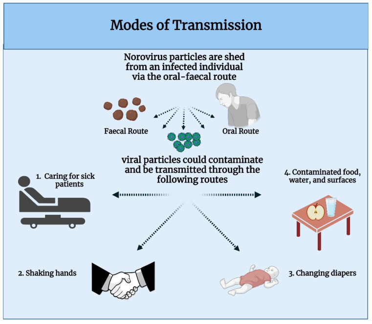
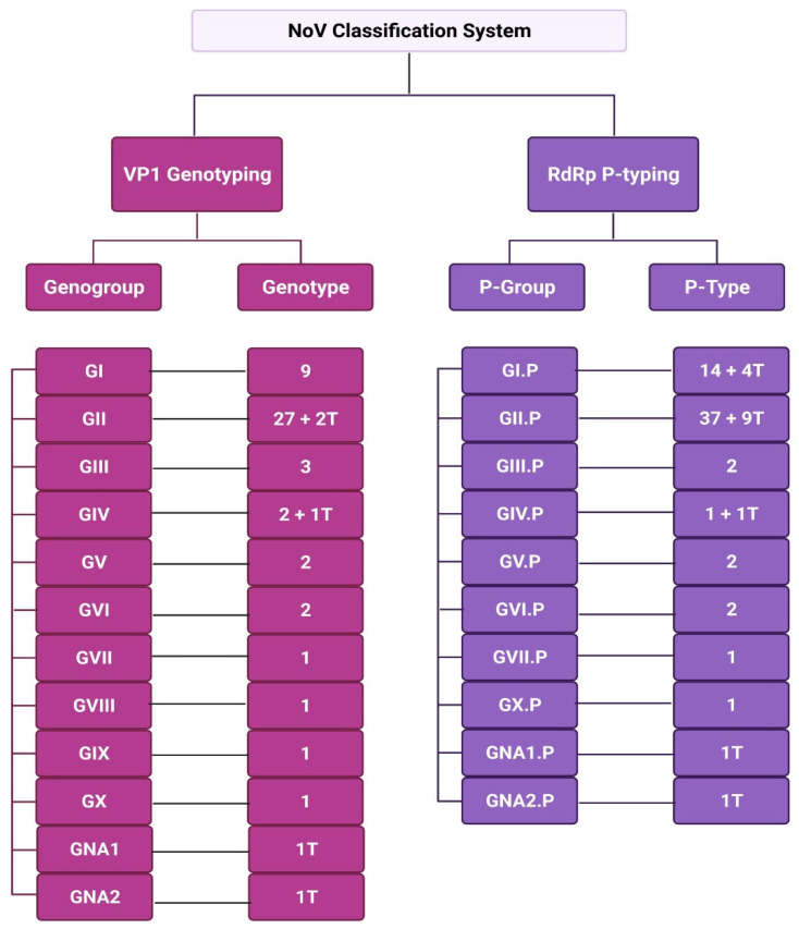
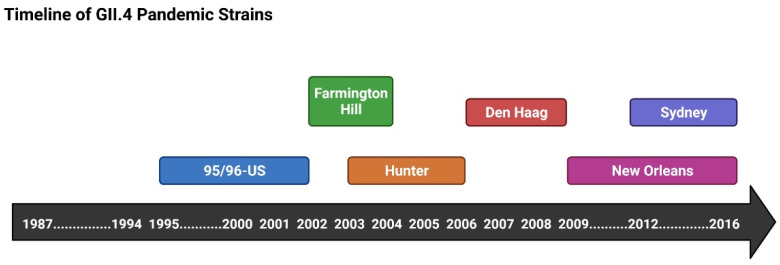
Norovirus (NoV) is an enteric non-enveloped virus which is the leading cause of gastroenteritis across all age groups. It is responsible for around 200,000 deaths annually and outbreaks are common in small communities such as educational and care facilities. 40% of all NoV outbreaks occur in long-term and acute-care facilities, forming the majority of outbreaks. Nosocomial settings set ideal environments for ease of transmission, especially due to the presence of immunocompromised groups. It is estimated to cost global economies around £48 billion a year, making it a global issue. NoV is transmitted via the faecal-oral route and infection with it results in asymptomatic cases or gastrointestinal disease. It has high mutational rates and this allows for new variants to emerge and be more resistant. The classification system available divides NoV into 10 genogroups and 49 genotypes based on whole amino acid sequencing of VP1 capsid protein and partial sequencing of RdRp, respectively. The most predominant genotypes which cause gastroenteritis in humans include GI.1 and GII.4, where GII.4 is responsible for more extreme clinical implications such as hospitalisation. In addition, GII.4 has been responsible for 6 pandemic strains, the last of which is the GII.4 Sydney (2012) variant. In recent years, the successful cultivation of HuNoV was reported in stem cell-derived human intestinal enteroids (HIEs), which promises to assist in giving a deeper understanding of its underlying mechanisms of infection and the development of more personalized control measures. There are no specific control measures against NoV, therefore common practices are used against it such as hand washing. No vaccine is available, but the HIL-214 candidate passed clinical phase 2b and shows promise.

Keywords: classification; control measures; epidemiology; genome; genotype; norovirus; outbreak prevention; pandemics and transmission.


Conflict of interest statement

The authors declare no conflict of interest.

Figures

Figure 1
Figure 1
NoV Genome Organisation and Translated Proteins. NoV is divided into ORF1-ORF4, where ORF4 is only present in MNV-1. ORF1 encodes for a structural polyprotein, which when cleaved by viral protease gives rise to 6 non-structural proteins. ORF2 encodes for major capsid protein VP1 and ORF2 encodes for minor capsid protein VP2, which both form the viral capsid. VF1 encoded for by ORF4 in MNV-1 has been shown to assist in enhancing viral infectivity. Made using Biorender.com, accessed on 18 October 2022.
Figure 2
Figure 2
NoV Viral Particle Structure. NoV capsid is formed of 180 VP1 proteins assembled into 90 dimers. The P2 domain of VP1 is the most surface exposed part of the capsid and is highly variable. This interface is thought to be the one which interacts with HBGAs. Made using Biorender.com, accessed on 18 October 2022.
Figure 3
Figure 3
HBGA and Receptor Engagement with MNV-1. HBGA act as attachment factors for MNV-1, where it improves infectivity by concentrating MNV-1 on the membrane. Recently, CD300lf, a protein belonging to the CD300 family, was identified as receptor which interacts on the surface of murine macrophages with MNV-1. It belongs to a family of cell death receptors and it has proven to be essential for viral ability to infect and replicate. Interaction with CD300lf results in the initiation of viral uptake via endocytosis, which is followed by uncoating and viral genome release. Made using Biorender.com, accessed on 18 October 2022.
Figure 4
Figure 4
NoV RNA replication model. NoV genome is a positive sense RNA strand, which is translated to give rise to NoV proteins. The replication of the positive RNA strand occurs through two initiation steps. De novo initiation occurs at the 3′ of viral genome resulting in the formation of an antisense RNA strand (-) using RNA-dependent RNA-polymerase (RdRp) NS7. This antisense strand is used as a template for the formation of a sense through VPg-primed initiation using RdRp again. Resulting genomic and subgenomic viral RNA undergoes encapsidation into virions, which are released from the cell. Made using Biorender.com, accessed on 18 October 2022.
Figure 5
Figure 5
Two models for subgenomic RNA formation. The premature termination model suggests that a termination signal within genomic RNA results in early termination during de novo initiation replication. On the other hand, the internal initiation suggests that a highly conserved RNA-stem loop upstream of ORF2 is responsible for early termination of de novo initiation replication. The short antisense RNA strands produced then undergo VPg-primed initiation to form positive sense subgenomic RNA. Image made in Biorender.com, accessed on 18 October 2022.
Figure 6
Figure 6
Transmission Modes of NoV reported by the CDC. Image made in Biorender.com, accessed on 18 October 2022.
Figure 7
Figure 7
A schematic overview of immune activation to NoV infection. Diagram indicated both Th1 bias immune activation to NoV infection (dominant immune activation), alongside the Th2 activation. Figure made in Biorender.com, accessed on 18 October 2022.
Figure 8
Figure 8
NoV Classification. NoV variants are classified on the basis of whole amino acid sequencing of VP1 capsid protein for assignment into genogroups and genotypes, while partial RdRp sequencing is used for assignment into P-groups and P-types. Currently there are 10 genogroups. Image generated using Biorender.com, accessed on 18 October 2022.
Figure 9
Figure 9
Timeline of GII.4 Pandemic Strains. The first GII.4 pandemic was reported in 1987 and was caused by the 95/96-US variant. Ever since, every 2–4 years a new variant emerged to take over. Since then, five more pandemic strains have emerged, and the Sydney (2012) variant has dominated ever since. Made using Biorender.com, accessed on 18 October 2022.

References

    1. Kapikian A.Z., Wyatt R.G., Dolin R., Thornhill T.S., Kalica A.R., Chanock R.M. Visualization by Immune Electron Microscopy of a 27-nm Particle Associated with Acute Infectious Nonbacterial Gastroenteritis. J. Virol. 1972;10:1075. doi: 10.1128/jvi.10.5.1075-1081.1972. - DOI - PMC - PubMed
    1. Chan M.C., Kwan H.S., Chan P.K. Structure and Genotypes of Noroviruses. Norovirus. 2017;1:51–63.
    1. Lopman B., Armstrong B., Atchison C., Gray J.J. Host, Weather and Virological Factors Drive Norovirus Epidemiology: Time-Series Analysis of Laboratory Surveillance Data in England and Wales. PLoS ONE. 2009;4:6671. doi: 10.1371/journal.pone.0006671. - DOI - PMC - PubMed
    1. Cubitt W.D., McSwiggan D.A., Moore W. Winter vomiting disease caused by calicivirus. J. Clin. Pathol. 1979;32:786–793. doi: 10.1136/jcp.32.8.786. - DOI - PMC - PubMed
    1. Lopman B.A., Reacher M.H., Vipond B., Sarangi J., Brown D.W. Clinical manifestation of norovirus gastroenteritis in health care settings. Clin. Infect. Dis. Off. Publ. IDSA. 2004;39:318–324. doi: 10.1086/421948. - DOI - PubMed

Publication types