. 2022 Oct 20;13(12):1575-1586.
doi: 10.1039/d2md00246a. eCollection 2022 Dec 14.

Revisiting a challenging p53 binding site: a diversity-optimized HEFLib reveals diverse binding modes in T-p53C-Y220C

Affiliations

Revisiting a challenging p53 binding site: a diversity-optimized HEFLib reveals diverse binding modes in T-p53C-Y220C

Jason Stahlecker et al. RSC Med Chem. .

Abstract

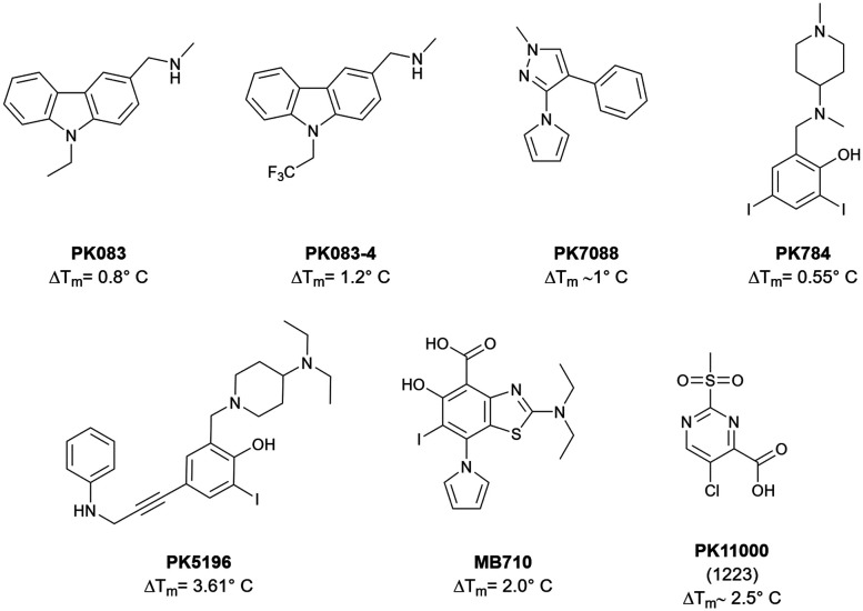
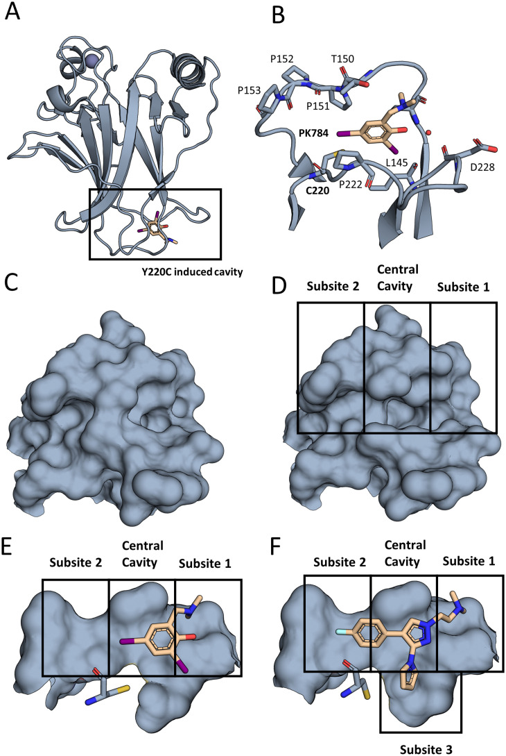
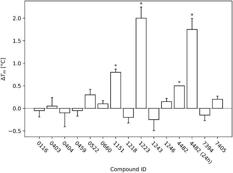
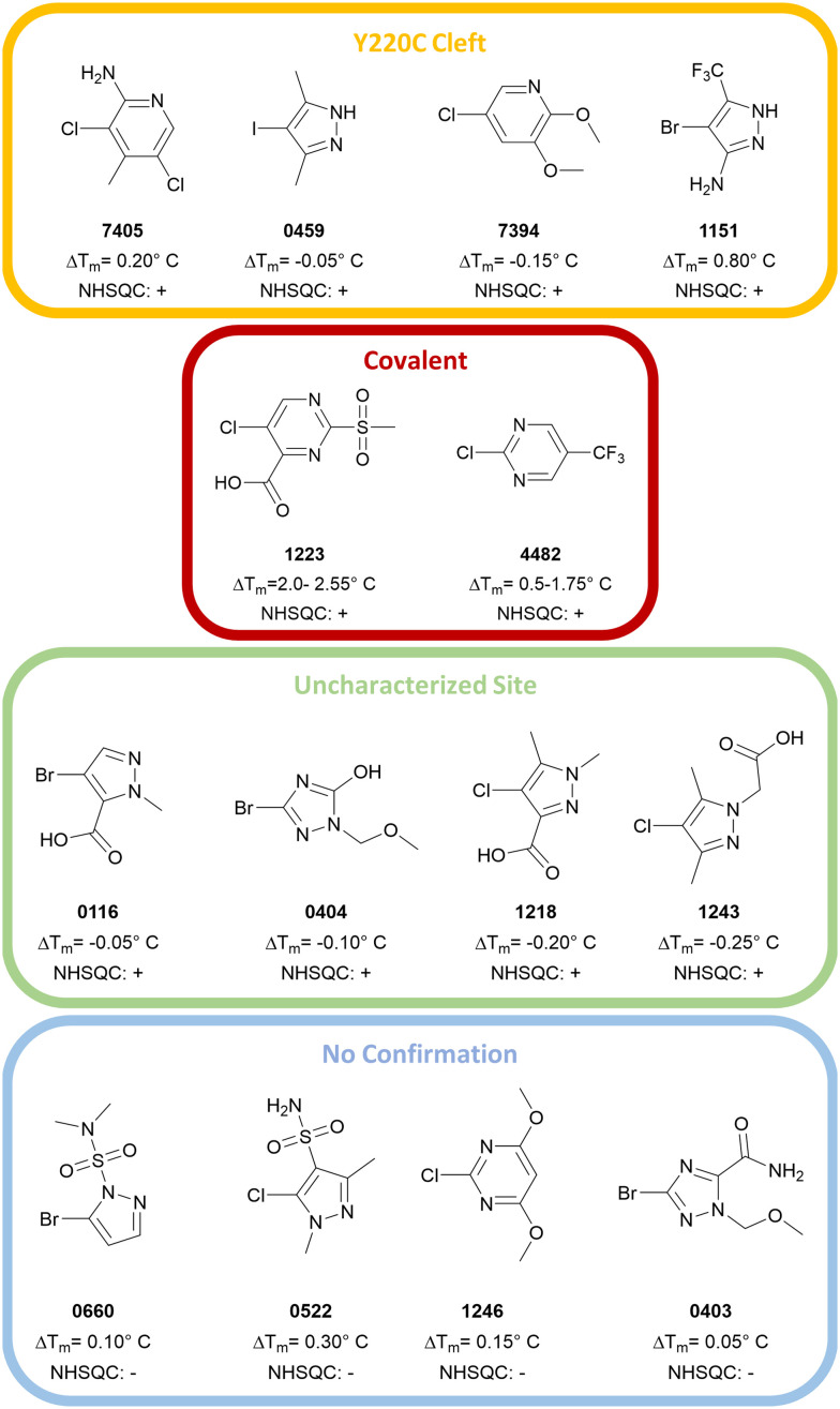
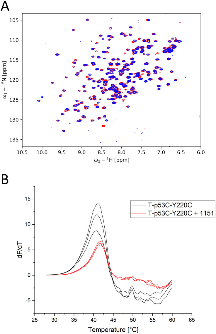
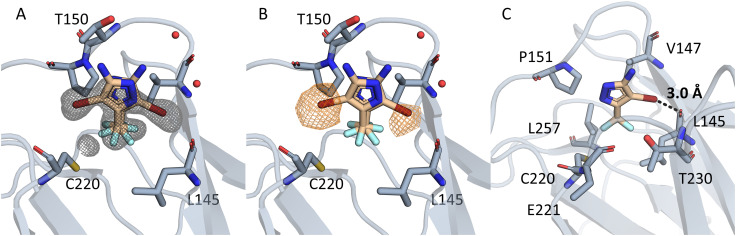
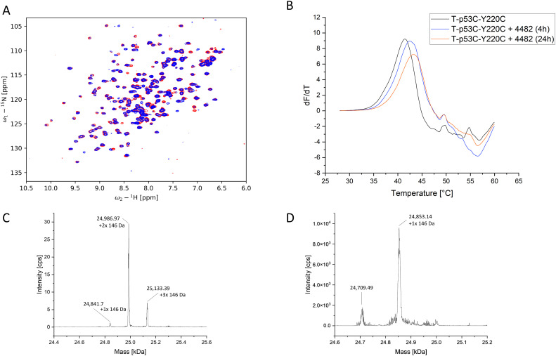
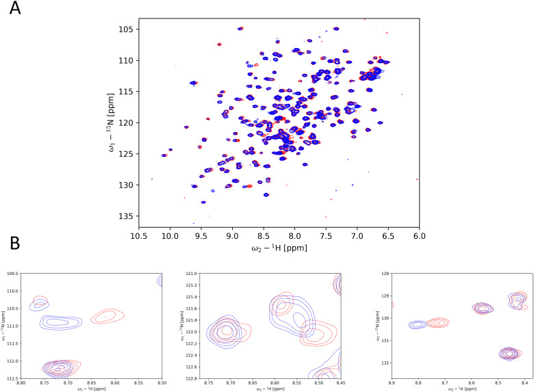
The cellular tumor antigen p53 is a key component in cell cycle control. The mutation Y220C heavily destabilizes the protein thermally but yields a druggable crevice. We have screened the diversity-optimized halogen-enriched fragment library against T-p53C-Y220C with STD-NMR and DSF to identify hits, which we validated by 1H,15N-HSQC NMR. We could identify four hits binding in the Y220C cleft, one hit binding covalently and four hits binding to an uncharacterized binding site. Compound 1151 could be crystallized showing a flip of C220 and thus opening subsite 3. Additionally, 4482 was identified to alkylate cysteines. Data shows that the diversity-optimized HEFLib leads to multiple diverse hits. The identified scaffolds can be used to further optimize interactions with T-p53C-Y220C and increase thermal stability.


Conflict of interest statement

There are no conflicts to declare.

Figures

Fig. 1
Fig. 1. An exemplary selection of published T-p53C-Y220C binders. Compounds PK083 and PK784 occupy the central cavity and subsite 1, while PK5196, PK7088 and MB710 additionally bind subsite 2. The pyrrole group of PK784 and MB710 also enables them to engage to the deep subsite 3. PK11000 showed covalent addition to multiple cysteines.
Fig. 2
Fig. 2. The Y220C mutation leads to a druggable cleft. (A) Overall view of T-p53C-Y220C bound to PK784 (4AGL). The location of the mutation is highlighted, leading to a druggable cleft. (B) PK784 engages in a halogen bond with L145. The other iodine points towards subsite 2 and can be used as vector for ligand growing. (C) Surface representation of Y220 and surrounding amino acids in T-p53C (1UOL). (D) The mutation opens the central cavity, connecting subsite 1 and subsite 2 (4AGL). (E) The lower part of the cleft is not targeted as C220 points towards the compound. (F) Upon binding of an electron rich group, e.g. pyrrole in PK7242, the cysteine is displaced enlarging subsite 3 and enabling targeting (3ZME).
Fig. 3
Fig. 3. The DSF screening results of all identified initial hits are displayed. The T-p53C-Y220C concentration was 8 μM and the ligand concentration 1 mM (ratio 1 : 125). The DSF runs were started after 30 min of incubation. All temperature changes that were significant (p < 0.05) compared to the reference (T-p53C-Y220C without ligand) of the respective run are marked with an asterisk. During the run of 4482, all three measurements showed exactly the same difference to the reference and therefore no error bar is displayed. Additionally, for 4482 another run with 24 h incubation is displayed, showing time dependent increase in stabilization.
Fig. 4
Fig. 4. Overview of the hits from the primary screens. Hits confirmed by 1H,15N-HSQC were categorized as either Y220C cleft binders (0459, 1151, 7394, 7405), covalent binders (1223/PK11000, 4482) or binders to an uncharacterized site (0116, 0404, 1218, 1243). The compounds 0660, 0522, 1246 and 0403 could not be confirmed by 1H,15N-HSQC.
Fig. 5
Fig. 5. (A) 1H,15N-HSQC of T-p53C-Y220C with 1151 at 2 mM (blue) and without addition of a ligand (red). Multiple peak shifts can be observed, all of which indicate Y220C cleft binding. (B) First derivative of the melting curves of T-p53C-Y220C and T-p53C-Y220C with 1151 of the initial screen (30 min incubation).
Fig. 6
Fig. 6. (A) Electron density is displayed in grey around the compound 1151 as an unbiased omit map contoured at 3σ. In chain A two binding poses can be seen. In both poses the CF3 group points towards C220 and engages in hydrophobic contacts. (B) The unbiased anomalous difference map is contoured at 4.5σ in orange. The signal shows that the bromine is located in two positions, but not towards C220. (C) The halogen engages in a halogen bond, with a distance of 3.0 Å and an angle of 153.4°. Residues in 4.5Å around the CF3 group are displayed.
Fig. 7
Fig. 7. (A) Flowchart of the effects on adduct-formation energy (ΔE) and Vmax by systematically adding or removing substituents based on 1151. The CF3 group has the largest effect on ΔE and Vmax. (B) ESP plot of 1151. (C) ESP plot of 1151 after removing the CF3 group. (D) ESP plot of 1151 after removing the CF3 and NH2 group.
Fig. 8
Fig. 8. (A) 1H,15N-HSQC of T-p53C-Y220C without compound (red) and 2 mM 4482 (blue). Multiple prominent peak shifts can be observed. (B) First derivative of the melting curve of T-p53C-Y220C and T-p53C-Y220C with 4482 after 4 h and 24 h incubation. (C) Deconvoluted ESI-MS spectrum of T-p53C-Y220C (50 μM) with 6.25 mM 4882 after 4 h incubation at 20 °C. The shift of the peaks is about 146 Da, which corresponds to the size of the attached 4482. (D) Deconvoluted ESI-MS spectrum of T-p53C-C124/182/277S (50 μM) with 6.25 mM 4482 after 4 h incubation at 20 °C. The major peak corresponds to the single alkylated protein.
Fig. 9
Fig. 9. Example of peak shifts of an uncharacterized binding site. (A) Overlay of peak shifts shown by compound 0116 at 2 mM (blue) in comparison to reference (no ligand) (red). (B) Typical peak shifts observed in the subgroup of the binders to an uncharacterized binding site.

References

    1. Lane D. P. Nature. 1992;358:15–16. doi: 10.1038/358015a0. - DOI - PubMed
    1. Vogelstein B. Lane D. Levine A. J. Nature. 2000;408:307–310. doi: 10.1038/35042675. - DOI - PubMed
    1. Joerger A. C. Fersht A. R. Annu. Rev. Biochem. 2008;77:557–582. doi: 10.1146/annurev.biochem.77.060806.091238. - DOI - PubMed
    1. Joerger A. C. Fersht A. R. Cold Spring Harbor Perspect. Biol. 2010;2:a000919. - PMC - PubMed
    1. Brown C. J. Lain S. Verma C. S. Fersht A. R. Lane D. P. Nat. Rev. Cancer. 2009;9:862–873. doi: 10.1038/nrc2763. - DOI - PubMed