[Preprint]. 2022 Dec 13:2022.12.12.520032.
doi: 10.1101/2022.12.12.520032.

Live-attenuated pediatric parainfluenza vaccine expressing 6P-stabilized SARS-CoV-2 spike protein is protective against SARS-CoV-2 variants in hamsters

Affiliations

Live-attenuated pediatric parainfluenza vaccine expressing 6P-stabilized SARS-CoV-2 spike protein is protective against SARS-CoV-2 variants in hamsters

Xueqiao Liu et al. bioRxiv. .

Update in

Abstract

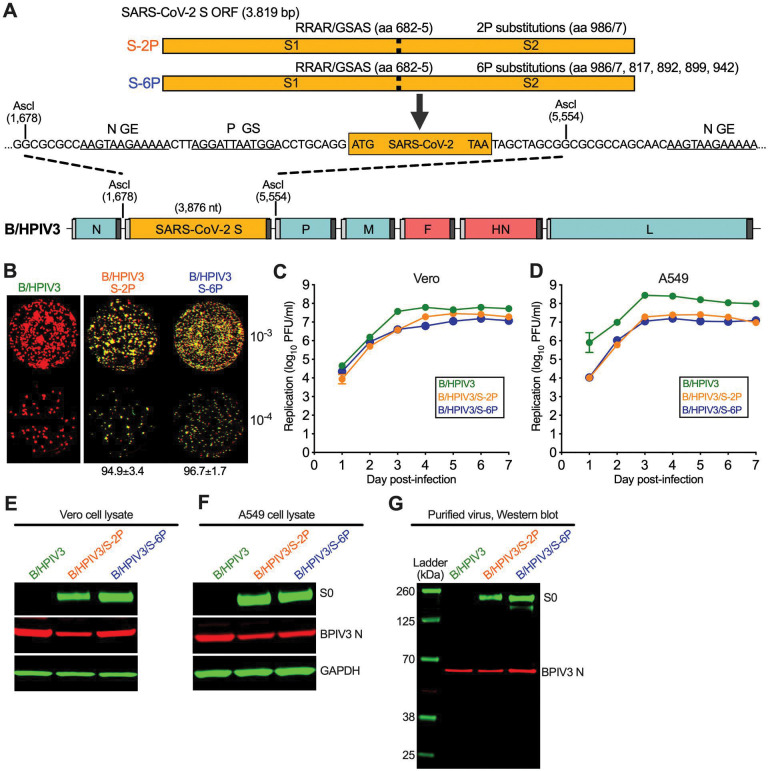
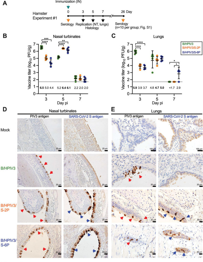
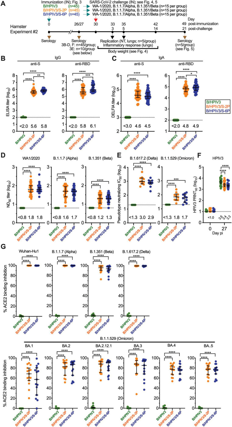
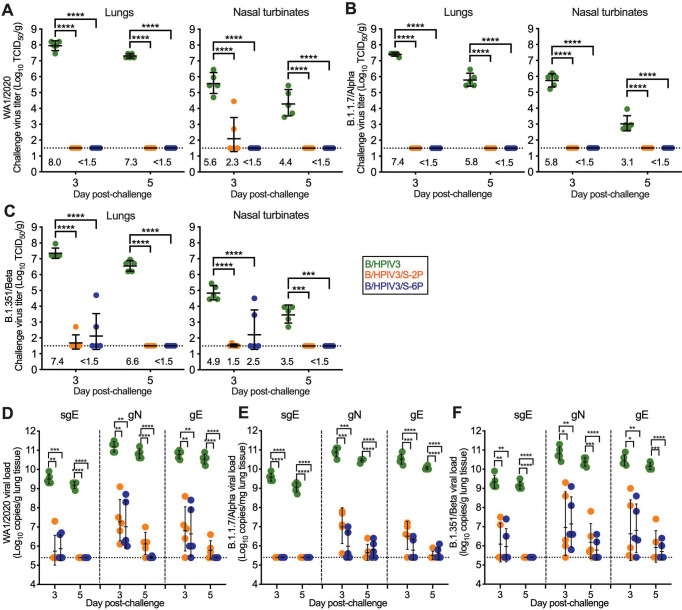
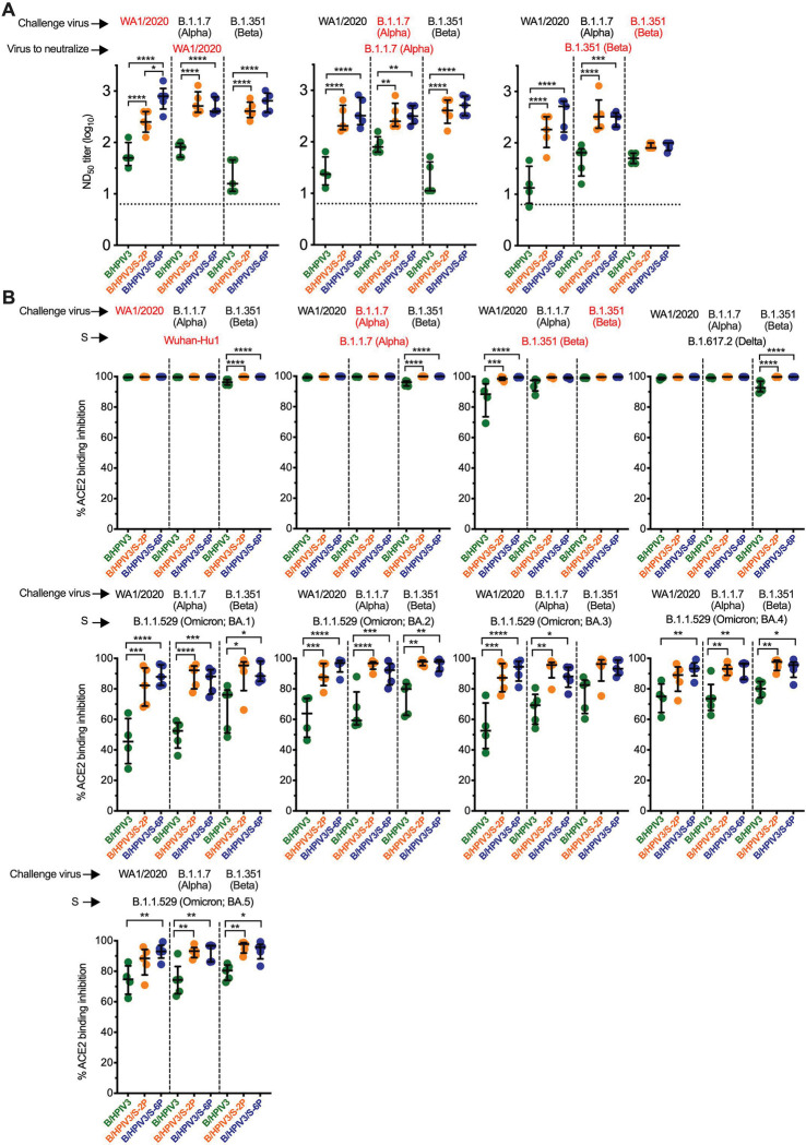
The pediatric live-attenuated bovine/human parainfluenza virus type 3 (B/HPIV3)-vectored vaccine expressing the prefusion-stabilized SARS-CoV-2 spike (S) protein (B/HPIV3/S-2P) was previously evaluated in vitro and in hamsters. To improve its immunogenicity, we generated B/HPIV3/S-6P, expressing S further stabilized with 6 proline mutations (S-6P). Intranasal immunization of hamsters with B/HPIV3/S-6P reproducibly elicited significantly higher serum anti-S IgA/IgG titers than B/HPIV3/S-2P; hamster sera efficiently neutralized variants of concern (VoCs), including Omicron variants. B/HPIV3/S-2P and B/HPIV3/S-6P immunization protected hamsters against weight loss and lung inflammation following SARS-CoV-2 challenge with the vaccine-matched strain WA1/2020 or VoCs B.1.1.7/Alpha or B.1.351/Beta and induced near-sterilizing immunity. Three weeks post-challenge, B/HPIV3/S-2P- and B/HPIV3/S-6P-immunized hamsters exhibited a robust anamnestic serum antibody response with increased neutralizing potency to VoCs, including Omicron sublineages. B/HPIV3/S-6P primed for stronger anamnestic antibody responses after challenge with WA1/2020 than B/HPIV3/S-2P. B/HPIV3/S-6P will be evaluated as an intranasal vaccine to protect infants against both HPIV3 and SARS-CoV-2.

Author summary: SARS-CoV-2 infects and causes disease in all age groups. While injectable SARS-CoV-2 vaccines are effective against severe COVID-19, they do not fully prevent SARS-CoV-2 replication and transmission. This study describes the preclinical comparison in hamsters of B/HPIV3/S-2P and B/HPIV3/S-6P, live-attenuated pediatric vector vaccine candidates expressing the "2P" prefusion stabilized version of the SARS-CoV-2 spike protein, or the further-stabilized "6P" version. B/HPIV3/S-6P induced significantly stronger anti-S serum IgA and IgG responses than B/HPIV3/S-2P. A single intranasal immunization with B/HPIV3/S-6P elicited broad systemic antibody responses in hamsters that efficiently neutralized the vaccine-matched isolate as well as variants of concern, including Omicron. B/HPIV3/S-6P immunization induced near-complete airway protection against the vaccine-matched SARS-CoV-2 isolate as well as two variants. Furthermore, following SARS-CoV-2 challenge, immunized hamsters exhibited strong anamnestic serum antibody responses. Based on these data, B/HPIV3/S-6P will be further evaluated in a phase I study.


Conflict of interest statement

DECLARATION OF INTERESTS

U.J.B., C.L., X.L, and C.LN. are inventors on the provisional patent application number 63/180,534, entitled “Recombinant chimeric bovine/human parainfluenza virus 3 expressing SARS-CoV-2 spike protein and its use”, filed by the United States of America, Department of Health and Human Services.

Figures

Figure 1.
Figure 1.. B/HPIV3 vectors expressing prefusion-stabilized versions of the SARS-CoV-2 S protein
(A) Map of the B/HPIV3 genome with the added SARS-CoV-2 S gene. BPIV3 genes are shown in blue, HPIV3 genes encoding the fusion and hemagglutinin-neuraminidase proteins are in red, and the S gene is in orange. Each gene, including the SARS-CoV-2 S gene, begins and ends with PIV3 gene start (GS) and gene end (GE) transcription signals (light and dark grey bars, respectively). The S gene encodes a prefusion-stabilized uncleaved (S-2P) version of the S protein, or a further stabilized version (S-6P) with 6 proline substitutions [6P; [21]], and was inserted into an AscI site between the BPIV3 N and P genes [17]. The stabilizing proline substitutions and four aa substitutions that ablate the furin cleavage site (RRAR to GSAS, aa 682–685) in the S-2P and S-6P proteins are indicated. (B) Stability of SARS-CoV-2 expression, analyzed by dual-staining immunoplaque assay. Virus stocks were titrated on Vero cells and analyzed by dual-staining immunoplaque assay essentially as described [17], using a goat hyperimmune antiserum against a recombinantly-expressed secreted form of S-2P protein and a rabbit hyperimmune antiserum against HPIV3 virions. HPIV3- and SARS-CoV-2 S-specific staining was pseudocolored in red and green, respectively; dual staining appeared as yellow. The percentage (± standard deviation) of yellow plaques is indicated at the bottom. (C, D) Multicycle replication of B/HPIV3 vectors on Vero and human lung epithelial A549 cells. Cells in 6-well plates were infected in triplicate with indicated viruses at a multiplicity of infection (MOI) of 0.01 PFU per cell and incubated at 32°C for a total of 7 days. At 24 h intervals, aliquots of culture medium were collected and flash-frozen for subsequent immunoplaque titration on Vero cells. (E, F, G) Viral proteins in infected cell lysates (E, F) and purified virions (G). Vero (E) or A549 (F) cells in 6-well plates were infected with B/HPIV3, B/HPIV3/S-2P or B/HPIV3/S-6P at an MOI of 1 and incubated at 32°C for 48 h. Cell lysates were prepared and analyzed by SDS-PAGE under denaturing and reducing conditions and Western blotting. The SARS-CoV-2 S protein was detected using the goat hyperimmune serum to S-2P protein, and the BPIV3 N protein was detected by a rabbit hyperimmune serum raised against sucrose-purified HPIV3, followed by immunostaining with infrared fluorophore labeled secondary antibodies and infrared imaging. Immunostaining for glyceraldehyde-3-phosphate dehydrogenase (GAPDH) is shown as a loading control. Images were acquired and analyzed using Image Studio software (LiCor) and are representatives of three independent experiments. (G) Vero-grown virus preparations of B/HPIV3, B/HPIV3/S-2P, and B/HPIV3/S-6P were purified by centrifugation through 30/60% sucrose gradients, and gently pelleted by centrifugation to remove sucrose. One μg of protein per lane was used for SDS-PAGE and Western blotting as described above. Ladder, molecular size marker.
Figure 2.
Figure 2.. Replication of B/HPIV3, B/HPIV3/S-2P, and B/HPIV3/S-6P in hamsters
(A) In Experiment #1, six-week-old golden Syrian hamsters in groups of 28 were inoculated intranasally with 5 log10 PFU of the indicated viruses. On days 3, 5, and 7, five animals per group were sacrificed and the viral titers in the nasal turbinates (NT) (B) and lungs (C) were determined by dual-staining immunoplaque assay. Titers from individual animals are represented by symbols, and geometric mean titers (GMT) and standard deviations are shown by lines. GMT values are indicated below the dotted line; the maximum mean peak titer irrespective of day for each group is in bold. The limit of detection (LOD) was 50 PFU/g of tissue (dotted line). *=P<0.05; **=P<0.01; ***=P<0.001; ****=P<0.0001 (Two-way ANOVA with Tukey multiple comparisons). (D, E) NT (D) and lung tissues (E) obtained on day 5 (n=2 animals per group and n=1 uninfected control animal) were processed for immunohistochemistry. Serial sections were immunostained for HPIV3 and SARS-CoV-2 antigen using hyperimmune antisera raised against HPIV3 virions and a secreted form of the S-2P protein, respectively. Areas with bronchial epithelial cells positive for HPIV3 and SARS-CoV-2 S antigens are marked by red and blue arrowheads, respectively (20 μm or 100 μm size bars are shown in the bottom right corners).
Figure 3.
Figure 3.. Immunogenicity of B/HPIV3, B/HPIV3/S-2P, and B/HPIV3/S-6P in hamsters
(A) In Experiment #2, six-week-old golden Syrian hamsters in groups of 45 were inoculated intranasally with 5 log10 PFU of the indicated viruses. On days 26 or 27, sera were obtained (n=45 per group) to determine IgG ELISA titers to a secreted form of the S-2P protein or to a fragment of the S protein (aa 328–531) containing SARS-CoV-2 receptor-binding domain (RBD) (B). (C) IgA titers to S-2P or the RBD were determined by dissociation-enhanced lanthanide time-resolved fluorescence (DELFIA-TRF) assay. (D) The 50% SARS-CoV-2 neutralizing titers (ND50) were determined on Vero E6 cells in live-virus SARS-CoV-2 neutralization assays performed at BSL3 using the vaccine-matched strain WA1/2020, USA/CA_CDC_5574/2020 (B.1.1.7/Alpha variant), and the USA/MD-HP01542/2021 (B.1.351/Beta variant). (E) Ten sera from each group were randomly selected for BSL2 neutralization assays to determine the 50% inhibitory concentration (IC50) titers to pseudoviruses bearing spike proteins from SARS-CoV-2 B.1.617.2/Delta or B.1.1.529/Omicron. (F) Sera (n=45 per group) were analyzed to determine the 60% plaque reduction neutralization titers (PRNT60) to HPIV3. The detection limits are indicated by dotted lines. (G) ACE2 binding inhibition assay, used as an alternative for a BSL3 live-virus neutralization assay. Heat-inactivated hamster sera were diluted 1:20 and added to duplicate wells of 96-well plates spot-coated with the indicated S proteins. The percent binding inhibition of sulfo-tag labelled ACE2 to S proteins of the indicated SARS-CoV-2 isolates by serum antibodies from immunized hamsters was determined by electrochemiluminescence (for additional antigens, see Fig. S2). Each hamster is represented by a symbol. Medians and interquartile ranges are indicated by lines. *=P<0.05; **=P<0.01; ***=P<0.001; ****=P<0.0001 (One-way ANOVA with Tukey multiple comparisons).
Figure 4.
Figure 4.. Protection of B/HPIV3, B/HPIV3/S-2P, and B/HPIV3/S-6P-immunized hamsters against weight loss and strong induction of inflammatory cytokines after vaccine-matched and heterologous SARS-CoV-2 challenge
Following immunization of 45 hamsters per group with B/HPIV3, B/HPIV3/S-2P, and B/HPIV3/S-6P in Experiment #2 (see Fig. 3A for schematic), groups were randomly subdivided into challenge groups of 15 animals. On day 30 after immunization, animals were challenged intranasally with 4.5 log10 TCID50 per animal of the vaccine-matched SARS-CoV-2 strain WA1/2020 (A, D), or representatives of the B.1.1.7/Alpha (B, E), or B.1.351/Beta (C, F) variants. (A-C) Weight changes from day −1 to day 16 post-challenge (pc), expressed as mean % body weight relative to the day 0 time point (n=15 hamsters per group from day −1 to day 3, n=10 hamsters per group on days 4 and 5, and 5 animals from day 6 through 16; in the B/HPIV3 empty-vector control/WA1/2020 challenge group, one animal reached the humane study endpoint of 25% weight loss and was euthanized on day 9; standard deviation of the mean is shown). *=P<0.05; **=P<0.01; ***=P<0.001; ****=P<0.0001 (Mixed-effects analysis with Tukey multiple comparisons). (D-F) Expression of inflammatory cytokines in lung tissues on days 3 and 5 post-challenge. Five animals per group were euthanized on indicated days, and tissues were collected. Total RNA was extracted from lung homogenates and cDNA was synthesized from 350 ng of RNA and analyzed by hamster-specific TaqMan assays. Relative gene expression of C-X-C motif chemokine ligand 10 (CXCL10) and of myxovirus resistance protein 2 (Mx2), a type 1 IFN-inducible antiviral gene, and interferon lambda (IFN-L) compared to the mean level of expression of unimmunized, unchallenged controls (dashed line). qPCR results were analyzed using the comparative threshold cycle (ΔΔCT) method, normalized to beta-actin. Each hamster is represented by a symbol. The medians with interquartile ranges are shown. *=P<0.05; **=P<0.01; ***=P<0.001 (Two-way ANOVA with Tukey multiple comparisons).
Figure 5.
Figure 5.. Protection of B/HPIV3, B/HPIV3/S-2P, and B/HPIV3/S-6P-immunized hamsters against SARS-CoV-2 challenge virus replication
As described in Figures 3 and 4, on day 30 after immunization with B/HPIV3 (green symbols), B/HPIV3/S-2P (orange symbols), and B/HPIV3/S-6P (blue symbols), 15 hamsters per group were challenged intranasally with 4.5 log10 TCID50 per animal of the vaccine-matched SARS-CoV-2 strain WA1/2020 (A, D), or representatives of the B.1.1.7/Alpha (B, E), or B.1.351/Beta (C, F) variants. On days 3 and 5 post-challenge, five animals per challenge virus group were euthanized, and SARS-CoV-2 challenge virus titers were determined in homogenates from lungs (A-C, left panels) and NT (right panels). GMTs are indicated above the x axes. (D-F) SARS-CoV-2 lung viral loads after challenge, expressed in log10 genome copies per g. To detect viral subgenomic E mRNA (sgE) RNA, indicative of replicating challenge virus, or genomic N (gN) or E (gE) RNA, indicative of the presence of SARS-CoV-2 challenge virus, cDNA was synthesized using total RNA extracted from lung homogenates as described above, and TaqMan qPCRs were performed (n=5 animals per time point). The GMTs and standard deviations are shown. *=P<0.05; **=P<0.01; ***=P<0.001; ****=P<0.0001 (Two-way ANOVA with Tukey multiple comparisons).
Figure 6.
Figure 6.. Serum antibody responses in B/HPIV3, B/HPIV3/S-2P, and B/HPIV3/S-6P-immunized hamsters three weeks after challenge with SARS-CoV-2 WA1/2020 or VoCs
As shown in Figure 3A, on day 30 after immunization with B/HPIV3 (green symbols), B/HPIV3/S-2P (orange symbols), and B/HPIV3/S-6P (blue symbols), hamsters were randomly assigned 3 subgroups, and challenged intranasally with 4.5 log10 TCID50 per animal of the vaccine-matched SARS-CoV-2 strain WA1/2020 or representatives of the B.1.1.7/Alpha or B.1.351/Beta variants. (A) On day 49 (21 days after challenge), sera from 5 animals from each challenge subgroup (shown at the top of the graphs) were collected, and serum neutralizing titers to the SARS-CoV-2 variant indicated in red were determined [WA1/2020 (left graph), B.1.1.7/Alpha (middle graph) or B.1.351/Beta (right graph)]. (B) The same sera were also evaluated in an ACE2 binding inhibition assay, used as an alternative for a BSL3 live-virus neutralization assay. The % ACE2 binding inhibition to ectodomains of S proteins from individual variant (indicated in red) in the presence of serum was calculated relative to ACE2 binding in the absence of serum. Each hamster is represented by a symbol. The median % inhibition and interquartile ranges are shown. The limit of detection is indicated by a dotted line. *=P<0.05; **=P<0.01; ***=P<0.001; ****=P<0.0001 (One-way ANOVA with Sidák’s multiple comparisons).

Similar articles

Cited by

References

    1. Martin B, DeWitt PE, Russell S, Anand A, Bradwell KR, Bremer C, et al. Characteristics, Outcomes, and Severity Risk Factors Associated With SARS-CoV-2 Infection Among Children in the US National COVID Cohort Collaborative. JAMA Netw Open. 2022;5(2):e2143151. Epub 20220201. doi: 10.1001/jamanetworkopen.2021.43151. - DOI - PMC - PubMed
    1. Cui X, Zhao Z, Zhang T, Guo W, Guo W, Zheng J, et al. A systematic review and meta-analysis of children with coronavirus disease 2019 (COVID-19). J Med Virol. 2021;93(2):1057–69. Epub 20200928. doi: 10.1002/jmv.26398. - DOI - PMC - PubMed
    1. Zachariah P, Johnson CL, Halabi KC, Ahn D, Sen AI, Fischer A, et al. Epidemiology, Clinical Features, and Disease Severity in Patients With Coronavirus Disease 2019 (COVID-19) in a Children’s Hospital in New York City, New York. JAMA Pediatr. 2020;174(10):e202430. Epub 20201005. doi: 10.1001/jamapediatrics.2020.2430. - DOI - PMC - PubMed
    1. Anderson EJ, Rouphael NG, Widge AT, Jackson LA, Roberts PC, Makhene M, et al. Safety and Immunogenicity of SARS-CoV-2 mRNA-1273 Vaccine in Older Adults. N Engl J Med. 2020;383(25):2427–38. Epub 20200929. doi: 10.1056/NEJMoa2028436. - DOI - PMC - PubMed
    1. Ewer KJ, Barrett JR, Belij-Rammerstorfer S, Sharpe H, Makinson R, Morter R, et al. T cell and antibody responses induced by a single dose of ChAdOx1 nCoV-19 (AZD1222) vaccine in a phase 1/2 clinical trial. Nat Med. 2021;27(2):270–8. Epub 20201217. doi: 10.1038/s41591-020-01194-5. - DOI - PubMed

Publication types

LinkOut - more resources