Review
. 2022 Nov 16;36(4):233-242.
doi: 10.1055/s-0042-1758205. eCollection 2022 Nov.

Approach to Complex Lower Extremity Reconstruction

Affiliations
Review

Approach to Complex Lower Extremity Reconstruction

David Cholok et al. Semin Plast Surg. .

Abstract

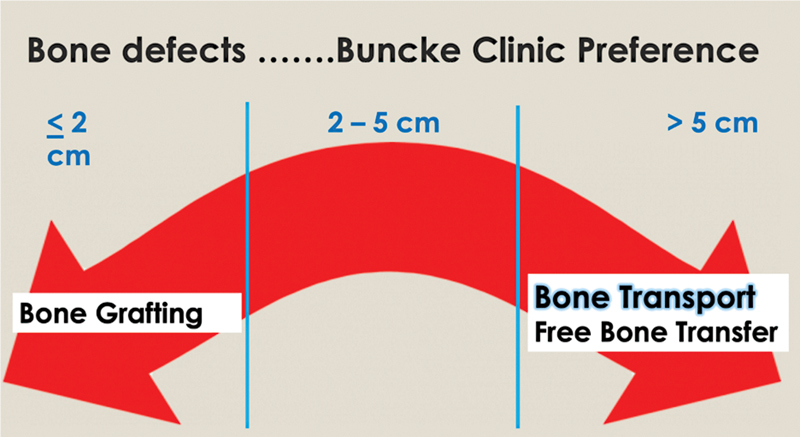
Composite injuries to the lower extremity from etiologies including trauma and infection present a complex dilemma for the reconstructive surgeon, and require multidisciplinary collaboration amongst plastic, vascular, and orthopaedic surgical specialties. Here we present our algorithm for lower-extremity reconstructive management, refined over the last decades to provide an optimized outcome for our patients. Reconstruction is predicated on the establishment of a clean and living wound, where quality of the wound-bed is prioritized over timing to soft-tissue coverage. Once established, soft-tissues and fractures are provisionally stabilized; our preference for definitive coverage is for microvascular free-tissue, due to the paucity of healthy soft-tissue available at the injury, and ability to avoid the zone of injury for microvascular anastomosis. Finally, definitive bony reconstruction is dictated by the length and location of long-bone defect, with a preference to utilize bone transport for defects longer than 5 cm.

Keywords: fractures; lower extremity; negative pressure wound therapy.


Conflict of interest statement

Conflict of Interest None declared.

Figures

Fig. 1
Fig. 1
The Buncke clinic, lower extremity reconstruction pathway.
Fig. 2
Fig. 2
A 57-year-old male with osteomyelitis of trimalleolar right ankle fracture, requiring hardware removal, debridement of soft tissue, and fixation with external fixator spanning the ankle. Pt underwent subsequent free-tissue transfer with latissimus dorsi and split thickness skin grafting. (Panel ( A ) demonstrates initial fixator placement and debridement. Panel ( B ) demonstrates soft-tissue reconstruction). (Source: Reproduced with permission of David W. Lowenberg, MD).
Fig. 3
Fig. 3
A progression of bone transport demonstrating defect, initial corticotomy, process of distraction, and final consolidation ( A ). Example of Hoffman external fixation bone transport frame ( B ). (Source: Reproduced with permission of David W. Lowenberg, MD).
Fig. 4
Fig. 4
A 51-year-old male involved in an All-Terrain Vehicle rollover accident with a closed right subtrochanteric femur fracture and a Type 3A open right tibial pylon fracture with shaft extension. He was treated with immediate rodding of his femur fracture and debridement with spanning external fixation of his tibia fracture. Ten days later his distal tibial was treated at this outside facility with open reduction and internal fixation with seven plates and screws and closure of the wounds. Over the next 7 days there was progressive wound breakdown and dehiscence with eventual exposed hardware and bone. He was then transferred to us for a higher level of care. The patient was taken to the OR with multiple serial debridement and removal of all hardware except the anterior periarticular rim plate which was not infected or exposed. All necrotic bones were removed and the dead space was managed with custom antibiotic beads ( Fig. 2A, B demonstrating initial fixation. 2C, D wound breakdown with exposed hardware). (Source: Reproduced with permission of David W. Lowenberg, MD).
Fig. 5
Fig. 5
With a healthy and “Living Wound” now present, it was then covered with a free rectus abdominus flap, the pre-constructed circular fixator modified to a bone transport frame, and bone transport initiated to fill a 5.5-cm segmental bone defect ( Fig. 3A, B demonstrating circular fixator placement. 3C, D fixator in place, with healed soft-tissue envelope). (Source: Reproduced with permission of David W. Lowenberg, MD).
Fig. 6
Fig. 6
At the conclusion of bone transport the docking site distally is freshened up and augmented with a flap elevation and limited posterior iliac crest bone grafting to hasten bone healing. Regenerate bone maturation is allowed to continue with the patient continuing to ambulate weight bearing as tolerated during this entire time frame. At this point there is now adequate bone healing ( Fig. 4A, B Transport of tibial segment. 4C,D Distal segment docked, with supplemented iliac crest bone grafting.) (Source: Reproduced with permission of David W. Lowenberg, MD).
Fig. 7
Fig. 7
Radiographs 18 months, and 4 years following frame removal. The patient has no pain, is working full time as a ranch manager, and walks 3.5 miles on average per day. Patient is 56-years old and continues to work full time as a ranch manager with no limitations (Fig. 5A, B at 18 months after frame removal. 5C, D at 4 years). (Source: Reproduced with permission of David W. Lowenberg, MD ).
Fig. 8
Fig. 8
Spectrum of most common treatments we generally perform depending on size of defect. Other treatments as listed in the text can also be utilized. (Source: Reproduced with permission of David W. Lowenberg, MD).

References

    1. Nance M L. 2012 National Trauma Data Bank Annual Report. American College of Surgeons. 2012:146.
    1. Georgiadis G M, Behrens F F, Joyce M J, Earle A S, Simmons A L. Open tibial fractures with severe soft-tissue loss. Limb salvage compared with below-the-knee amputation. J Bone Joint Surg Am. 1993;75(10):1431–1441. - PubMed
    1. Lowenberg D W, Buntic R F, Buncke G M, Parrett B M. Long-term results and costs of muscle flap coverage with Ilizarov bone transport in lower limb salvage. J Orthop Trauma. 2013;27(10):576–581. - PubMed
    1. Bosse M J, MacKenzie E J, Kellam J F. An analysis of outcomes of reconstruction or amputation after leg-threatening injuries. N Engl J Med. 2002;347(24):1924–1931. - PubMed
    1. Laughlin R T, Smith K L, Russell R C, Hayes J M. Late functional outcome in patients with tibia fractures covered with free muscle flaps. J Orthop Trauma. 1993;7(02):123–129. - PubMed