Review
. 2022 Nov 16;36(4):260-273.
doi: 10.1055/s-0042-1758691. eCollection 2022 Nov.

Approach to Lymphedema Management

Affiliations
Review

Approach to Lymphedema Management

Walter C Lin et al. Semin Plast Surg. .

Abstract

Millions of people worldwide suffer from lymphedema. In developed nations, lymphedema most commonly stems secondarily from oncologic treatment, but may also result from trauma. More recently, lymphedema has been identified in patients after gender-affirmation phalloplasty reconstruction. Regardless of the etiology, the underlying pathophysiology involves blockage of lymphatic flow, resulting in lymph stasis, thus triggering a cascade of inflammation culminating in fibrosis and adipose deposition. Recent technical advances led to the refinement of physiologic and reductive surgeries-including lymphovenous anastomosis and free functional lymphatic transfer, which collectively encompass a variety of flap procedures including lymph node transfer, lymph channel transfer, and lymphatic system transfer. This article provides a summary of our approach in the assessment and management of the lymphedema patient, including detailed intraoperative photography and imaging, in addition to advanced technical considerations in physiologic reconstruction.

Keywords: FFLT; LYST; VLCT; VLNT; VLVT; free functional lymphatic transfer; lymphedema; lymphovenous anastomosis; lymphovenous bypass; supermicrosurgery; vascularized lymph node transfer.


Conflict of interest statement

Conflict of Interest None declared.

Figures

Fig. 1
Fig. 1
Donor site lymphedema and cellulitis in a radial forearm phalloplasty donor extremity.
Fig. 2
Fig. 2
Lymphatic mapping ( A ) intraoperatively after radial forearm flap harvest ( B ) demonstrating lymphatic disruption and leakage with indocyanine green (ICG) mapping intraoperatively. ( C ) Severe dermal backflow in a separate postoperative patient demonstrates lymphatic obstruction at the interface with the skin graft.
Fig. 3
Fig. 3
Indocyanine green (ICG) lymphatic mapping for preservation at the time of radial forearm flap design.
Fig. 4
Fig. 4
Severe hand cellulitis in lymphedema requiring inpatient intravenous (IV) antibiotics.
Fig. 5
Fig. 5
Feedback loop between fluid stasis, inflammation, and worsened lymphatic function.
Fig. 6
Fig. 6
Indocyanine green (ICG) lymphatic mapping showing linear intact channels along the dorsal hand.
Fig. 7
Fig. 7
Flow proximally shows diffusion and leaking, suggestive of lymphatic vessel damage.
Fig. 8
Fig. 8
Diffusion and leakage of indocyanine green (ICG) demonstrates dermal backflow due to damage and fibrosis of the lymphatic channels, and confirms lymphedema.
Fig. 9
Fig. 9
Lower extremity lymphedema in gynecologic cancer-related lymphedema. Note transition from linear flow to dermal backflow.
Fig. 10
Fig. 10
International Society of Lymphology staging system of disease severity with associated treatment options, including physiologic (lymphovenous anastomosis [LVA], vascularized lymph node transfer [VLNT], free functional lymphatic transfer) and reductive (liposuction, excision) techniques.
Fig. 11
Fig. 11
Preop and 1-month postop demonstration of lymphovenous anastomosis bypassing focal lymphatic blockage. ( A , B ) Preoperative indocyanine green (ICG) lymphatic mapping showing complete blockage and severe dermal backflow at the ankle. ( C , D ) View of lymphovenous anastomosis (LVA) located just inferior to the scabbed wound 1 month postop, with ICG lymphatic mapping showing restoration of lymphatic flow via the LVA into a superficial venule, with decreased dermal backflow.
Fig. 12
Fig. 12
Example of a linear lymphatic transitioning to dermal backflow, demonstrating focally identifiable lymphatic blockage.
Fig. 13
Fig. 13
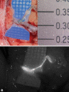
( A ) Comparative size between a penny and 10–0 nylon suture with superfine needle. ( B ) Scaled comparison with a lymphovenous anastomosis. Each blue square indicates 1 mm.
Fig. 14
Fig. 14
Example of ( A ) preoperative lymphedema and ( B ) early improvement 3 weeks after microvascular vascularized lymph node transfer, likely due to direct lymphatic drainage through the flap.
Fig. 15
Fig. 15
Steps during end-to-end lymphovenous anastomosis. ( A ) The lymphatic is identified with isosulfan blue dye, adjacent to a superficial venule. ( B ) The lymphatic is divided and anastomosed to the end of a side branch of the venule. ( C ) Indocyanine green (ICG) confirms patent lymphatic flow without leakage. ( D ) Isosulfan blue dye similarly confirms patency without leakage.
Fig. 16
Fig. 16
End-to-side lymphovenous anastomosis (LVA) demonstrating passage of lymphatic fluid into the venous system. ( A ) Patency viewed with isosulfan blue. ( B ) Patency viewed by indocyanine green (ICG).
Fig. 17
Fig. 17
Lymphatic mapping and indocyanine green (ICG) tracing showing abrupt transition to dermal backflow, indicating an ideal candidate site for lymphovenous anastomosis (LVA).
Fig. 18
Fig. 18
Side-to-end lymphovenous anastomosis (LVA), from the side of the lymphatic (blue) to the end of the vein.
Fig. 19
Fig. 19
Side-to-side lymphovenous anastomosis (LVA) from the smaller lymphatic to the larger diameter vein.
Fig. 20
Fig. 20
Supraclavicular vascularized lymph node transfer (VLNT) with proximal artery and vein (right), and distal artery and vein (left) with anastomoses to the radial artery and radial vena comitans.
Fig. 21
Fig. 21
Close up of vascularized lymph node transfer (VLNT) proximal artery and vein. The inflow arterial anastomosis is end-to-side to the radial artery. The outflow venous anastomosis is end-to-end to a venous side branch of a radial vena comitans.
Fig. 22
Fig. 22
Close up of vascularized lymph node transfer (VLNT) distal artery and vein. The outflow arterial anastomosis is end-to-side to the radial artery. The inflow venous anastomosis is end to side to a radial vena comitans.

References

    1. Dayan J H, Ly C L, Kataru R P, Mehrara B J. Lymphedema: pathogenesis and novel therapies. Annu Rev Med. 2018;69:263–276. - PubMed
    1. Cormier J N, Askew R L, Mungovan K S, Xing Y, Ross M I, Armer J M. Lymphedema beyond breast cancer: a systematic review and meta-analysis of cancer-related secondary lymphedema. Cancer. 2010;116(22):5138–5149. - PubMed
    1. Gillespie T C, Sayegh H E, Brunelle C L, Daniell K M, Taghian A G. Breast cancer-related lymphedema: risk factors, precautionary measures, and treatments. Gland Surg. 2018;7(04):379–403. - PMC - PubMed
    1. Achouri A, Huchon C, Bats A S, Bensaid C, Nos C, Lécuru F.Complications of lymphadenectomy for gynecologic cancer Eur J Surg Oncol 2013390181–86.(EJSO) - PubMed
    1. Szczesny G, Olszewski W L. The pathomechanism of posttraumatic edema of lower limbs: I. The effect of extravasated blood, bone marrow cells, and bacterial colonization on tissues, lymphatics, and lymph nodes. J Trauma. 2002;52(02):315–322. - PubMed