

Case Reports
doi: 10.1016/j.radcr.2022.11.039.
eCollection 2023 Feb.
A giant parapharyngeal space lipoma
Affiliations
- PMID: 36561546
- PMCID: PMC9763680
- DOI: 10.1016/j.radcr.2022.11.039
Item in Clipboard
Case Reports
A giant parapharyngeal space lipoma
Radiol Case Rep.
.
Abstract
Parapharyngeal space (PPS) lipomas are incredibly uncommon. Prestyloid or poststyloid compartments are the only locations for PPS lipomas. Liposarcoma is a crucial differential to rule out. In order to treat PPS lipomas, the required radiological tests, including magnetic resonance imaging, a biopsy of the lesion if that is available, and lipoma removal surgery are all necessary. We intended to describe a unique giant PPS lipoma that affects both prestyloid and poststyloid compartments.
Keywords: Giant; Lipoma; Parapharyngeal space.
© 2022 The Authors. Published by Elsevier Inc. on behalf of University of Washington.
Figures

(A) B-mode ultrasonography and (B) color-Doppler mode ultrasonography revealed a hypoechoic mass (arrow) with well-demarcated margin on the right submandibular region.

Magnetic resonance imaging revealed that the mass (arrow) was hyperintense on (A) axial T1-weighted imaging and (B) coronal T2-weighted imaging but hypointense on (C) fat-suppressed T2-weighted imaging.
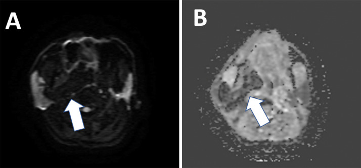
Diffusion-weighted imaging revealed that the diffusivity of the mass (arrow) was restricted.

Axial (A) and coronal (B) fat-suppressed T1-weighted imaging with contrast enhancement revealed that the mass (arrow) did not absorb contrast agent.

Microscopic image showing sheets made of mature adipocytes (H&E staining, magnification ×100 (A), × 400 (B)).
References
-
- Carrau RL, Johnson JT, Myers EN. Management of tumors of the parapharyngeal space. Oncology (Williston Park) 1997;11(5):633–640. discussion 640, 642. PMID: 9159790. - PubMed
Publication types
LinkOut - more resources
Full Text Sources
