

Association Between Coronary Artery Calcium Score and Bone Mineral Density in Type 2 Diabetes Mellitus with Different Visceral Fat Area
- PMID: 36561919
- PMCID: PMC9766512
- DOI: 10.2147/DMSO.S392152
Association Between Coronary Artery Calcium Score and Bone Mineral Density in Type 2 Diabetes Mellitus with Different Visceral Fat Area
Abstract
Purpose: The relationship between coronary artery calcification and bone mineral density (BMD) in T2DM is still unclear. The aim of this study is to analyze the association between coronary artery calcium score (CACs) and BMD in T2DM with different visceral fat area (VFA), and further to explore the clinical significance of CACs in predicting osteoporosis in T2DM patients.
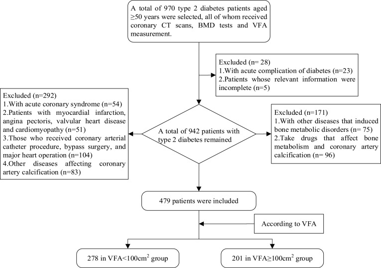
Patients and methods: A total of 479 T2DM patients aged ≥50 years were included. Agatston was applied to calculate CACs to evaluate the degree of coronary artery calcification. Dual-energy X-ray absorptiometry (DXA) was used to measure BMD. According to VFA, all subjects were divided into VFA <100cm2 and VFA ≥100cm2 group. Adjusted regression analysis was performed to analyze the association between CACs and BMD. ROC curve was used to analyze the optimal cut-off value of CACs for screening osteoporosis.
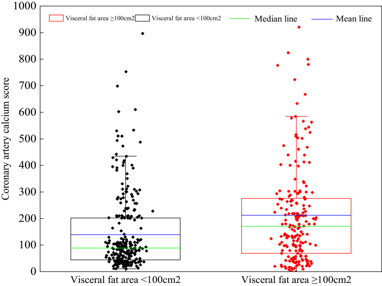
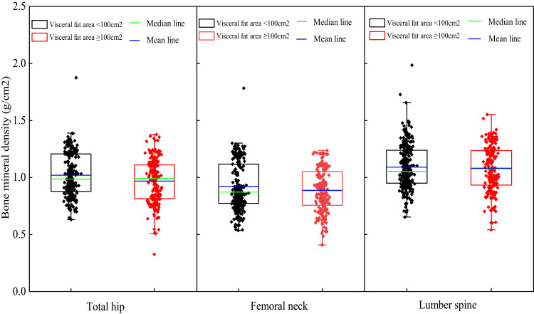
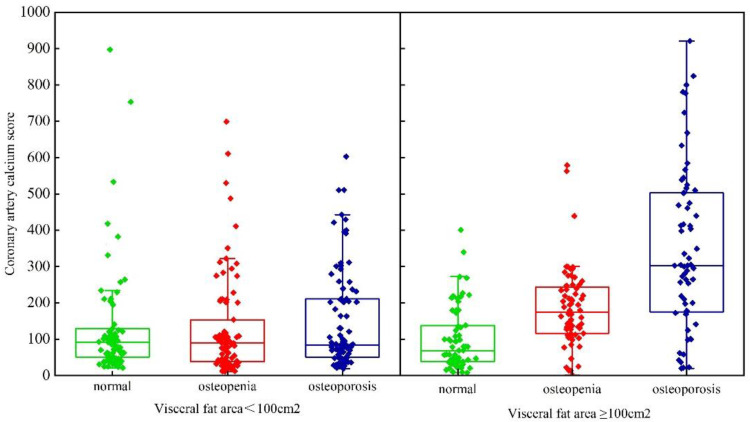
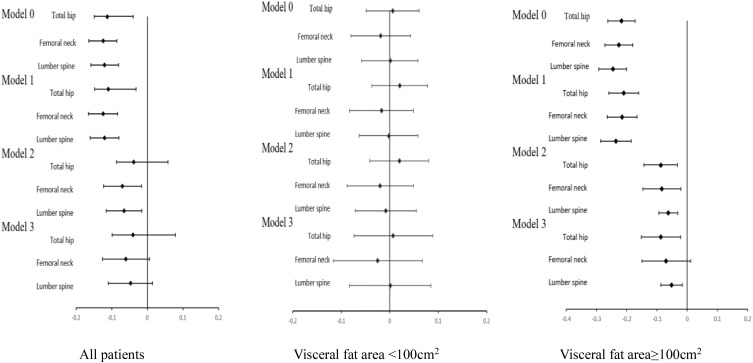
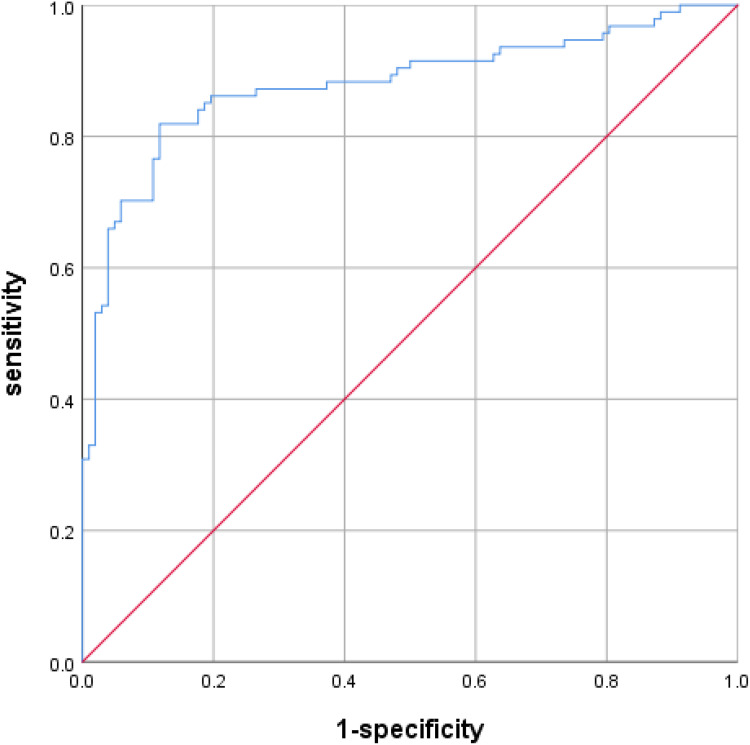
Results: The baseline showed that in VFA ≥100cm2 group, CACs increased significantly than that in VFA <100cm2 group (212.1±195.9 vs 139.3±141.8, p<0.001) and total hip BMD decreased obviously (0.968±0.19 vs 1.021±0.184, p=0.01). After multivariable adjustment, CACs was not significantly associated with BMD in all patients (p>0.05). However, CACs was negatively associated with BMD of total hip and lumbar spine in patients with VFA ≥100cm2 (total hip β=-0.087 p=0.01; lumbar spine β=-0.052 p=0.005), but not VFA <100cm2. ROC curve analysis showed that the optimal cut-off value of CACs for screening osteoporosis was 191.505.
Conclusion: The present study implied that associations between CACs and BMD varied by the visceral fat deposition. It is critical to evaluate the condition of visceral fat accumulation for exploring the complex interplay of coronary artery calcification and BMD in T2DM patients. It may be of some clinical value for CACs in predicting osteoporosis in T2DM with visceral obesity.
Keywords: bone mineral density; coronary artery calcium score; type 2 diabetes mellitus; visceral fat area.
© 2022 Yang et al.
Conflict of interest statement
The authors report no conflicts of interest in this work.
Figures
Similar articles
-
Relationship between body composition and bone mineral density in postmenopausal women with type 2 diabetes mellitus.BMC Musculoskelet Disord. 2022 Oct 3;23(1):893. doi: 10.1186/s12891-022-05814-8. BMC Musculoskelet Disord. 2022. PMID: 36192772 Free PMC article.
-
Sex Differences in the Association Between Bone Mineral Density and Coronary Artery Disease in Patients Referred for Cardiac Computed Tomography.J Clin Densitom. 2021 Jan-Mar;24(1):55-66. doi: 10.1016/j.jocd.2019.09.003. Epub 2019 Sep 24. J Clin Densitom. 2021. PMID: 31668962
-
Differences in association of bone mineral density with coronary artery calcification in men and women: the Rancho Bernardo Study.Menopause. 2005 Nov-Dec;12(6):691-8. doi: 10.1097/01.gme.0000184422.50696.ef. Epub 2005 Nov 8. Menopause. 2005. PMID: 16278612
-
Association between bone mineral density and coronary plaque burden in patients with coronary artery disease: a cross-sectional study using quantitative computed tomography.Coron Artery Dis. 2024 Mar 1;35(2):105-113. doi: 10.1097/MCA.0000000000001316. Epub 2023 Dec 29. Coron Artery Dis. 2024. PMID: 38164995
-
Visceral adiposity and inflammatory bowel disease.Int J Colorectal Dis. 2021 Nov;36(11):2305-2319. doi: 10.1007/s00384-021-03968-w. Epub 2021 Jun 9. Int J Colorectal Dis. 2021. PMID: 34104989 Review.
Cited by
-
Nonlinear association between liver fat content and lumbar bone mineral density in overweight and obese individuals: evidence from a large-scale health screening data in China.Endocrine. 2025 May;88(2):446-456. doi: 10.1007/s12020-025-04168-0. Epub 2025 Jan 27. Endocrine. 2025. PMID: 39869295 Free PMC article.
-
Correlation between visceral fat area and cardiac valve calcification in hemodialysis patients.Front Cardiovasc Med. 2025 Apr 10;12:1574649. doi: 10.3389/fcvm.2025.1574649. eCollection 2025. Front Cardiovasc Med. 2025. PMID: 40276255 Free PMC article.
-
Association between visceral fat and bone mineral density in perimenopausal women.PeerJ. 2025 Feb 13;13:e18957. doi: 10.7717/peerj.18957. eCollection 2025. PeerJ. 2025. PMID: 39959823 Free PMC article.
-
Risk factors for osteoporosis in the elderly and predictive value of age, body mass index, and visceral fat area.Nutr Res Pract. 2025 Jun;19(3):375-385. doi: 10.4162/nrp.2025.19.3.375. Epub 2025 Jan 8. Nutr Res Pract. 2025. PMID: 40496047 Free PMC article.
References
-
- Kurabayashi M. Vascular calcification - pathological mechanism and clinical application - role of vascular smooth muscle cells in vascular calcification. Clin Calcium. 2015;25(5):661–669. - PubMed
LinkOut - more resources
Full Text Sources
