. 2023 Feb;49(2):32.
doi: 10.3892/or.2022.8469. Epub 2022 Dec 23.

Combined inhibition of ACLY and CDK4/6 reduces cancer cell growth and invasion

Affiliations

Combined inhibition of ACLY and CDK4/6 reduces cancer cell growth and invasion

Brian Christopher Velez et al. Oncol Rep. 2023 Feb.

Abstract

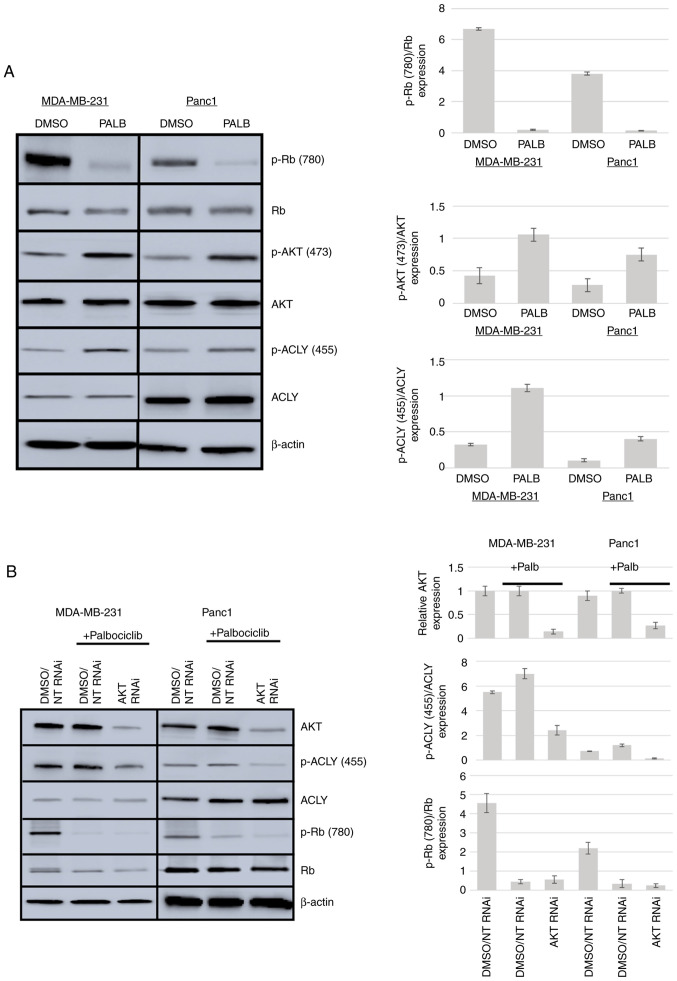
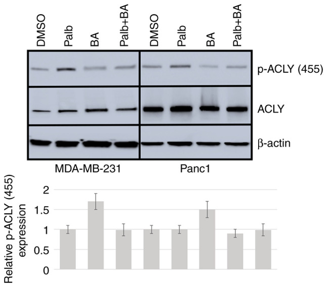
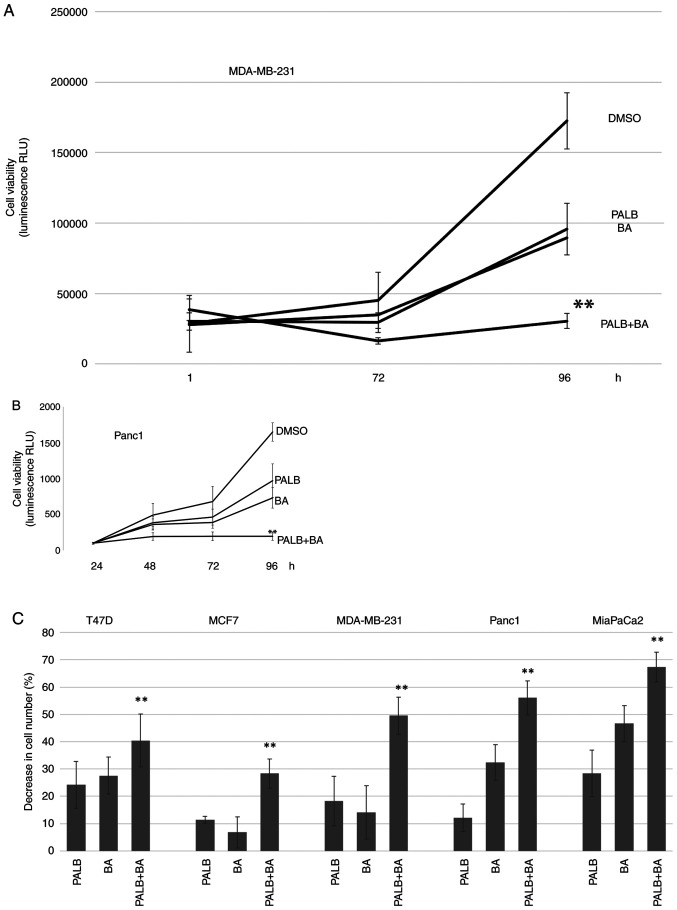
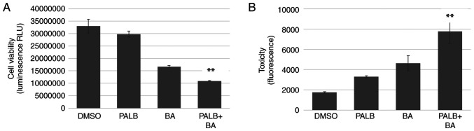
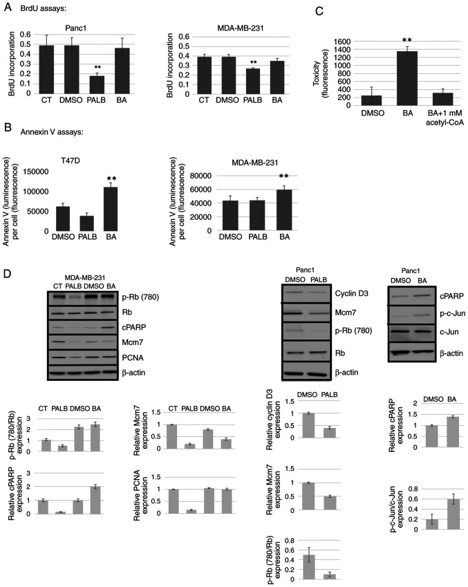
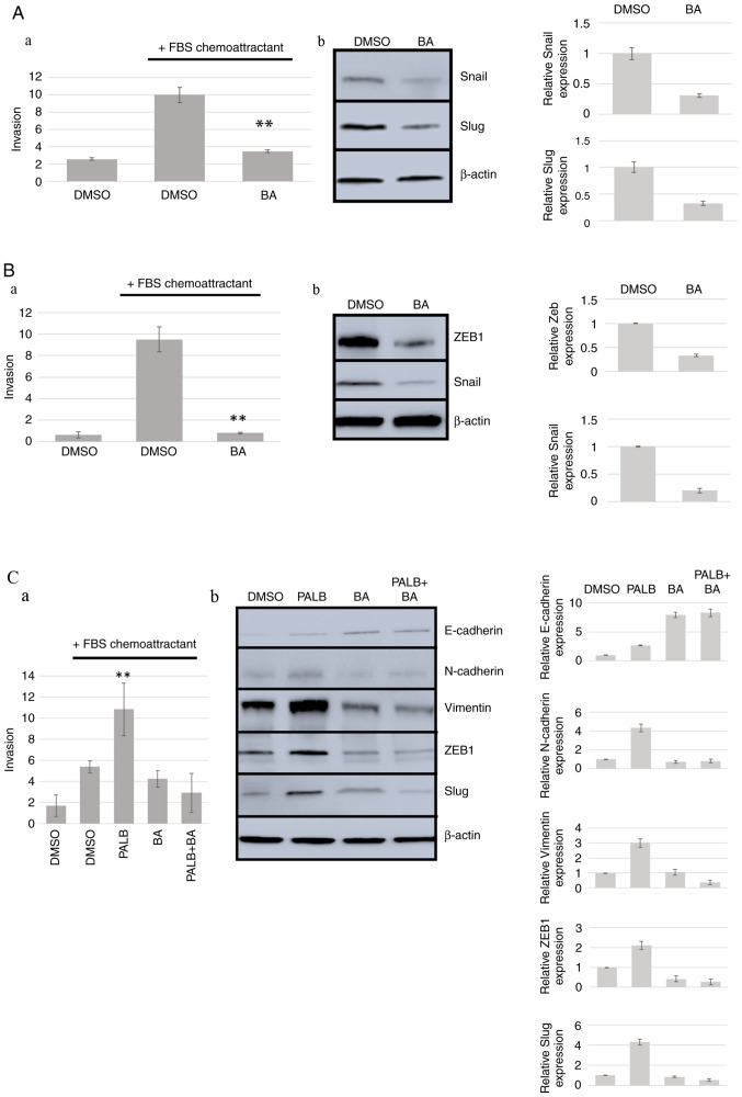
The use of small molecule kinase inhibitors, which target specific enzymes that are overactive in cancer cells, has revolutionized cancer patient treatment. To treat some types of breast cancer, CDK4/6 inhibitors, such as palbociclib, have been developed that target the phosphorylation of the retinoblastoma tumor suppressor gene. Acquired resistance to CDK4/6 inhibitors may be due to activation of the AKT pro‑survival signaling pathway that stimulates several processes, such as growth, metastasis and changes in metabolism that support rapid cell proliferation. The aim of the present study was to investigate whether targeting ATP citrate lyase (ACLY), a downstream target of AKT, may combine with CDK4/6 inhibition to inhibit tumorigenesis. The present study determined that ACLY is activated in breast and pancreatic cancer cells in response to palbociclib treatment and AKT mediates this effect. Inhibition of ACLY using bempedoic acid used in combination with palbociclib reduced cell viability in a panel of breast and pancreatic cancer cell lines. This effect was also observed using breast cancer cells grown in 3D cell culture. Mechanistically, palbociclib inhibited cell proliferation, whereas bempedoic acid stimulated apoptosis. Finally, using Transwell invasion assays and immunoblotting, the present study demonstrated that ACLY inhibition blocked cell invasion, when used alone or in combination with palbociclib. These data may yield useful information that could guide the development of future therapies aimed at the reduction of acquired resistance observed clinically.

Keywords: ATP citrate lyase; CDK4/6; bempedoic acid; palbociclib; resistance.


Conflict of interest statement

The authors declare that they have no competing interests.

Figures

Figure 1.
Figure 1.
Palb activation of ACLY is dependent on AKT. (A) MDA-MB-231 and Panc1 cells were treated with 0.1% DMSO or 1 µM P for 96 h. Cellular protein was quantified using Bradford assays and equal amounts of protein were separated by SDS-PAGE. (B) MDA-MB-231 or Panc1 cells were treated with 0.1% DMSO and NT RNA as a control. Cells treated with 1 µM Palb for 96 h were also subjected to AKT knockdown. Protein analysis, western blotting and antibodies utilized was as described in the Materials and methods. Results shown were repeated twice, and Palb, palbociclib; NT, non-targeting; ACLY, ATP citrate lyase; Rb, retinoblastoma; RNAi, RNA interference; p, phosphorylated.
Figure 2.
Figure 2.
BA reverses Palb-induced activation of ACLY. MDA-MB-231 and Panc1 cells were treated with 0.1% DMSO as a control, 1 µM Palb, 25 µM BA or the combination of Palb + BA for 96 h followed by western blotting. Results shown were repeated twice. BA, bempedoic acid; Palb, palbociclib; ACLY, ATP citrate lyase; p, phosphorylated.
Figure 3.
Figure 3.
The combination of Palb and BA reduces cell viability further than either drug alone. (A) MDA-MB-231 or (B) Panc1 cells were treated with 0.1% DMSO, 1 µM Palb, 25 µM BA or the combination of Palb + BA for 96 h. Cell viability was determined in MDA-MB-231 cells at 1, 72 and 96 h and in Panc1 cells at 24, 48, 72 and 96 h after drug addition, using the RealTime-Glo MT Cell Viability assay that measures luminescence or RLU. Experiments were repeated twice with n=6 Error bars represent standard deviation of the mean. One-way ANOVA (of 96-h values) with Tukey's post hoc test was used to determine statistical significance. **P<0.01 compared with Palb or BA treatment alone. (C) A panel of breast and pancreatic cancer cell lines were used in cell viability experiments. The CellTiter-Fluor Cell Viability Assay was used on cells after treatment with 0.1% DMSO, 1 µM P or 25 µM BA or the combination P + BA for 96 h. The average percent decrease in cell number compared with controls is shown and error bars represent standard deviation of the mean. Experiments were performed on each cell type three times with n=6. **P<0.01 compared with Palb or BA treatment alone. Palb, palbociclib; BA, bempedoic acid; RLU, relative light units.
Figure 4.
Figure 4.
The combination of Palb and BA reduces viability of breast cancer cells grown in 3D cell culture. MDA-MB-231 cells were grown in 3D cell culture and treated with 0.1% DMSO, Palb (1 µM), BA (25 µM) or the combination P + BA for 96 h. (A) Cells in Matrigel formed spheres in 7 days and then were subjected to treatments. Cell viability was measured using CellTiter-Glo 3D Cell Viability Assay. (B) Cells were plated in ultra-low attachment plates that facilitate 3D sphere formation. Cell toxicity after treatment was measured using the CellTox Green Cytotoxicity Assay using Fluorescence (Ex:485 nm Em:520 nm). Experiments were repeated twice with n=8. Error bars represent standard deviation of the mean. One-way ANOVA with Tukey's post hoc test was used to determine statistical significance **P<0.01 compared with Palb or BA treatment alone. Palb, palbociclib; BA, bempedoic acid.
Figure 5.
Figure 5.
Palb inhibits proliferation and BA induces apoptosis. MDA-MB-231, Panc1 and T47D cells were used to analyze the mechanistic effects of P (1 µM) or BA (25 µM). Culture medium or 0.1% D were used as controls. (A) The BrdU cell proliferation assay was performed after drug treatments for 72 h. (B) The RealTime-Glo Annexin V assay was utilized to measure apoptosis 48 h after addition of drug and presented as Annexin V (absorbance)/cell number (fluorescence). Cell number was determined using the CellTiter-Fluor Cell Viability Assay. (C) Target validation of BA. The product of the ACLY enzyme (acetyl-CoA) was shown to reverse toxicity induced by BA using the CellTox Green Cytotoxicity Assay. Error bars display standard deviation of the mean of n=8. Statistical analysis was performed using one-way ANOVA with Tukey's post hoc test. **P<0.01 compared with all other treatments. (D) Western blotting was performed on cells treated for 96 h as aforementioned using antibodies to p-Rb (780), total Rb, apoptosis markers cPARP and p-c-Jun and the proliferation markers Mcm7, cyclin D3 and PCNA. β-actin was used as a loading control. Results shown were repeated twice. CT, control; p, phosphorylated; cPARP, cleaved poly (ADP-ribose) polymerase; PCNA, proliferating cell nuclear antigen; BA, bempedoic acid; Palb, palbociclib; ACLY, ATP citrate lyase; BrdU, 5-bromo-2-deoxyuridine; Rb, retinoblastoma; Mcm7, minichromosome maintenance complex component 7.
Figure 6.
Figure 6.
BA inhibits invasion in various cancer cell types. (Aa) HT1080 cells were treated with 0.1% DMSO or 25 µM BA, followed by assays as described in the Materials and methods. Statistical analysis was performed using unpaired Student's t-test. **P<0.01 compared with DMSO control in the presence of FBS. (Ab) Detection of the mesenchymal markers Snail and Slug by western blotting. Results shown were repeated twice. (Ba) Panc1 cells were treated with 0.1% DMSO or 25 µM BA, followed by assays as described in the Materials and methods. Statistics were performed as in Aa. (Bb) Detection of the mesenchymal markers Snail and ZEB1 through western blotting. (Ca) MDA-MB-231 cells were treated with 0.1% D or 1 µM P for 96 h followed by invasion assays. One-way ANOVA with Tukey's post hoc test was used for statistical analysis. **P<0.01 compared with all other treatments. Western blotting analysis was performed in (Cb) using antibodies against markers of the epithelial phenotype (E-cadherin) or the mesenchymal phenotype (N-cadherin, Vimentin, ZEB1 and Slug). β-actin was used as a loading control. Results shown were repeated twice. ZEB1, zinc finger E-box-binding homeobox 1; BA, bempedoic acid; Palb, palbociclib.

References

    1. Mittnacht S. The retinoblastoma protein-from bench to bedside. Eur J Cell Biol. 2005;84:97–107. doi: 10.1016/j.ejcb.2004.12.012. - DOI - PubMed
    1. Asghar U, Witkiewicz AK, Turner NC, Knudsen ES. The history and future of targeting cyclin-dependent kinases in cancer therapy. Nat Rev Drug Discov. 2015;14:130–146. doi: 10.1038/nrd4504. - DOI - PMC - PubMed
    1. Guarducci C, Bonechi M, Boccalini G, Benelli M, Risi E, Di Leo A, Malorni L, Migliaccio I. Mechanisms of resistance to CDK4/6 inhibitors in breast cancer and potential biomarkers of response. Breast Care (Basel) 2017;12:304–308. doi: 10.1159/000484167. - DOI - PMC - PubMed
    1. Hamilton E, Infante JR. Targeting CDK4/6 in patients with cancer. Cancer Treat Rev. 2016;45:129–138. doi: 10.1016/j.ctrv.2016.03.002. - DOI - PubMed
    1. McCartney A, Migliaccio I, Bonechi M, Biagioni C, Romagnoli D, De Luca F, Galardi F, Risi E, De Santo I, Benelli M, et al. Mechanisms of resistance to CDK4/6 inhibitors: Potential implications and biomarkers for clinical practice. Front Oncol. 2019;9:666. doi: 10.3389/fonc.2019.00666. - DOI - PMC - PubMed

MeSH terms

Substances