Review
. 2023 Feb:66:103006.
doi: 10.1016/j.scr.2022.103006. Epub 2022 Dec 20.

Restoring vision and rebuilding the retina by Müller glial cell reprogramming

Affiliations
Review

Restoring vision and rebuilding the retina by Müller glial cell reprogramming

Devansh Agarwal et al. Stem Cell Res. 2023 Feb.

Abstract

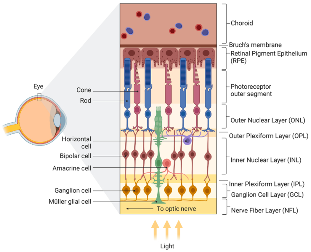
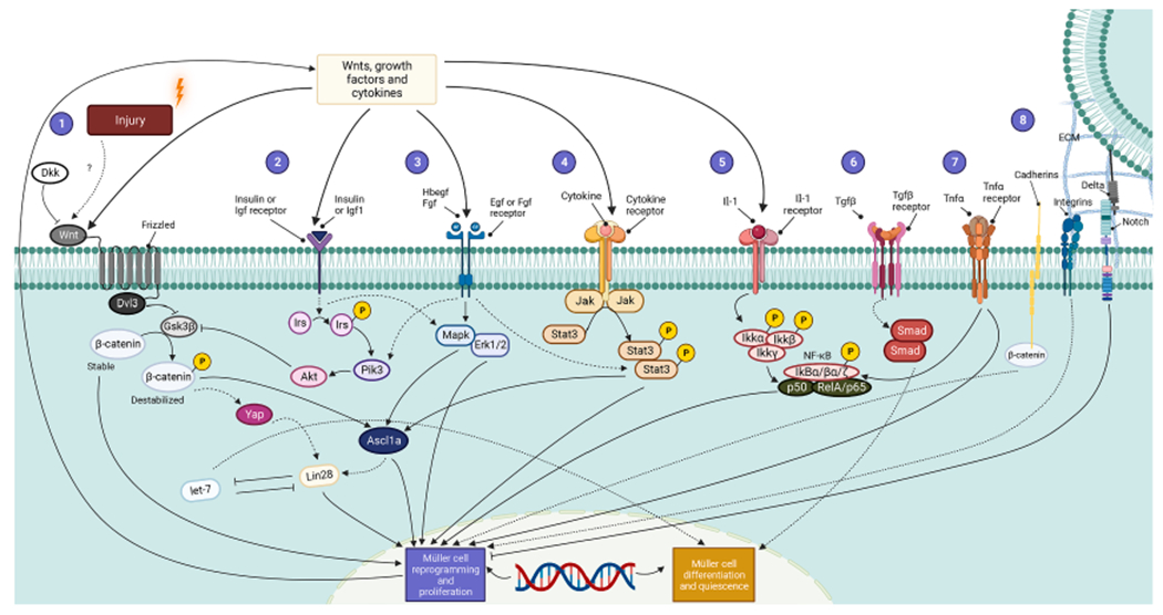
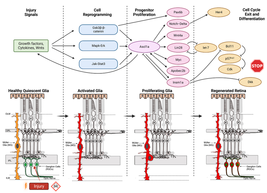
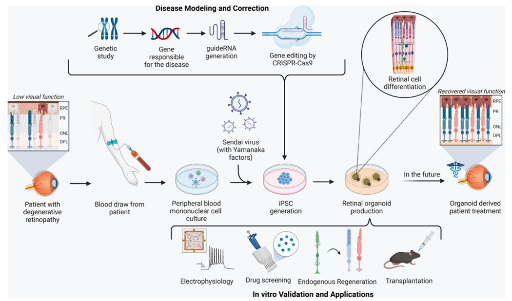
Müller glia are non-neuronal support cells that play a vital role in the homeostasis of the eye. Their radial-oriented processes span the width of the retina and respond to injury through a cellular response that can be detrimental or protective depending on the context. In some species, protective responses include the expression of stem cell-like genes which help to fuel new neuron formation and even restoration of vision. In many lower vertebrates including fish and amphibians, this response is well documented, however, in mammals it is severely limited. The remarkable plasticity of cellular reprogramming in lower vertebrates has inspired studies in mammals for repairing the retina and restoring sight, and recent studies suggest that mammals are also capable of regeneration, albeit to a lesser degree. Endogenous regeneration, whereby new retinal neurons are created from existing support cells, offers an exciting alternative approach to existing tissue transplant, gene therapy, and neural prosthetic approaches being explored in parallel. This review will highlight the role of Müller glia during retinal injury and repair. In the end, prospects for advancing retinal regeneration research will be considered.

Keywords: Cellular reprogramming; Müller glia; Retinal regeneration.


Conflict of interest statement

Declaration of Competing Interest The authors declare that they have no known competing financial interests or personal relationships that could have appeared to influence the work reported in this paper.

Figures

Fig. 1.
Fig. 1.
Structure of the Human Retina. The human retina has four nuclear layers: the outer, inner, ganglion cell, and retinal pigment epithelium layers (ONL, INL, GCL, RPE). Rod and cone photoreceptor (PR) cells, which are responsible for transducing light into electrical signals, are found in the outer retina between the retinal pigment epithelial (RPE) layer and the outer plexiform layer (OPL) where they connect with bipolar cells (BC) in the inner retina. Electrochemical signals then propagate through bipolar cells of the INL towards the inner plexiform layer (IPL) where they synapse with retinal ganglion cells (RGCs). The INL has horizontal and amacrine cell interneurons whose role is to modulate and refine vision through lateral inhibition and/or neuromodulation. RGC axons bundle emanating from the retinal ganglion cell layer (RGC) form the optic nerve which leaves the back of the eye, passes through the optic chiasm and connects with either the lateral geniculate nucleus (LGN) or to a lesser extent the superior colliculus (SC). From there nerve impulses connect to the visual cortex. In addition to neurons, two other types of cells found in the retina include Müller glia (MG) and RPE cells, both of which support the neurons of the retina. MG serve as the principle supporting glia of the eye and have nuclei in the INL, but cell bodies that span all layers of the retina. RPE cells absorb light while remaining in contact with PRs and serve a critical role in recycling pigmentation of PRs.
Fig. 2.
Fig. 2.
Signaling Pathways for Müller Cell Reprogramming and Proliferation. Regeneration is a highly regulated, multistep process that requires the expression of specific genetic programs at each stage and is influenced by the combined activity of secretory Wnt glycoproteins, inflammatory cytokines, and growth factors. (1) Wnts bound to Frizzled receptors activate Dishevelled Segment Polarity Protein 3 (Dvl3) to inhibit Gsk3β, which normally destabilizes β-catenin. A stabilized β-catenin is required for the proper linking of E-Cadherin to cytoskeletal structures to provide proper cell adhesion as well as gene expression leading to Müller cell reprogramming and retinal progenitor formation. β-catenin is a dual-function molecule that participates in cell adhesion to maintain structural integrity and polarity and as a transcriptional regulator that modulates gene expression; both contribute to MG dedifferentiation and progenitor formation. β-catenin can induce Yap/Taz (Hippo) signaling which can indirectly activate Lin28 to coax Müller glia de-differentiation. In addition, Wnt signaling is inhibited by its antagonist Dickkopf (Dkk). (2) Simultaneously, insulin and Igf1 signal through Insulin Receptor Substrate (IRS), Pik3, and Akt to inhibit Gsk3β. (3) Next, fibroblast growth factor (Fgf) and Hbegf bind to their receptors and control Egfr and mitogen-activated Mapk–Erk signaling that also end in reprogramming of Müller glia and cell division. (4) Meanwhile, cytokines acting via their respective receptors stimulate Jak proteins that phosphorylate Stat molecules to stimulate retinal regeneration by promoting gliogenesis. (5) Inhibition of NF-kB enhances Müller glia proliferation while its activation suppresses proliferating progenitor glia in a manner coordinated by microglia. (6) Furthermore, Tgf-β acts through the Smad pathway to coax Müller cells to exit quiescence, (7) interfering microRNA let-7, and RNA binding protein Lin28 are negatively regulated by each other and (8) Delta-Notch signaling complement modulated by the extracellular matrix (ECM); collectively these confer stem cell-like properties to Müller cells and stimulate retinal progenitor formation. Solid lines trace the pathways directly involved in Müller cell-mediated retinal regeneration, whereas dashed lines indicate the indirect ones.
Fig. 3.
Fig. 3.
Transcriptional Signaling Cascades in Retinal Regeneration. Injury signals are transduced by growth factors, cytokines, and Wnts that impinge upon the Gsk3ββ-catenin, Mapk-Erk, and Jak-Stat signaling pathways which enable activation and reprogramming of Müller Glia. These pathways stimulate the injury-dependent expression of Ascl1a that participates in progenitor proliferation by regulating the Notch-Delta, Wnt4a, Lin28, Apobec2b, Insm1a genes. Ascl1a also controls factors that inhibit neural progenitor formation and help in cell cycle exit/differentiation such as microRNA let-7, Notch, Insm1a, Dkk, p57, etc.
Fig. 4.
Fig. 4.
Generation and Application of Retinal Organoids. To generate retinal organoids, a blood draw is conducted in a patient with degenerative retinopathy, and peripheral blood mononuclear cells are extracted from the specimen. Sendai virus is used to reprogram the cells to form iPSCs which are later differentiated into 3D retinal organoids. CRISPR-Cas9 mediated gene editing can be utilized to model or correct disease states during the creation of organoids which can be validated by electrophysiology techniques or applied in drug screening, endogenous regeneration, and transplantation studies. These organoid-based approaches can potentially accelerate the development of treatments for retinal degeneration in the future.

References

    1. Bernardos RL, Barthel LK, Meyers JR, Raymond PA, 2007. Late-stage neuronal progenitors in the retina are radial Muller glia that function as retinal stem cells. J. Neurosci 27 (26), 7028–7040. 10.1523/JNEUROSCI.1624-07.2007. - DOI - PMC - PubMed
    1. Blackshaw S, Sanes JR, 2021. Turning lead into gold: reprogramming retinal cells to cure blindness. J. Clin. Investig 131 (3) 10.1172/JCI146134. - DOI - PMC - PubMed
    1. Cepko CL, Austin CP, Yang X, Alexiades M, Ezzeddine D, 1996. Cell fate determination in the vertebrate retina. PNAS 93 (2), 589–595. 10.1073/pnas.93.2.589. - DOI - PMC - PubMed
    1. Daiger SP, Sullivan LS, Bowne SJ, Rossiter BJF, 1996. RetNet - Retinal Information Network. The University of Texas Health Science Center.
    1. Del Rio-Tsonis K, Tsonis PA, 2003. Eye regeneration at the molecular age. Dev. Dyn 226 (2), 211–224. 10.1002/dvdy.10224. - DOI - PubMed

Publication types