. 2022 Dec 22;185(26):4887-4903.e17.
doi: 10.1016/j.cell.2022.11.029.

Chimeric efferocytic receptors improve apoptotic cell clearance and alleviate inflammation

Affiliations

Chimeric efferocytic receptors improve apoptotic cell clearance and alleviate inflammation

Sho Morioka et al. Cell. .

Abstract

Our bodies turn over billions of cells daily via apoptosis and are in turn cleared by phagocytes via the process of "efferocytosis." Defects in efferocytosis are now linked to various inflammatory diseases. Here, we designed a strategy to boost efferocytosis, denoted "chimeric receptor for efferocytosis" (CHEF). We fused a specific signaling domain within the cytoplasmic adapter protein ELMO1 to the extracellular phosphatidylserine recognition domains of the efferocytic receptors BAI1 or TIM4, generating BELMO and TELMO, respectively. CHEF-expressing phagocytes display a striking increase in efferocytosis. In mouse models of inflammation, BELMO expression attenuates colitis, hepatotoxicity, and nephrotoxicity. In mechanistic studies, BELMO increases ER-resident enzymes and chaperones to overcome protein-folding-associated toxicity, which was further validated in a model of ER-stress-induced renal ischemia-reperfusion injury. Finally, TELMO introduction after onset of kidney injury significantly reduced fibrosis. Collectively, these data advance a concept of chimeric efferocytic receptors to boost efferocytosis and dampen inflammation.

Keywords: BAI1; CHEF; ELMO1; TIM4; acute kidney injury; apoptosis; chimeric receptor; efferocytosis; inflammatory diseases.


Conflict of interest statement

Declaration of interests The authors declare no competing financial interests.

Figures

Figure 1.
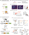
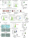
Figure 1.. Chimeric efferocytosis receptor boosts phagocytosis.
(A) Schematic of BELMO. The cytoplasmic tail of BAI1 was truncated to delete the natural ELMO-binding helix region, and the truncated BAI1 was directly fused to the C-terminal domain of ELMO that is sufficient to interact with Dock180 and activate Rac1. (B) Cell surface expression of BELMO was confirmed using BAI1 antibody. (C) LR73 cells were co-cultured with CypHer5E-labeled apoptotic Jurkats for 2 h and phagocytosis measured by flow cytometry. Data are represented as box and whiskers. ***p < 0.001, n=5. (D) Efferocytosis of multiple corpses by BELMO+ LR73 cells was assessed using CypHer5E-labeled apoptotic thymocytes as targets. Scale Bar = 20 µm. (E) Control or BELMO+ LR73 cells were tested for efferocytosis using as targets CypHer5E-labeled live or apoptotic Jurkat cells (2hr). ***p < 0.001. n.s., Not significant. n=4. (F) Apoptotic Jurkat cells were incubated with or without Annexin V before use in efferocytosis. A dominant negative Rac1 (N17) was used to test whether Rac1 is required for efferocytosis. Control or BELMO+ LR73 cells were treated with 1 µM of Cytochalasin D for 1 h, and tested in efferocytosis. ***p < 0.001. n=4. (G) Schematic of BELMO 6M mutation within a region of ELMO that disrupts Dock180 binding. LR73 cells expressing BELMO or BELMO-6M were co-cultured with CypHer5E-labeled apoptotic Jurkat cells for the indicated times. Data are shown as % engulfment and mean fluorescence intensity (MFI), an indirect measure of corpse burden indicated by apoptotic cell-derived fluorescence per phagocyte. Data are represented as mean ± SD. ***p < 0.001. n=4.
Figure 2.
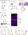
Figure 2.. Tissue-specific BELMO expression promotes efferocytosis in vivo
(A and B) Schematic for expression of BELMO in zebrafish and laser injury. At 3 dpf, slc1a3b-eGFP reporter zebrafish larvae were treated with 10 µM doxycycline to induce BELMO and mCherry in slc1a3b-positive glia for 24 h (A). Injury was created using a laser and the glia imaged every 12 minutes for 10 h. BELMO+ glia were identified by the expression of nuclear mCherry (B). (C) BELMO+ glia (eGFP+ along with their morphology) have larger phagocytic cups. Quantification of the size of phagocytic cups is shown on the right. ***p < 0.001. n= 22 phagocytic cups (BELMO-negative) and n =45 phagocytic cups for BELMO+ from 8 larvae. Scale bar = 10 µm. (D) Via live imaging, the time for a phagocytic cup to be taken up into a cell was measured from initiation of phagosome formation to corpse internalization. Duration and corresponding size of the phagocytic cup (left) and duration normalized for size of the phagocytic cup (right). ***p < 0.001. n= 22 phagocytic cups (BELMO-negative) and n =45 phagocytic cups for BELMO+ from 8 larvae. (E) Schematic for generation of BELMOTg mice. (F and G) BELMOTg mice were crossed with Cx3cr1-cre mice. Peritoneal macrophages were isolated and incubated with CypHer5E-labeled apoptotic Jurkat cells and efferocytosis assessed. Data are represented as mean ± SD. n=4 (F). CypHer5E labeled apoptotic Jurkat cells were injected intraperitoneally, and 15 min later, the engulfment by CD11b+ F4/80hi macrophages was assessed. n=5 (G). ***p < 0.001.
Figure 3.
Figure 3.. BELMO attenuates DSS-induced colitis.
(A) BELMO boosts apoptotic cell uptake in colonic epithelial cells. BELMO expressing HCT116 cells were co-cultured with CypHer5E-labeled apoptotic Jurkat cells for 2hr and assessed for efferocytosis. ***p < 0.001. n=5. (B-F) BELMO expression in intestinal epithelial cells (IEC) dampens DSS-induced colitis. BELMOTg mice were crossed with Villin-cre mice for IEC expression of BELMO. Mice were treated with 3% DSS, monitored for 7 days, and analyzed for the indicated parameters (B). On day 5, colon samples were collected and analyzed by H&E staining and histological scoring (C). Colon length on day 7 (D). On day 3, colon samples were collected and cell death was analyzed by TUNEL staining. 10 sections at 200 µm intervals per colon were counted for TUNEL positive cells (E). mRNA was isolated from the colons and expression level of cytokines analyzed by qPCR (F). ***p < 0.001. *p < 0.05. n=5. Scale bars 200 µm.
Figure 4.
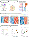
Figure 4.. CHEF ameliorates drug-induced hepatotoxicity and nephrotoxicity.
(A) BELMO boosts apoptotic cell uptake in primary hepatocytes. BELMOTg mice were crossed with Alb-cre mice to induce BELMO expression in hepatocytes. Primary hepatocytes were isolated and incubated with CypHer5E-labeled apoptotic Jurkat cells for 2h and efferocytosis determined. ***p < 0.001. n=5. (B and C) BELMO dampens diethylnitrosamine (DEN)-induced hepatotoxicity. Mice were treated with 100 mg/kg of DEN (intraperitoneal) (B). 48h later, liver samples were collected and analyzed by TUNEL staining and DNase II-mediated DNA cleavage, the latter indicative of efferocytic phagocytes. n=8. Liver damage was analyzed using an ALT activity assay. Untreated control and BELMO mice: n=4; DEN treated control: n=9; DEN treated BELMO mice: n=10 (C). ***p < 0.001. Scale bars 200 µm (D) BELMO boosts apoptotic cell uptake in primary kidney tubular epithelial cells (TEC). BELMOTg mice were crossed with PEPCK-cre to induce TEC specific expression of BELMO. Primary TECs were isolated and assessed for efferocytosis with CypHer5E-labeled apoptotic Jurkat cells ***p < 0.001. n=4. (E and F) BELMO alleviates cisplatin-induced nephrotoxicity. Schematic of the cisplatin-induced nephrotoxicity model (E). 48h after cisplatin treatment (20 mg/kg body weight), kidney samples were collected and analyzed by TUNEL staining (cell death) and efferocytic phagocytes. n=4. Plasma samples were collected and assessed for creatinine in circulation. n=6. After cisplatin treatment (25 mg/kg body weight), the viability of mice was monitored over 7 days. Control: n=5; BELMO: n=8 (F). ***p < 0.001. **p < 0.005. Scale bars 200 µm
Figure 5.
Figure 5.. Protein folding modulators regulate efferocytosis.
(A) Transcriptomic analysis of BELMO expressing phagocytes. Control or BELMO+ LR73 cells were incubated with apoptotic human Jurkat cells for 2h. The unbound/free apoptotic cells were removed by washing and LR73 cells were cultured for an additional 2h, RNAseq was performed. (B) Venn diagram illustrating the shared and distinct genes between control and BELMO+ LR73 cells induced during efferocytosis. Adjusted p value < 0.05, and Log2Foldchange > 0.3 or < −0.3. (C) Gene ontology analysis reveals various upregulated (red) or downregulated (blue) gene programs in BELMO+ cells. The bidirectional plots represent normalized enrichment score. Protein folding/ER function gene sets were manually curated from a public gene cluster (GO:0034976). (D) BELMO+ phagocytes upregulate genes involved in protein folding and ER function. Heat maps represent differentially expressed genes before and after efferocytosis. Adjusted p <0.05. (E-G) Control and BELMO LR73 cells were pretreated with thapsigargin (Tg) for 1h. Data are represented as mean ± SD. n=4 (E), 100 µM BiP inhibitor (epigallocatechin gallate, EGCG) for 1h. n=4 (F) or 5 µM 4-Phenylbutric acid for 6h. n=4 (G). Cells were co-cultured with CypHer5E-labeled apoptotic Jurkat cells and assessed for efferocytosis ***p < 0.001. *p < 0.05.
Figure 6.
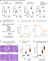
Figure 6.. BELMO ameliorates the defective ER proteostasis associated with acute kidney injury.
(A) siRNA-targeting of BiP, Dnajc3, Atp2a3 and Vimp suppress efferocytosis. ***p < 0.001. **p < 0.01. *p < 0.05. n=4. (B) BELMO reduces ischemia reperfusion (IRI)-induced acute kidney injury. BELMOTg PEPCK-cre mice were subjected to bilateral IRI injury for 26 min or 29 min. After removal of the clamps, the kidneys were allowed to reperfuse for the indicated times. (C) After IRI, blood samples were collected and plasma creatinine was analyzed over 3 days. Data are represented as mean ± SEM. ***p < 0.001. n > 10. (D) BELMO improves mouse viability after ischemia reperfusion-induced acute kidney injury. After IRI (29 min), mice were monitored over 7 days for mortality. *p < 0.05. Control mice: n=8; BELMO transgenic mice: n=10. (E) Kidney samples were collected, and histological samples analyzed by H&E staining (at least 10 mice per condition). Scale bar 200 µm. (F) BELMO increases BiP expression during acute kidney injury. 24h after IRI surgery, kidneys were collected for mRNA extraction. Expression of Bip was analyzed via qPCR. *p < 0.05. Sham mice: n=4; IRI mice: n=6. (G) Plasma creatinine was measured 24h after bilateral kidney IRI. 30 mg/kg BiP inhibitor (HA15) was intraperitoneally administered 1 day before and immediately after surgery. ***p < 0.001. Untreated sham mice: n=4; HA15 treated sham mice: n=4; untreated IRI mice: n=8; HA15 treated IRI mice: n=6.
Figure 7.
Figure 7.. AAV-transduced TELMO ameliorates kidney disease progression.
(A) Schematic of TELMO generation. (B) Cell surface expression of TELMO confirmed by TIM4 antibody and flow cytometry. TELMO boosts efferocytosis. LR73 cells expressing TELMO, TELMO 6M, TIM4, and TIM4 lacking its intracellular domain were co-cultured with (C) CypHer5E-labeled apoptotic Jurkat cells and efferocytosis assessed. ***p < 0.001. n=4. (D-G) Adeno-associated virus (AAV)-mediated delivery ameliorates chronic kidney disease induced by ischemia reperfusion. Schematic of IRI model used (D). Left kidney was clamped and simultaneously AAV9-TELMO-GFP or control virus was introduced via renal vein injection; 25 min later, the clamp was removed. 14d later, the contralateral right kidney was removed. The left kidney function was evaluated 24h later. After AAV injection, TELMO expression was measured on day 2 and day 7 by GFP+ cells via flow cytometry on day 7, GFP positive cells were assessed for SLC34A1, a tubular epithelial cell marker expression (E). Blood samples were collected for plasma creatinine quantification. ***p < 0.001. n=5; no nephrectomy control: n=4 (F). Kidney fibrosis was visualized by Masson Trichrome staining. Scale bar 200 µm (G). (H) Cell surface expression of TELMO 4A. (I) TELMO 4A-expressing assessed for efferocytosis ***p < 0.001. n=4. (J) Left kidney was clamped and simultaneously AAV9-TELMO 4A, ELMO1 (532–727)-CAAX-GFP or control virus was introduced via renal vein injection; 25min later, the clamp was removed. 14d later, the contralateral right kidney was removed and, after 24h, blood samples collected for plasma creatinine quantification. n=5; no nephrectomy control: n=4. (K) Kidney samples were collected on day 7 following AAV9-TELMO-GFP or control virus introduction. Expression of Bip was measured via qPCR. Vehicle: n=7; TELMO: n=6 (H). Data are represented as box and whiskers. ***p < 0.001.

References

    1. Gregory CD, and Pound JD (2010). Microenvironmental influences of apoptosis in vivo and in vitro. Apoptosis 15, 1029–1049. 10.1007/s10495-010-0485-9. - DOI - PubMed
    1. Elliott MR, and Ravichandran KS (2016). The Dynamics of Apoptotic Cell Clearance. Dev Cell 38, 147–160. 10.1016/j.devcel.2016.06.029. - DOI - PMC - PubMed
    1. Boada-Romero E, Martinez J, Heckmann BL, and Green DR (2020). The clearance of dead cells by efferocytosis. Nat Rev Mol Cell Biol 21, 398–414. 10.1038/s41580-020-0232-1. - DOI - PMC - PubMed
    1. Morioka S, Maueroder C, and Ravichandran KS (2019). Living on the Edge: Efferocytosis at the Interface of Homeostasis and Pathology. Immunity 50, 1149–1162. 10.1016/j.immuni.2019.04.018. - DOI - PMC - PubMed
    1. Lemke G (2019). How macrophages deal with death. Nature Reviews Immunology 19, 539–549. 10.1038/s41577-019-0167-y. - DOI - PMC - PubMed

Publication types

Substances