. 2023 Jan 9;41(1):164-180.e8.
doi: 10.1016/j.ccell.2022.12.002. Epub 2022 Dec 22.

Modulation of RNA splicing enhances response to BCL2 inhibition in leukemia

Affiliations

Modulation of RNA splicing enhances response to BCL2 inhibition in leukemia

Eric Wang et al. Cancer Cell. .

Abstract

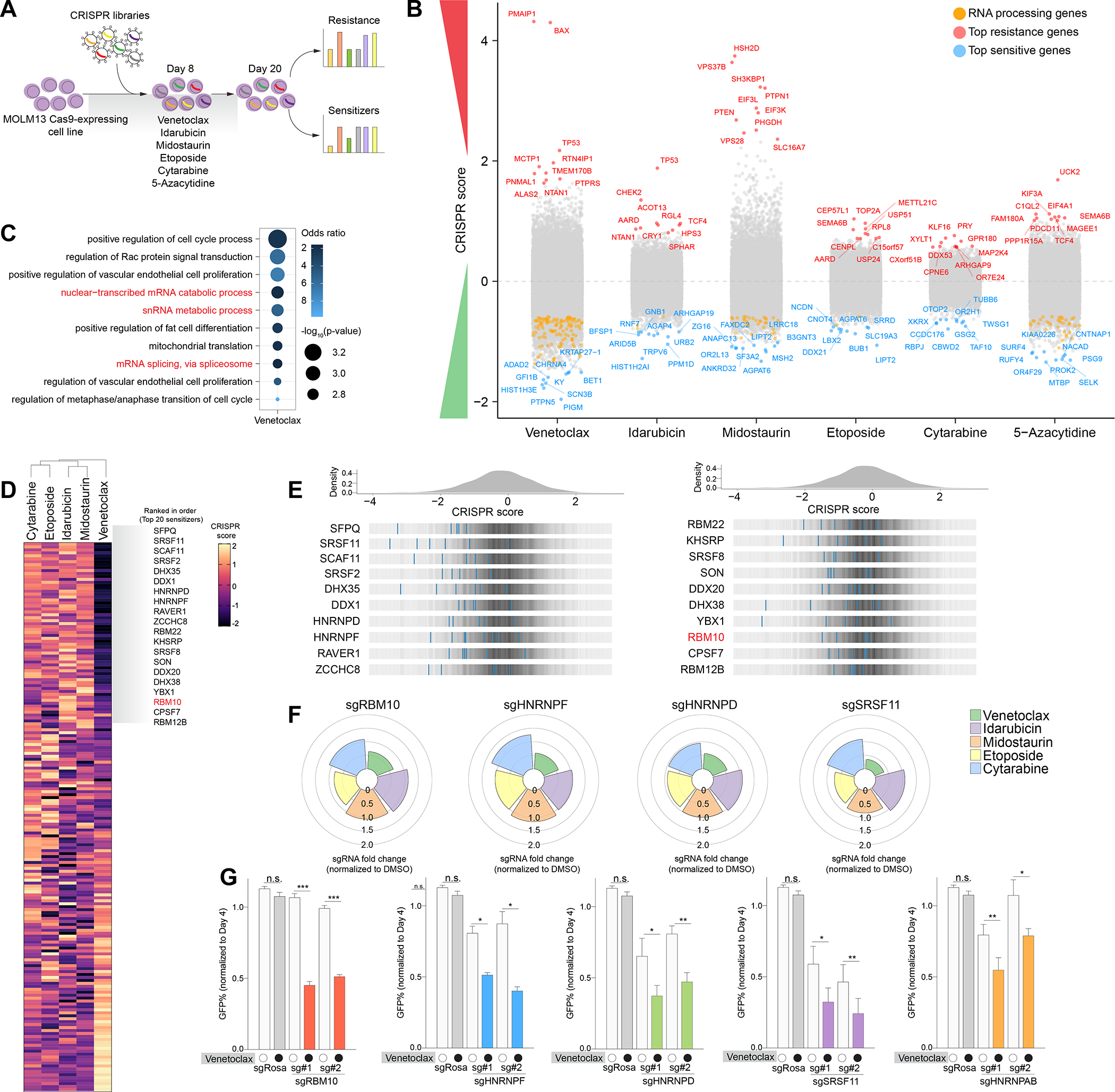
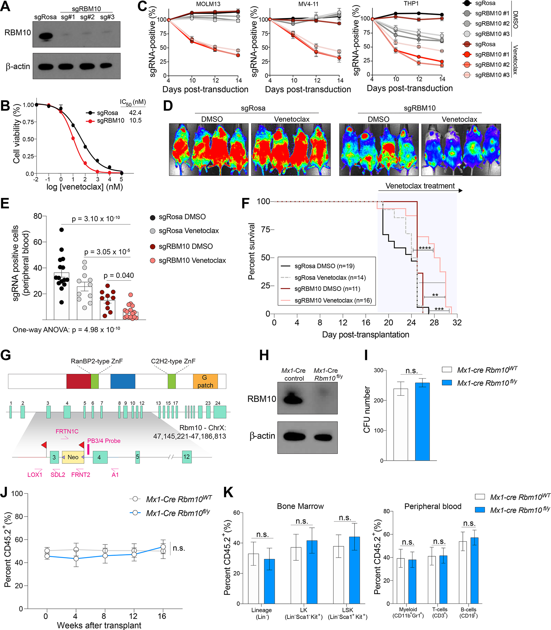
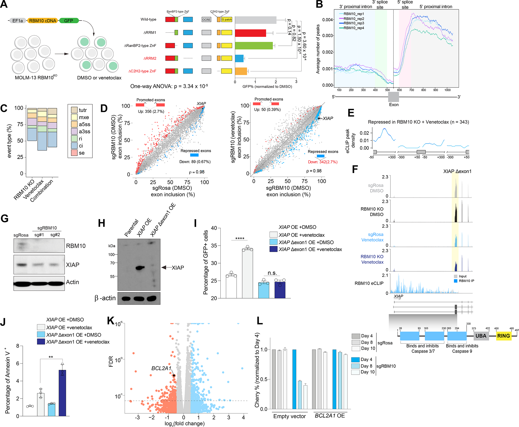
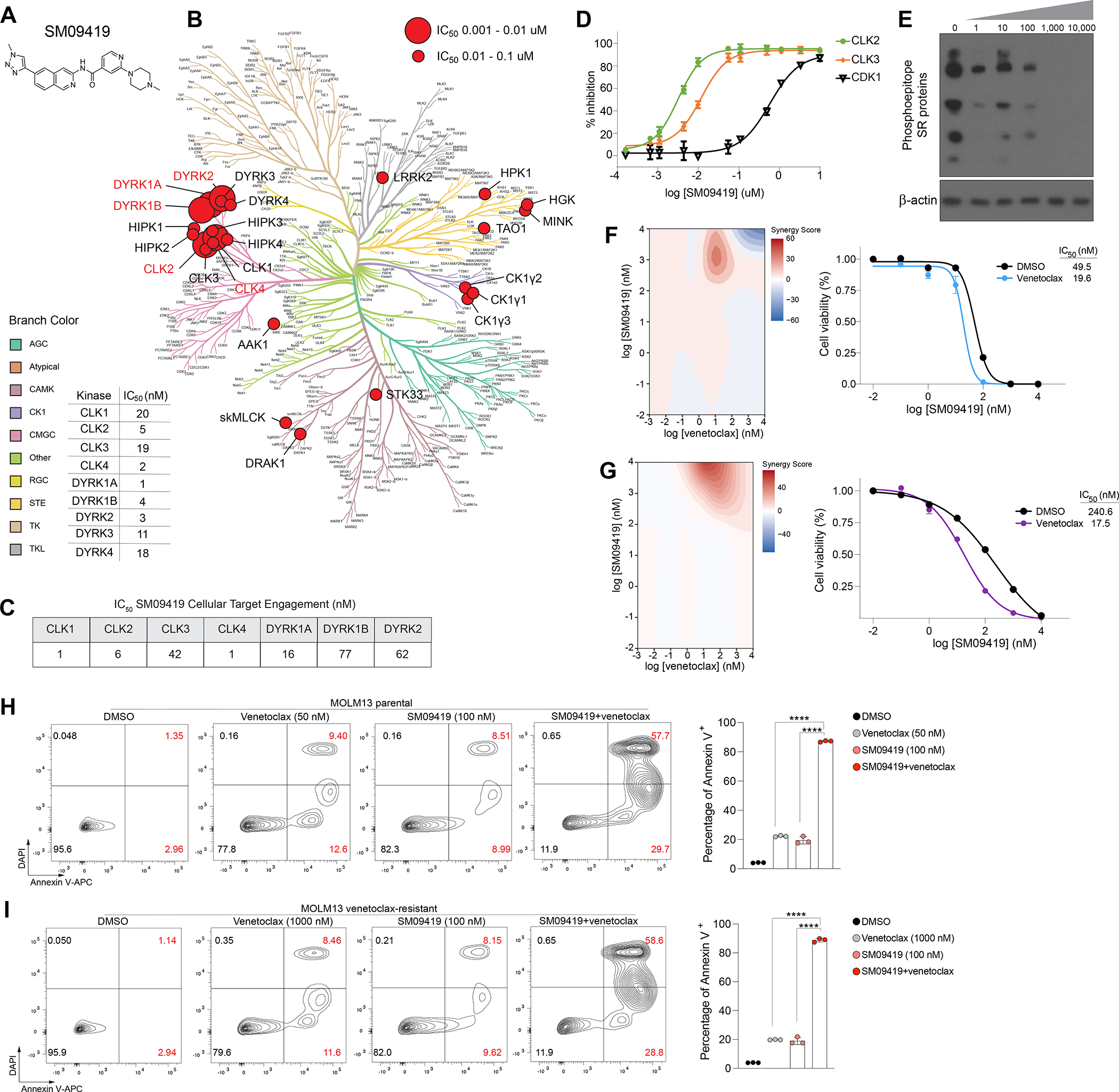
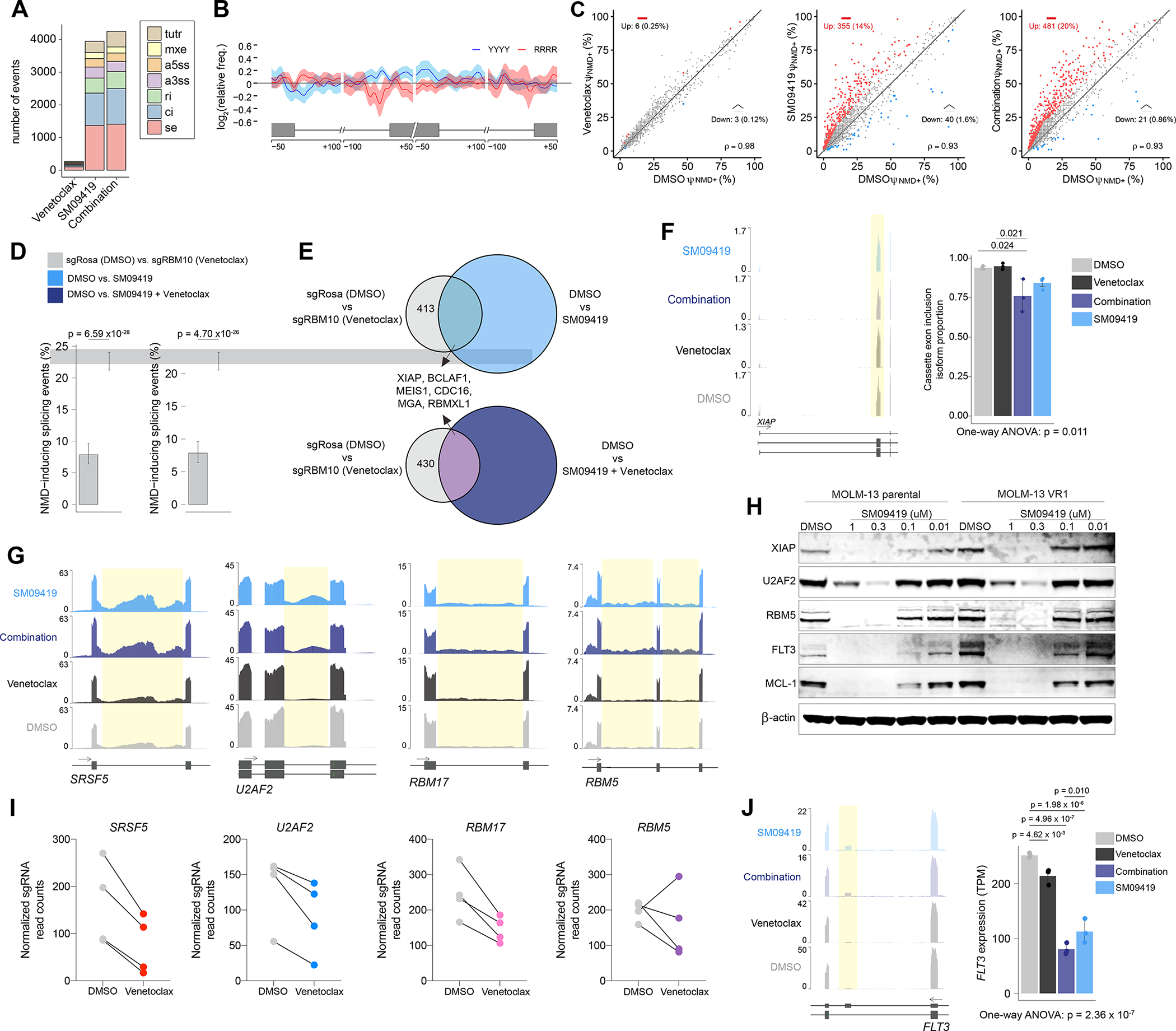
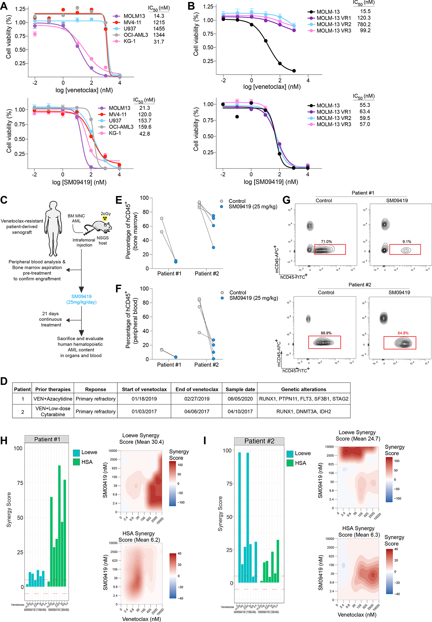
Therapy resistance is a major challenge in the treatment of cancer. Here, we performed CRISPR-Cas9 screens across a broad range of therapies used in acute myeloid leukemia to identify genomic determinants of drug response. Our screens uncover a selective dependency on RNA splicing factors whose loss preferentially enhances response to the BCL2 inhibitor venetoclax. Loss of the splicing factor RBM10 augments response to venetoclax in leukemia yet is completely dispensable for normal hematopoiesis. Combined RBM10 and BCL2 inhibition leads to mis-splicing and inactivation of the inhibitor of apoptosis XIAP and downregulation of BCL2A1, an anti-apoptotic protein implicated in venetoclax resistance. Inhibition of splicing kinase families CLKs (CDC-like kinases) and DYRKs (dual-specificity tyrosine-regulated kinases) leads to aberrant splicing of key splicing and apoptotic factors that synergize with venetoclax, and overcomes resistance to BCL2 inhibition. Our findings underscore the importance of splicing in modulating response to therapies and provide a strategy to improve venetoclax-based treatments.

Keywords: BCL2; CLK; DYRK; RBM10; RNA splicing; XIAP; acute myeloid leukemia; venetoclax.


Conflict of interest statement

Declaration of interests E.M., E.C., M.J., C.B., C.-C.M., and D.M.B. are employees of Biosplice Therapeutics. O.A.-W. has served as a consultant for H3B Biomedicine, Foundation Medicine Inc., Merck, Prelude Therapeutics, and Janssen and is on the Scientific Advisory Board of Envisagenics Inc., AIChemy, Harmonic Discovery Inc., and Pfizer Boulder. O.A.-W. has received prior research funding from H3B Biomedicine, Nurix Therapeutics, and LOXO Oncology unrelated to the current manuscript. The remaining authors declare no competing interests.

Figures

Figure 1.
Figure 1.. Mapping genomic determinants of AML drug response and synthetic lethal relationship between RNA splicing factors and venetoclax sensitivity.
(A) Schematic of genome-wide CRISPR screens in MOLM-13 AML cells treated with a panel of clinically approved AML drugs. (B) Manhattan plot depicting top 10 genes that sensitizes (blue) or confer resistance (red) in individual CRISPR drug screens. Orange dots represent RNA processing genes. CRISPR score represents the log2 (fold-change) values of sgRNAs normalized to DMSO. (C) Gene ontology (GO) enrichment analysis of top sensitizers in the venetoclax screen. (D) Clustered heatmap of results of the RNA-binding protein-focused CRISPR drug screens in MOLM-13 AML cells treated with drugs. CRISPR score represents log2 fold change of sgRNAs normalized to DMSO. (E) Histogram of CRISPR scores for all sgRNAs in the venetoclax screen in (D). Values represent the log2 (fold-change) values of sgRNAs normalized to DMSO. The blue lines represent individual sgRNAs targeting the indicated genes among the top splicing factor candidates. (F) Polar plots of top synergistic splicing factors identified in (D) treated with various AML drugs. The height of the wedge corresponds to the sgRNA fold change normalized to DMSO. (G) Competition-based assay in MOLM-13 cells 10 days post-transduction with top 2 sgRNAs targeting each splicing factor or non-targeting sgRosa control (n=3 per condition, mean+SEM) treated with 50 nM venetoclax. Statistical analysis was performed using unpaired Student’s t test by Prism GraphPad (*p < 0.05, **p < 0.01, ***p < 0.001, n.s., not significant). See also Figure S1 and Table S1.
Figure 2.
Figure 2.. RBM10 loss enhances BCL2 inhibition in AML cells but is dispensable for normal hematopoiesis.
(A) Western blot of CRISPR-mediated knockout of RBM10 in MOLM-13 cells. (B) Dose-response curves of sgRBM10 or sgRosa treated with indicated venetoclax concentrations on the x-axis and cell viability on the y-axis at 48 hours. IC50 values were calculated from technical triplicates per experiment, error bars represent SEM. (C) Competition proliferation assays of sgRBM10 or non-targeting sgRosa in human AML cell line expressing Cas9 and treated with 50 nM of venetoclax or DMSO (n=3 biological replicates per time point and condition, mean+SEM). (D) Bioluminescent imaging of mice transplanted with MOLM-13 cells transduced with sgRBM10 or sgRosa and treated daily with venetoclax (100 mg/kg) or vehicle control. Representative images of 4 mice per condition is shown. Images were taken 4 days post-treatment. (E) Flow cytometry analysis of GFP-positive sgRNA-expressing indicated by y-axis MOLM-13 cells in peripheral blood at day 6 post-treatment. Statistical analysis was performed using One-way ANOVA with post-hoc testing as indicated (number of mice used in each group is indicated in F, mean+SEM). (F) Kaplan-Meier survival curves of mice transplanted with MOLM-13 cells transduced with sgRBM10 or sgRosa and treated daily with venetoclax (100 mg/kg) or vehicle. The p values were determined using a log-rank Mantel-Cox test (**p < 0.01, ***p < 0.001, n.s., not significant). (G) Schematic depiction of the targeting strategy to generate Rbm10 cKO mice. The Rbm10 allele was deleted by targeting exon 3 that resulted in a frameshift following excision. Two LoxP sites flanking exon 3 and an Frt-flanked neomycin selection cassette were inserted in the downstream intron. (H) Western blot of Rbm10 in bone marrow mononuclear cells from Mx1-cre Rbm10fl/y (Rbm10 cKO) or Mx1-cre control 7 days after polyinosinic-polycytidylic acid (pIpC) treatment. (I) Total number of colony-forming units (CFU) from bone marrow cells of Mx1-cre Rbm10fl/y (Rbm10 cKO) or Mx1-cre control mice following 7 days of culture (n=6, mean+SEM). The p values were determined by unpaired student t test. n.s., not significant. (J) Percentage of CD45.2+ cells in peripheral blood over the course of 4 months competitive transplantation (n=6 for Mx1-cre control and n=7 mice for Rbm10 cKO, mean+SEM). (K) Percentage of CD45.2+ of hematopoietic stem and progenitor cells in the bone marrow (left) and mature immune cells in the peripheral blood (right) (n=6 for Mx1-cre control and n=7 mice for Rbm10 cKO, mean+SEM). See also Figure S2.
Figure 3.
Figure 3.. Impact of RBM10 on RNA binding, RNA splicing, and response to venetoclax.
(A) Competition-based assay of RBM10 KO in MOLM-13 cells and transduced with RBM10 cDNA wild-type (WT) or individual mutant (lacking RNA-binding domains) RBM10 cDNA and treated with venetoclax (50 nM) or DMSO at 48 hours (n=3, mean+SEM). The p-values were determined by One-way ANOVA with post-hoc testing. (B) Metaintron plots of average number indicated by y-axis of RBM10 peaks mapped to intronic regions flanking exons in MOLM-13 cells (n=4 eCLIP replicates). This plot is exon-centered (500–600 bp) on the x-axis. Enhanced crosslinking and immunoprecipitation (eCLIP) was performed in 4 replicates. (C) Percentage of treatment-responsive (RBM10 KO, venetoclax, or RBM10 KO and venetoclax) differentially spliced event types: cassette exons (SE), alternative 5’ ss exon (A5E), alternative 3’ ss exon (A3E), mutually exclusive exons (MXE), retained intron (RI), constitutive intron (CI), and tandem 3’ UTR (TUTR) from RNA-seq (n=3 per condition). (D) Scatter plot of cassette exons (SE) promoted (red circles) or repressed (blue circles) in MOLM-13 cells transduced with sgRosa (y-axis) or sgRBM10 (x-axis) treated with DMSO or venetoclax RNA-seq (n=3 per condition). ρ denotes Spearman’s rank correlation coefficient. (E) RBM10 splicing map generated by integrating RBM10 KO splicing changes from RNA-seq and RBM10 eCLIP binding sites. (F) RNA-seq and eCLIP (bottom) coverage plots of XIAP Δexon1 in MOLM-13 cells with RBM10 KO or non-targeting sgRNAs treated with DMSO or venetoclax. Yellow shadow depicts exon exclusion event in RBM10KO venetoclax-treated MOLM-13 cells overlapped with functional protein domains of XIAP. (G) Western blotting of XIAP after 50 nM venetoclax treatment of MOLM-13 cells with sgRosa or sgRBM10 for 48 hrs. (H) Western blotting of XIAP protein levels after ectopic overexpression of XIAP full-length (FL) or XIAP Δexon1. (I) Competition-based assay of XIAP full-length (FL) or XIAP Δexon1 linked to GFP reporter after 24 hrs of venetoclax treatment (n=3, mean+SEM). Y-axis denotes GFP positive cells. (J) Annexin V staining of XIAP full-length (FL) or XIAP Δexon1 after 24 hrs of venetoclax treatment (n=3, mean+SEM). Y-axis denotes Annexin V positive cells. Statistical analysis was performed using unpaired Student’s t test by Prism GraphPad (*p < 0.05, **p < 0.01, n.s., not significant). (K) Volcano plot of differentially expressed genes (DEGs) upon RBM10KO venetoclax-treated MOLM-13 cells compared to DMSO RNA-seq (n=3 per condition). (L) Competition-based assay measuring Cherry-expressing sgRBM10 or sgRosa cells transduced with overexpression (OE) of BCL2A1 cDNA or empty vector GFP-positive cells in MOLM-13 cells treated with 50 nM venetoclax for 48 hours (n=3, mean+SEM). Y-axis denotes mCherry positive cells. See also Figure S3 and Tables S2–4.
Figure 4.
Figure 4.. Pharmacologic inhibition of splicing-dependent kinases synergizes with venetoclax. (A) Structure of SM09419 selectivity.
(B) Kinase dendrogram of SM09419. Kinases with IC50 values of 0.01 to 0.1 μM are indicated by small red circle, whereas larger red circles represent more potent IC50 values with 0.001 to 0.01 μM. (C) NanoBRET target engagement assay of CLK1-4, DYRK1A/B, and DYRK2 upon 24 hrs of SM09419 treatment. (D) Inhibition of CLK kinases (CLK2 and CLK3) and CDK1 kinase (n=3). IC50 values were determined from dose response curves. Y-axis denotes the percent inhibition for CLK2, CLK3, and CDK1 (n=3, mean+SEM). (E) Western blot of phosphorylated SR proteins treated with increasing concentration of SM09419 for 48 hrs in MOLM-13 cells. (F) 2D synergy plots using Zero interaction potency (ZIP) model (left) and dose-response curves (right) of SM09419 and venetoclax combination at various concentration treated for 48 hrs in MOLM-13 (n=3, mean+SEM) and (G) KG-1 cells (n=3, mean+SEM). The presence of synergy was determined using the SynergyFinder computational package and the ZIP synergy index in which red signifies synergism and blue is antagonism. A positive synergy score is the percent more cell death than expected. IC50 values were calculated from technical triplicates per experiment. (H) Annexin V staining (left) and quantification (right) of MOLM-13 parental and (I) venetoclax-resistant cell lines treated with SM09419, venetoclax, or the combination at 48 hrs post-treatment (n=3, mean+SEM). Y-axis denotes percent of Annexin V positive cells. Statistical analysis was performed using unpaired Student’s t test by Prism GraphPad (****p < 0.0001). See also Figure S4–5.
Figure 5.
Figure 5.. SM09419 promotes mis-splicing of key oncogenic pathways in AML.
(A) Total number of splicing changes observed after SM09419 (100 nM), venetoclax (10 nM), or combination of SM09419 (100 nM) and venetoclax (10 nM) treatment for 48 hours RNA-seq (n=3 per condition). Cassette exons (SE), alternative 5’ ss exon (A5E), alternative 3’ ss exon (A3E), mutually exclusive exons (MXE), retained intron (RI), constitutive intron (CI), and tandem 3’ UTR (TUTR). (B) Spatial distribution of pyrimidines-rich (YYYY) and purine-rich (RRRR) motifs comparing sequence enrichment of excluded exons (n = 674) against included exons (n = 370) in SM0419-treated (100 nM) MOLM-13 cells. (C) Scatter plot of NMD-inducing retained intron (RI) events (red circles) in MOLM-13 cells treated with venetoclax (left), SM09419 (middle) or the combination of venetoclax and SM09419 (right) RNA-seq in triplicates for each condition. (D) Percentage of NMD-inducing events indicated on the y-axis in RBM10 KO venetoclax (compared to non-targeting sgRosa) and SM09419, or SM09419+venetoclax (compared to DMSO) RNA-seq in triplicates for each condition (mean+SEM). (E) Venn diagram of NMD-inducing events in RBM10 KO venetoclax (compared to non-targeting sgRosa) and SM09419, or SM09419+venetoclax (compared to DMSO). (F) RNA-seq coverage plot (left) and mean PSI of XIAP cassette exon inclusion isoform (n=3 per condition, mean+SEM). (G) RNA-seq coverage plots of the splicing factors SRSF5, U2AF2, RBM17, and RBM5 in MOLM-13 cells. Yellow regions represent retained intron events in each of the genes. (H) Western blotting of XIAP, U2AF2, RBM5, FLT3, MCL-1, and actin in MOLM-13 parental or venetoclax-resistant (VR1) cells treated with varying concentration of SM09419 for 24 hrs. (I) Normalized sgRNA counts of top splicing factors from RNA-binding protein CRISPR screen that synergized with venetoclax treatment in MOLM-13 cells. (J) RNA-seq coverage plots (left) and gene expression (right) plots for FLT3 mRNA (n=3 per condition, mean+SEM). p-values were determined by One-way ANOVA with post-hoc testing as indicated. See also Figure S6 and Tables S5–6.
Figure 6.
Figure 6.. SM09419 circumvents therapeutic resistance to venetoclax.
(A) Dose-response curves of human AML cell lines treated with various concentrations of venetoclax (top) or SM09419 (bottom). IC50 values were calculated from technical triplicates per experiment, error bars represent SEM. (B) Dose-response curves of venetoclax-resistant MOLM-13 cells treated with different concentrations of venetoclax (top) and SM09419 (bottom) as indicated by x-axis (n=3, mean+SEM). Cell viability is denoted on the y-axis. (C) Schematic of patient-derived xenograft (PDXs) generation and treated daily with SM09419 (25 mg/kg, QD, PO) or vehicle. (D) Diagnosis, treatment regimen and genetic characteristics of AML patient-derived xenograft samples. (E) Percentage of human CD45+ (hCD45+) cells in bone marrow and (F) peripheral blood of PDXs following 3-weeks of SM09419 treatment. (G) Representative flow-cytometry plots of hCD45+ and mouse CD45+ (mCD45+) in bone marrow from PDXs treated daily with 25 mg/kg SM09419 after 3-weeks. (H) Synergy scores (Loewe and HSA) (left) and 2D synergy plots (right) from ex vivo cultured patient #1 and (I) patient #2 samples treated with venetoclax, SM09419 or the combination after 48 hours.

References

    1. Ferrara F, and Schiffer CA (2013). Acute myeloid leukaemia in adults. Lancet 381, 484–495. 10.1016/S0140-6736(12)61727-9. - DOI - PubMed
    1. Short NJ, Konopleva M, Kadia TM, Borthakur G, Ravandi F, DiNardo CD, and Daver N (2020). Advances in the Treatment of Acute Myeloid Leukemia: New Drugs and New Challenges. Cancer Discov 10, 506–525. 10.1158/2159-8290.CD-19-1011. - DOI - PubMed
    1. Ganzel C, Sun Z, Cripe LD, Fernandez HF, Douer D, Rowe JM, Paietta EM, Ketterling R, O’Connell MJ, Wiernik PH, et al. (2018). Very poor long-term survival in past and more recent studies for relapsed AML patients: The ECOG-ACRIN experience. Am J Hematol 93, 1074–1081. 10.1002/ajh.25162. - DOI - PMC - PubMed
    1. Breems DA, Van Putten WL, Huijgens PC, Ossenkoppele GJ, Verhoef GE, Verdonck LF, Vellenga E, De Greef GE, Jacky E, Van der Lelie J, et al. (2005). Prognostic index for adult patients with acute myeloid leukemia in first relapse. J Clin Oncol 23, 1969–1978. 10.1200/JCO.2005.06.027. - DOI - PubMed
    1. Nechiporuk T, Kurtz SE, Nikolova O, Liu T, Jones CL, D’Alessandro A, Culp-Hill R, d’Almeida A, Joshi SK, Rosenberg M, et al. (2019). The TP53 Apoptotic Network Is a Primary Mediator of Resistance to BCL2 Inhibition in AML Cells. Cancer Discov 9, 910–925. 10.1158/2159-8290.CD-19-0125. - DOI - PMC - PubMed

Publication types

MeSH terms

Substances