. 2023 Feb;299(2):102822.
doi: 10.1016/j.jbc.2022.102822. Epub 2022 Dec 21.

Elucidation of ubiquitin-conjugating enzymes that interact with RBR-type ubiquitin ligases using a liquid-liquid phase separation-based method

Affiliations

Elucidation of ubiquitin-conjugating enzymes that interact with RBR-type ubiquitin ligases using a liquid-liquid phase separation-based method

Ryota Hayashida et al. J Biol Chem. 2023 Feb.

Abstract

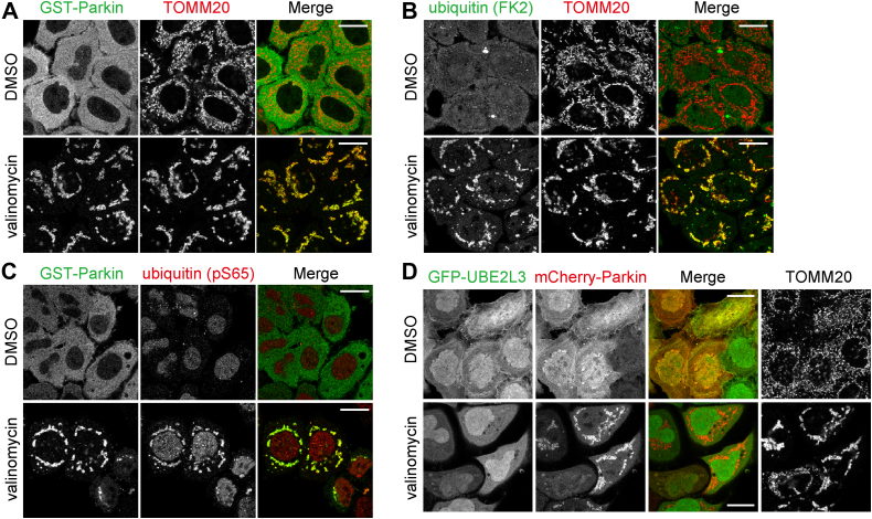
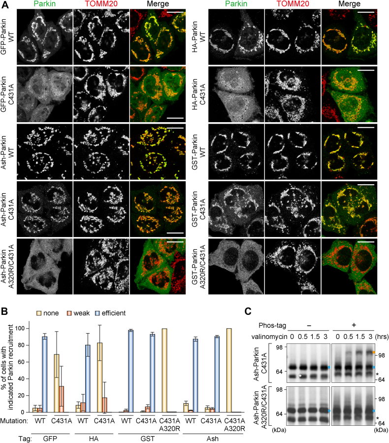
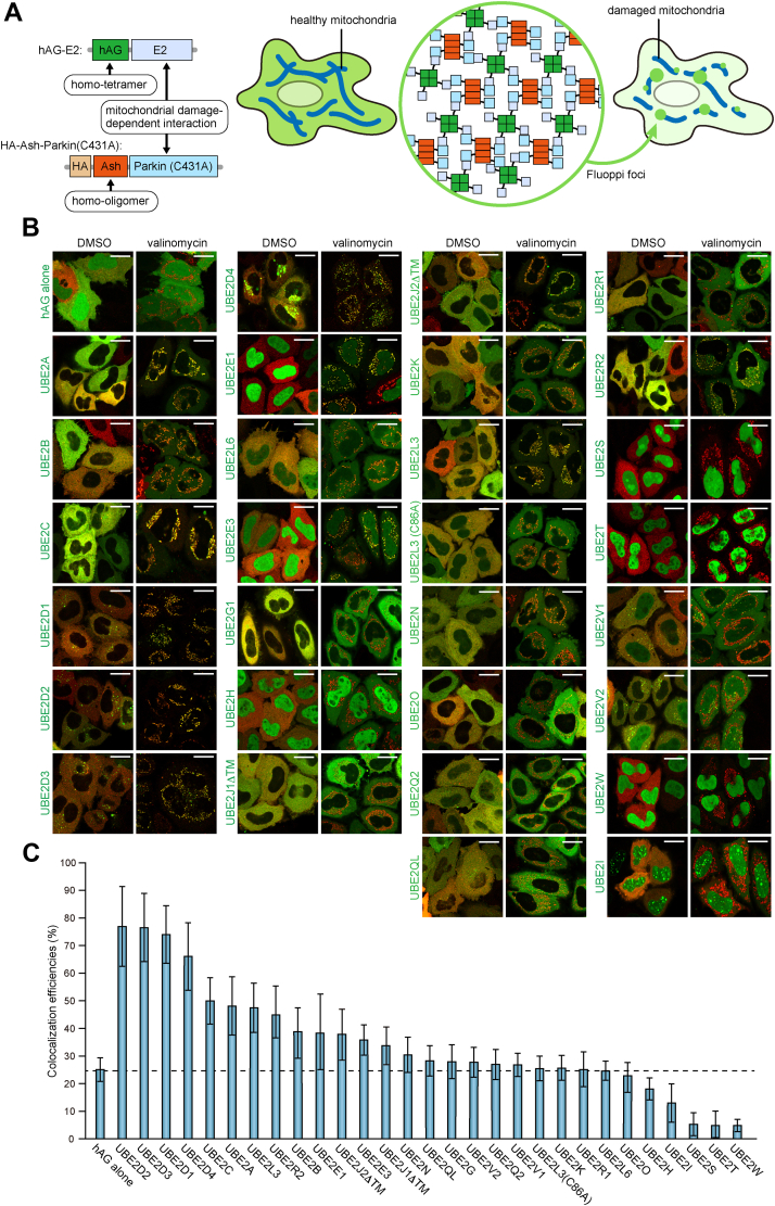
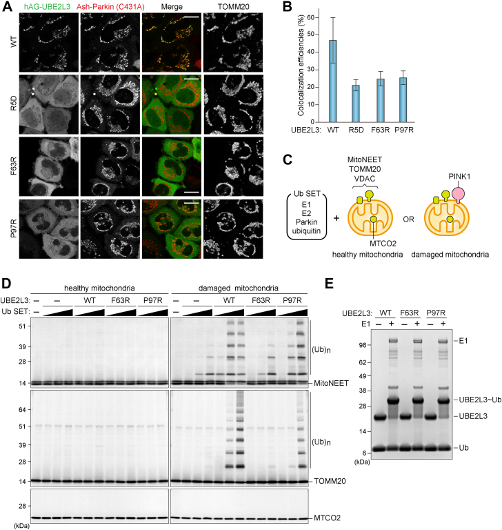
RING-between RING (RBR)-type ubiquitin (Ub) ligases (E3s) such as Parkin receive Ub from Ub-conjugating enzymes (E2s) in response to ligase activation. However, the specific E2s that transfer Ub to each RBR-type ligase are largely unknown because of insufficient methods for monitoring their interaction. To address this problem, we have developed a method that detects intracellular interactions between E2s and activated Parkin. Fluorescent homotetramer Azami-Green fused with E2 and oligomeric Ash (Assembly helper) fused with Parkin form a liquid-liquid phase separation (LLPS) in cells only when E2 and Parkin interact. Using this method, we identified multiple E2s interacting with activated Parkin on damaged mitochondria during mitophagy. Combined with in vitro ubiquitination assays and bioinformatics, these findings revealed an underlying consensus sequence for E2 interactions with activated Parkin. Application of this method to other RBR-type E3s including HOIP, HHARI, and TRIAD1 revealed that HOIP forms an LLPS with its substrate NEMO in response to a proinflammatory cytokine and that HHARI and TRIAD1 form a cytosolic LLPS independent of Ub-like protein NEDD8. Since an E2-E3 interaction is a prerequisite for RBR-type E3 activation and subsequent substrate ubiquitination, the method we have established here can be an in-cell tool to elucidate the potentially novel mechanisms involved in RBR-type E3s.

Keywords: PINK1; Parkin; RBR-type E3; mitochondria; mitophagy; ubiquitin.


Conflict of interest statement

Conflict of interest The authors declare that they have no conflicts of interest with the contents of this article.

Figures

Figure 1
Figure 1
UBE2L3 is not stably associated on mitochondria during Parkin-driven mitophagy. HeLa cells stably expressing GST-Parkin (AC) or HeLa cells expressing GFP-UBE2L3 and mCherry-Parkin (D) were treated with dimethyl sulfoxide (DMSO) or valinomycin for 3 h and immunostained with anti-GST, TOMM20, ubiquitin (FK2), and ubiquitin (pS65) antibodies. Bars represent 10 μm. GST, glutathione-S-transferase.
Figure 2
Figure 2
Oligomerized Parkin can translocate to mitochondria in the absence of the ubiquitin amplification positive-feedback loop.A, HeLa cells expressing the indicated Parkin were treated with valinomycin for 3 h and then immunostained. Bars represent 10 μm. B, the percentage of cells in (A) exhibiting mitochondria with translocated Parkin. Error bars represent mean ± SD of >100 cells counted in each of three independent experiments. C, HeLa cells expressing Ash-Parkin (C431A or A320R/C431A) were treated with valinomycin for the indicated times. Total cell lysates were analyzed by phos-tag PAGE and immunoblotted with an anti-Parkin antibody (51). Unphosphorylated and phosphorylated Parkin are indicated by light blue and orange dots, respectively. Asterisks denote truncated Parkin species. Ash, Assembly helper.
Figure 3
Figure 3
Fluoppi analysis reveals intracellular interactions between various E2 enzymes and Parkin on dysfunctional mitochondria.A, schematic representation of Parkin–E2 Fluoppi. hAG-E2 forms a homotetramer, and HA-Ash-Parkin (C431A) forms a homooligomer. When mitochondrial damage induces the interaction between Parkin and E2, Fluoppi foci will be formed on the damaged mitochondria. B, HeLa cells expressing Ash-Parkin (C431A) (red) and the indicated hAG-tagged E2 enzymes (green) were treated with dimethyl sulfoxide (DMSO) or valinomycin for 3 h and then immunostained with an anti-Parkin antibody. Bars represent 10 μm. The same images are shown in Figs. S2 and S3 as merge along with the separated hAG-E2 and HA-Ash-Parkin images and TOMM20 signals. C, the efficiency of colocalization in (A) between various hAG-tagged E2 enzymes and TOMM20 after 3 h valinomycin treatment. Error bars represent mean ± SD of >50 cells counted in two independent experiments. Ash, Assembly helper; HA, hemagglutinin.
Figure 4
Figure 4
UBE2L3 mutants defective for Parkin interactions do not mediate mitochondrial ubiquitination.A, HeLa cells expressing Ash-Parkin (C431A) and hAG-UBE2L3 (WT, R5D, F63R, or P97R) were treated with valinomycin for 3 h and then immunostained. Bars represent 10 μm. B, the efficiency of colocalization in (A) between the various hAG-UBE2L3 constructs and TOMM20 after 3 h valinomycin treatment. Error bars represent mean ± SD of >50 cells counted in two independent experiments. C, schematic diagram of in vitro mitochondrial ubiquitination. Ubiquitin (Ub) SET contains recombinant E1 (UBA1), E2, Parkin, and ubiquitin. Healthy mitochondria and damaged mitochondria were isolated from HeLa cells treated with dimethyl sulfoxide (DMSO) and valinomycin for 6 h, respectively. MitoNEET, TOMM20, and VDAC localize on the outer membrane, whereas MTCO2 is in the matrix. PINK1 accumulates only on the outer membrane of damaged mitochondria. D, healthy and damaged mitochondria were incubated with or without various concentrations of Ub SET at 32 °C for 30 min and immunoblotted with the indicated antibodies. (Ub)n denotes polyubiquitin chains. E, recombinant UBE2L3 was incubated with ubiquitin in the presence or the absence of E1 at 32 °C for 30 min. Samples were analyzed by SDS-PAGE under nonreducing conditions followed by Coomassie brilliant blue staining. Ash, Assembly helper.
Figure 5
Figure 5
In vitro mitochondrial ubiquitination assay identifies E2s involved in Parkin activation.A, the indicated recombinant E2s were expressed in bacterial cells and purification using a nickel–nitrilotriacetic acid column. A ubiquitin (Ub) conjugation assay was performed by incubating the recombinant E2s and Ub with or without E1 at 32 °C for 30 min. Samples were analyzed by SDS-PAGE under nonreducing conditions followed by Coomassie brilliant blue staining. Light blue and orange dots denote E2 alone and Ub-conjugated E2, respectively. B, mitochondria isolated from valinomycin-treated HeLa cells were incubated with various concentrations of Ub SET (Fig. 4C) containing the indicated E2s at 32 °C for 30 min. Mitochondrial ubiquitination was analyzed by immunoblotting with anti-TOMM20, VDAC, and MTCO2 antibodies. (Ub)n denotes polyubiquitin chains.
Figure 6
Figure 6
Consensus sequence in E2s is critical for interacting with activated Parkin.A, three regions in UBE2L3 that are situated within 5 Å of Parkin were selected based on the UBE2L3–Parkin structure (Protein Data Bank ID: 6N13). B, changes in protein binding affinity (ΔAffinity) after Ala mutation of the target residue. Values were calculated using BioLuminate alanine scanning as implemented by Schrödinger Release 2021-4 (Schrödinger, LLC). C, multiple sequence alignment of E2s with high and low ubiquitination. The three Parkin proximal regions are indicated. The E2s having >60% sequence identity are removed.
Figure 7
Figure 7
Interactions between UBE2L3 and HOIP captured with the Fluoppi assay.A, HeLa cells expressing HA-Ash-HOIP (C885A) and hAG-UBE2L3 (WT, C86A, or F63R) were treated with dimethyl sulfoxide (DMSO) (NT) or tumor necrosis factor alpha (TNF-α) for 10 min and then immunostained with an anti-HA antibody. Bars represent 10 μm. B, the percentage of cells in (A) with foci consisting of HA-Ash-HOIP and hAG-UBE2L3. Error bars represent mean ± SD of >100 cells counted in each of three independent experiments. C, HeLa cells expressing HA-Ash-HOIP (C885A), hAG-UBE2L3, and FLAG-NEMO were treated with TNF-α for 10 min and then immunostained with anti-HA and anti-FLAG antibodies. Insets represent magnified images of the areas indicated. Bars represent 10 μm. Ash, Assembly helper; HA, hemagglutinin.
Figure 8
Figure 8
HHARI and TRIAD1 interactions captured with the Fluoppi assay.A, HeLa cells expressing hAG constructs (hAG alone, hAG-UBE2L3 WT, or P97R) and HA-Ash-HHARI C357A were immunostained with an anti-HA antibody. To inhibit neddylation, cells were treated overnight with 1 μM MLN4924 and then immunostained. Nuclei were stained with 4′,6-diamidino-2-phenylindole (DAPI). Bars represent 10 μm. B, the percentage of cells in (A) with foci. CA denotes the HHARI C357A mutant. Error bars represent mean ± SD of >100 cells counted in each of three independent experiments. C, HeLa cells expressing the hAG constructs and HA-Ash-TRIAD1 C310A were immunostained with an anti-HA antibody. Bars represent 10 μm. D, the percentage of cells in (C) with foci. CA denotes the TRIAD1 C310A mutant. Error bars represent mean ± SD of >100 cells counted in each of three independent experiments. E, total cell lysates prepared from HeLa cells expressing hAG constructs (hAG alone or hAG-UBE2L3) and HA-Ash-RBR constructs (HHARI or TRIAD1) were immunoblotted with anti-HA, anti-AG, and anti-TOMM20 antibodies. Orange dots represent ubiquitinated HHARI and TRIAD1. F, HeLa cells expressing hAG-UBE2L3, V5-NEDD8, and HA-Ash-HHARI C357A or HA-Ash-TRIAD1 C310A were immunostained with anti-HA and V5 antibodies. Bars represent 10 μm; bars in magnification panels represent 2 μm. G, inhibition of neddylation was confirmed by immunoblotting. HeLa cells expressing HA-Ash-HHARI or HA-Ash-TRIAD1 were treated with or without MLN4924 overnight. Total cell lysates were immunoblotted with anti-CUL1, anti-CUL4, and anti-TOMM20 antibodies. Ash, Assembly helper; HA, hemagglutinin.

References

    1. Wenzel D.M., Lissounov A., Brzovic P.S., Klevit R.E. UBCH7 reactivity profile reveals parkin and HHARI to be RING/HECT hybrids. Nature. 2011;474:105–108. - PMC - PubMed
    1. Trempe J.F., Sauve V., Grenier K., Seirafi M., Tang M.Y., Menade M., et al. Structure of parkin reveals mechanisms for ubiquitin ligase activation. Science. 2013;340:1451–1455. - PubMed
    1. Duda D.M., Olszewski J.L., Schuermann J.P., Kurinov I., Miller D.J., Nourse A., et al. Structure of HHARI, a RING-IBR-RING ubiquitin ligase: autoinhibition of an Ariadne-family E3 and insights into ligation mechanism. Structure. 2013;21:1030–1041. - PMC - PubMed
    1. Stieglitz B., Rana R.R., Koliopoulos M.G., Morris-Davies A.C., Schaeffer V., Christodoulou E., et al. Structural basis for ligase-specific conjugation of linear ubiquitin chains by HOIP. Nature. 2013;503:422–426. - PMC - PubMed
    1. Dove K.K., Klevit R.E. RING-between-RING E3 ligases: emerging themes amid the variations. J. Mol. Biol. 2017;429:3363–3375. - PMC - PubMed

Publication types

MeSH terms