. 2023 Feb 23;141(8):930-944.
doi: 10.1182/blood.2022017419.

Proximity proteomics identifies septins and PAK2 as decisive regulators of actomyosin-mediated expulsion of von Willebrand factor

Affiliations

Proximity proteomics identifies septins and PAK2 as decisive regulators of actomyosin-mediated expulsion of von Willebrand factor

Sammy El-Mansi et al. Blood. .

Abstract

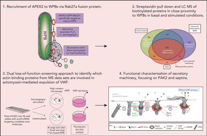
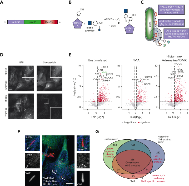
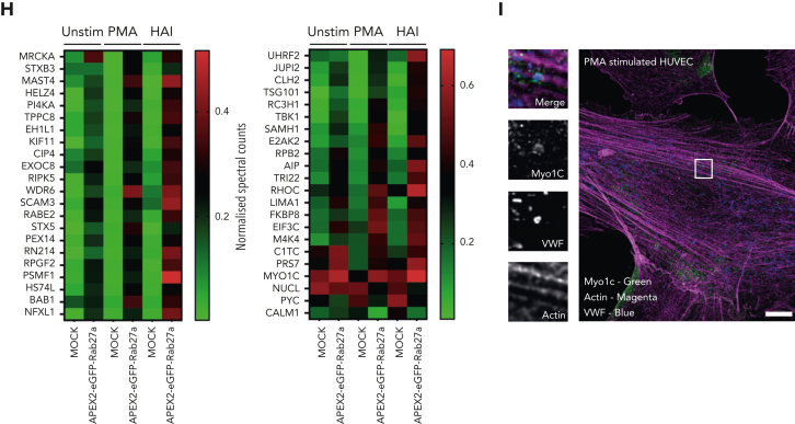
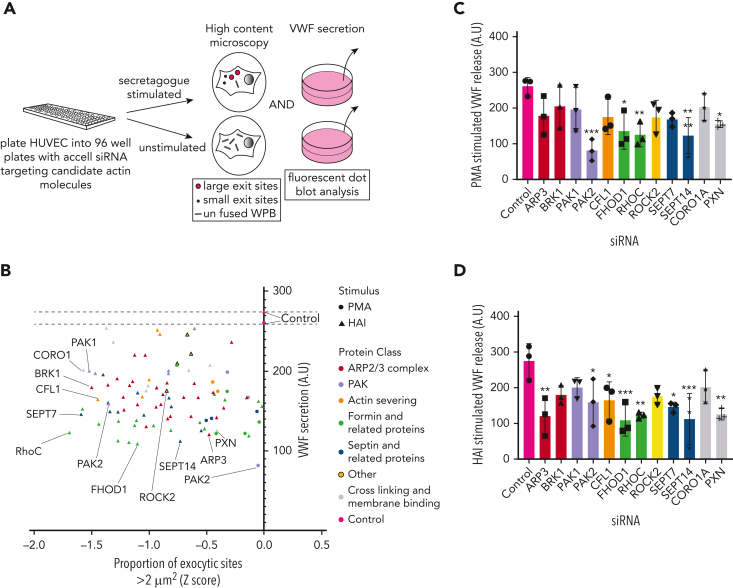
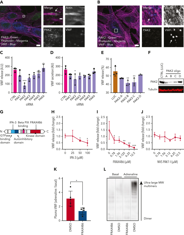
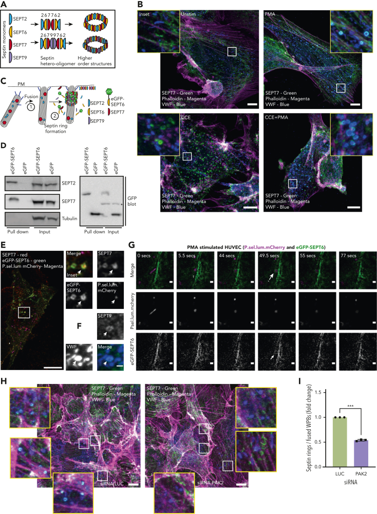
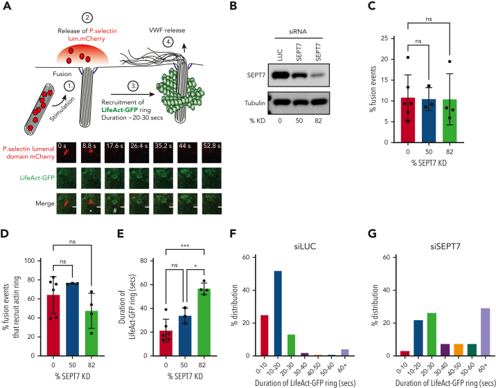
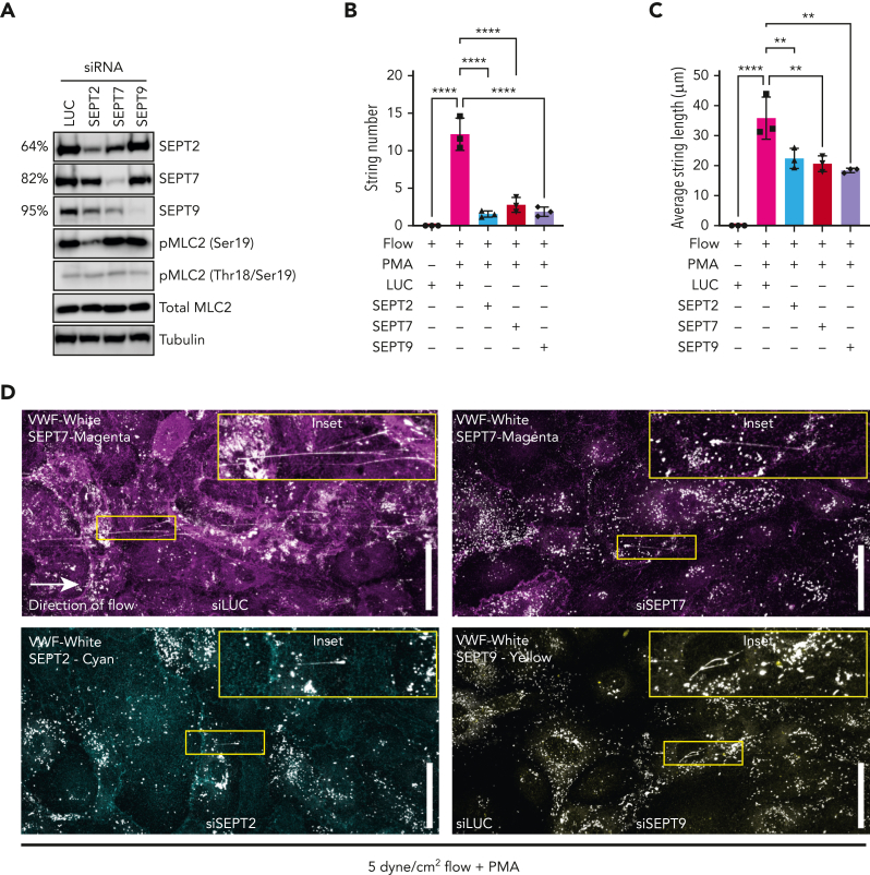
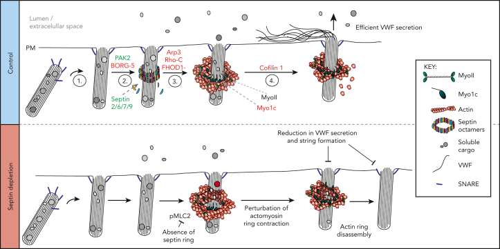
In response to tissue injury, within seconds the ultra-large glycoprotein von Willebrand factor (VWF) is released from endothelial storage organelles (Weibel-Palade bodies) into the lumen of the blood vasculature, where it leads to the recruitment of platelets. The marked size of VWF multimers represents an unprecedented burden on the secretory machinery of endothelial cells (ECs). ECs have evolved mechanisms to overcome this, most notably an actomyosin ring that forms, contracts, and squeezes out its unwieldy cargo. Inhibiting the formation or function of these structures represents a novel therapeutic target for thrombotic pathologies, although characterizing proteins associated with such a dynamic process has been challenging. We have combined APEX2 proximity labeling with an innovative dual loss-of-function screen to identify proteins associated with actomyosin ring function. We show that p21 activated kinase 2 (PAK2) recruits septin hetero-oligomers, a molecular interaction that forms a ring around exocytic sites. This cascade of events controls actomyosin ring function, aiding efficient exocytic release. Genetic or pharmacological inhibition of PAK2 or septins led to inefficient release of VWF and a failure to form platelet-catching strings. This new molecular mechanism offers additional therapeutic targets for the control of thrombotic disease and is highly relevant to other secretory systems that employ exocytic actomyosin machinery.


Conflict of interest statement

Conflict-of-interest disclosure: The authors declare no competing financial interests.

Figures

None
Graphical abstract
Figure 1.
Figure 1.
Proximity proteomics reveals novel WPB-associated machinery. (A) Schematic representation of the APEX2-eGFP-Rab27a construct used to transfect HUVECs. (B) Biotinylation reaction catalyzed following the addition of hydrogen peroxide and biotin tyramide. (C) The APEX2-eGFP-Rab27a construct is recruited to mature WPBs, allowing spatiotemporally-specific biotin labeling on nearby proteins. The addition of stimulants allows detection of proteins with putative roles in basal and regulated granule release. (D) Confocal LSM of HUVECs in the presence or absence of biotin tyramide. Biotinylated proteins are detected using AF647-conjugated streptavidin and colocalized with the enhanced green fluorescent protein (eGFP)−tagged fusion protein. (E) Scatter graphs depicting the most significant (paired t test) and upregulated proteins as compared to mock-transfected control HUVECs in unstimulated, PMA-stimulated, and HAI-stimulated cells. Kinesin-1 heavy chain (KINH) (green) was used to validate the data set. (F) IF analyses of HUVECs illustrate KIF5B (KINH) localizing to one end of WPBs under resting conditions. Scale bar, 10 μm. (G) Venn diagram of mass spectrometry data sets illustrates the number of proteins in close proximity to WPBs under all conditions, exclusively under basal conditions or following stimulation. (H) Heat map depiction of the average normalized spectral counts of the 44 stimulation-associated proteins in mock and APEX2-eGFP-Rab27a groups (in the presence or absence of PMA/HAI). (I) Myosin-1c (green) is present at points of WPB fusion in PMA-stimulated HUVECs. Scale bar, 10 μm.
Figure 1.
Figure 1.
Proximity proteomics reveals novel WPB-associated machinery. (A) Schematic representation of the APEX2-eGFP-Rab27a construct used to transfect HUVECs. (B) Biotinylation reaction catalyzed following the addition of hydrogen peroxide and biotin tyramide. (C) The APEX2-eGFP-Rab27a construct is recruited to mature WPBs, allowing spatiotemporally-specific biotin labeling on nearby proteins. The addition of stimulants allows detection of proteins with putative roles in basal and regulated granule release. (D) Confocal LSM of HUVECs in the presence or absence of biotin tyramide. Biotinylated proteins are detected using AF647-conjugated streptavidin and colocalized with the enhanced green fluorescent protein (eGFP)−tagged fusion protein. (E) Scatter graphs depicting the most significant (paired t test) and upregulated proteins as compared to mock-transfected control HUVECs in unstimulated, PMA-stimulated, and HAI-stimulated cells. Kinesin-1 heavy chain (KINH) (green) was used to validate the data set. (F) IF analyses of HUVECs illustrate KIF5B (KINH) localizing to one end of WPBs under resting conditions. Scale bar, 10 μm. (G) Venn diagram of mass spectrometry data sets illustrates the number of proteins in close proximity to WPBs under all conditions, exclusively under basal conditions or following stimulation. (H) Heat map depiction of the average normalized spectral counts of the 44 stimulation-associated proteins in mock and APEX2-eGFP-Rab27a groups (in the presence or absence of PMA/HAI). (I) Myosin-1c (green) is present at points of WPB fusion in PMA-stimulated HUVECs. Scale bar, 10 μm.
Figure 2.
Figure 2.
Dual siRNA subscreen identifies which actin-binding proteins play a role in VWF release. (A) Schematic representation of the loss-of-function screening approach used. HUVECs were transfected with siRNA-targeting proteins (and their associated effectors) identified through proximity proteomics. Transfected cells were stimulated with PMA or HAI and either fixed in the presence of an anti-VWF antibody for analysis by confocal LSM to quantify WPB exit site size or the supernatants were collected for quantification of VWF release by fluorescent dot blot. (B) A scatter plot was generated that depicted only the proteins whose reduction in expression led to a decrease in VWF release or proportion of exit sites less than 2 μm2 in comparison to control siRNA. The targets with most prominent effect from each protein class have been annotated. The effect of depletion of shortlisted candidates on (C) PMA-stimulated and (D) HAI-stimulated VWF release. ∗P < .05, ∗∗P < .01, ∗∗∗P < .005 (one-way analysis of variance [ANOVA] with Dunnett multiple comparison).
Figure 3.
Figure 3.
PAK2 signaling regulates VWF release from endothelial cells. (A) Confocal analysis localizes PAK2 (green) to the cytoplasm and at the end of dorsal actin stress fibers (magenta) in HUVECs. (B) PAK2 (green) is not present on the actin ring (magenta) that forms at exocytosis (inset). Scale bars, 10 μm (full size) and 1 μm (inset); brightness and contrast increased for clarity. (C-D) The effect of siRNA depletion of the 6 PAK isoforms on (C) PMA-stimulated and (D) HAI-stimulated VWF secretion. (E) PAK2 depletion reduced PMA stimulated VWF release in a dose-dependent fashion. HUVECs were independently transfected with 4 different siRNA oligonucleotides targeting PAK2. VWF secretion was determined by fluorescent dot blot and PAK2 protein abundance determined by western blotting. (F) Western blotting of HUVEC lysate detected as single band at the estimated size of 61 kDa. (G) Schematic representation of PAK2 structure and targets for pharmacological inhibition. (H) Pharmacological inhibition of the autoregulatory domain of PAK2 with 25 to 50 μM IPA-3 prevents VWF release. (I) Targeting the catalytic kinase domain of PAK2 with FRAX486 inhibits VWF release (0.78-12.5 μM). (J) The specific PAK1 inhibitor NVS PAK1.1 has no effect on VWF release at 12.5 μM. ∗P < .05, ∗∗P < .01, ∗∗∗P < .005 (one-way ANOVA with Dunnett multiple comparison). (K-L) Effect of FRAX486 administration on adrenaline-stimulated VWF secretion in vivo. C57 black WT mice were administered FRAX486 or an equivalent volume of DMSO. Tail vein bleeds were performed to assess basal VWF levels. Intraperitoneal injection of adrenaline (0.5 mg/kg) was used to stimulate VWF secretion from the murine vasculature (DMSO, n = 5; FRAX486, n = 4). After 30 minutes, mice were sacrificed and the plasma was isolated. Plasma VWF levels and multimer composition were assessed by near-infrared dot blot (K) and multimer gel (L). MW, molecular weight. ∗P < .05.
Figure 4.
Figure 4.
Septin rings are recruited to WPBs post fusion in an actin-independent but PAK2-dependent process. (A) To form higher-order structures such as rings, septin (SEPT) monomers from different subfamilies (eg, SEPT2/6/7/9) must form hetero-oligomers (6mers or 8mers). (B) SEPT7 is recruited to WPBs following stimulation. IF analyses of SEPT7 (green), VWF (blue), and actin (magenta) in HUVEC cultures stimulated with the presence or absence 100 ng/mL PMA and/or 1 μM CCE. Scale bar, 10 μm. (C) Schematic representation of the approach used to image septins in live endothelial cells. GFP-tagged SEPT6 interacts with endogenous SEPT7 upon ring formation. (D) GFP pull-down of control GFP and eGFP-SEPT6−transfected cells detects the association of the eGFP-SEPT6 transgene product with endogenous SEPT2 and SEPT7. Beta tubulin was absent in both eGFP control and eGFP-SEPT6 pull-down samples. ∗Cross-reactivity. (E) Confocal microscopy of fixed HUVECs transfected with P.sel.lum.mCherry (magenta) and eGFP-SEPT6 (green) and probed for SEPT7 (red) by IF. Images show that eGFP-SEPT6 is incorporated into rings that are positive for SEPT7 staining. Scale bars, 10 μm. (F) SEPT9 is localized to fused WPBs following stimulation with PMA. (G) Live cell imaging of PMA (100 ng/mL)−stimulated HUVECs indicates that SEPT6-eGFP rings form post fusion. Brightness and contrast increased for clarity. †WPB fusion. ∗Septin ring formation; 0.5- to 6-μm Z stacks acquired every 5 seconds for 10 minutes. Scale bars, 1 μm. (H) HUVECs were transfected with LUC control (left panel) or PAK2-targeted siRNA (right panel) and probed for SEPT7 (green), VWF (blue), and actin (magenta). Scale bar, 10 μm. (I) Quantification of the number of SEPT7 associated with VWF as a proportion of the total number of rounded (fused) WPBs. A total of 15 images per condition from three independent experiments (ratio paired t test). ∗∗∗P < .005.
Figure 5.
Figure 5.
siRNA-mediated depletion of SEPT7 perturbs actin ring kinetics during WPB exocytosis. (A) WPB fusion was quantified by change in morphology (rod>round) and loss of mCherry signal, whereas actin ring recruitment was assessed by the presence or absence of LifeAct-GFP signal. White asterisk (∗) indicates change in morphology from rod>round. Number sign (#) indicates recruitment of LifeAct GFP ring. HUVECs were co-transfected (2 rounds) with either 300 pM LUC or independent SEPT7-targeted siRNAs and both LifeAct-GFP and p.sel.lum.mCherry. (B) Western blotting was used to determine KD efficiencies of approximately 50% and 82% (averaged from 3 independent experiments). (C) SEPT7 knockdown did not affect vesicle fusion. (D) Quantification of actin ring formation as a percentage of total fusion events. (D-E) Quantification of the duration of the “lifetime” of actin ring (LifeAct-GFP signal) in control and SEPT7 KD cells. ∗∗P < .01, ∗∗∗P < .005. One-way ANOVA with Tukey multiple-comparison test. Frequency distribution (%) of actin ring duration in luciferase siRNA (F) and SEPT7 siRNA-treated cells (84% KD efficiency) (G). Cell numbers for live cell imaging. LUC control, 46 cells; SEPT7, 25 cells (50% KD), and SEPT7, 30 cells (82% KD).
Figure 6.
Figure 6.
Septin depletion reduces efficiency of VWF string release. HUVECs were depleted of SEPT2, SEPT7, and SEPT9 using siRNA and VWF string secretion under flow assessed by immunofluorescence. (A) Western blotting confirmed effective target protein depletion in HUVECs treated with 300 pM SEPT2/7/ siRNA. MLC2 phosphorylation at ser19 was reduced in SEPT2 KD cells. (B) Depletion of SEPT/7/9 inhibited VWF string formation in HUVECs exposed to 5 dynes/cm2. (C) On average, SEPT2/7/9 depletion resulted in shorter VWF strings bound to the endothelial cell surface. Data presented as mean from each independent experiment (n = 3). One-way ANOVA with Tukey multiple comparison test. (D) Confocal microscopy images (0.5 μm Z stacks) were acquired using identical settings. Scale bar, 50 μm. Representative immunofluorescence images of septin and VWF staining in siLUC (top left panel), siSEPT7 (top right panel), siSEPT2 (bottom left panel), and siSEPT9 (bottom right panel) treated HUVECs.
Figure 7.
Figure 7.
Schematic representation of a working model depicting machinery associated with actomyosin-dependent expulsion of VWF from endothelial cells. (1) Upon stimulation, WPBs are trafficked to the cell surface where fusion with the plasma membrane occurs. (2) Septin rings are recruited to fused WPBs (independently of actin), where they likely transfer myosins (eg, NMII or Myo1C) or their activators. This process is PAK2 dependent. (3) Actin rings are formed by rearrangement of cortical actin or de novo nucleation. Possible candidates from our screen include FHOD1 and Arp3. Actin ring formation itself could displace the septin ring. (4) The actin ring contracts/compresses with the aid of NMII and cofilin 1. Myo1c may play a role in tethering actin to the WPB membrane. Proteins in red and green were identified through proximity proteomics and loss of function screening approaches. Proteins in black were identified through previous research. (Bottom panel) Disrupting the formation of the septin ring during WPB exocytosis results in prolonged actin ring dynamics as well as an inhibition of VWF secretion and string formation. SEPT2 depletion reduces the phosphorylation status of MLC2. This results in actin rings contracting more slowly or failing to contract before being disassembled. Interfering with actin ring formation or function results in less efficient VWF secretion.

Comment in

  • CSI: Weibel-Palade bodies.
    Bierings R, Voorberg J. Bierings R, et al. Blood. 2023 Feb 23;141(8):820-821. doi: 10.1182/blood.2022019268. Blood. 2023. PMID: 36821184 No abstract available.

References

    1. Nightingale TD, White IJ, Doyle EL, et al. Actomyosin II contractility expels von Willebrand factor from Weibel-Palade bodies during exocytosis. J Cell Biol. 2011;194(4):613–629. - PMC - PubMed
    1. Erent M, Meli A, Moisoi N, et al. Rate, extent and concentration dependence of histamine-evoked Weibel-Palade body exocytosis determined from individual fusion events in human endothelial cells. J Physiol. 2007;583(Pt 1):195–212. - PMC - PubMed
    1. McCormack JJ, Lopes da Silva M, Ferraro F, Patella F, Cutler DF. Weibel-Palade bodies at a glance. J Cell Sci. 2017;130(21):3611–3617. - PubMed
    1. De Ceunynck K, De Meyer SF, Vanhoorelbeke K. Unwinding the von Willebrand factor strings puzzle. Blood. 2013;121(2):270–277. - PubMed
    1. Leebeek FW, Eikenboom JC. Von Willebrand's Disease. N Engl J Med. 2016;375(21):2067–2080. - PubMed

Publication types