Review
. 2023 Feb;49(1):e12875.
doi: 10.1111/nan.12875.

Arteriolar neuropathology in cerebral microvascular disease

Affiliations
Review

Arteriolar neuropathology in cerebral microvascular disease

Chuo Fang et al. Neuropathol Appl Neurobiol. 2023 Feb.

Abstract

Cerebral microvascular disease (MVD) is an important cause of vascular cognitive impairment. MVD is heterogeneous in aetiology, ranging from universal ageing to the sporadic (hypertension, sporadic cerebral amyloid angiopathy [CAA] and chronic kidney disease) and the genetic (e.g., familial CAA, cerebral autosomal dominant arteriopathy with subcortical infarcts and leukoencephalopathy [CADASIL] and cerebral autosomal recessive arteriopathy with subcortical infarcts and leukoencephalopathy [CARASIL]). The brain parenchymal consequences of MVD predominantly consist of lacunar infarcts (lacunes), microinfarcts, white matter disease of ageing and microhaemorrhages. MVD is characterised by substantial arteriolar neuropathology involving ubiquitous vascular smooth muscle cell (SMC) abnormalities. Cerebral MVD is characterised by a wide variety of arteriolar injuries but only a limited number of parenchymal manifestations. We reason that the cerebral arteriole plays a dominant role in the pathogenesis of each type of MVD. Perturbations in signalling and function (i.e., changes in proliferation, apoptosis, phenotypic switch and migration of SMC) are prominent in the pathogenesis of cerebral MVD, making 'cerebral angiomyopathy' an appropriate term to describe the spectrum of pathologic abnormalities. The evidence suggests that the cerebral arteriole acts as both source and mediator of parenchymal injury in MVD.

Keywords: cerebral angiomyopathy; cerebral arterioles; cerebral microvascular disease; neuropathology; smooth muscle cells.


Conflict of interest statement

The author(s) declared no potential conflicts of interest with respect to the research, authorship and/or publication of this article.

Figures
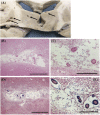
FIGURE 1
FIGURE 1
Arteriolar neuropathology in hypertension. (A) Lacunar infarcts (arrows) in the basal ganglia and thalamus of an autopsy specimen. (B, C) Hyalinosis with a ‘glassy’ or hyalinised appearance of the arteriolar wall associated with infarcts in these sections (hematoxylin & eosin [H&E]) and characterised by vessel wall thickening. (D, E) Profound collagen replacement (blue) and smooth muscle cell (SMC) loss and degeneration in the arteriolar wall with hypertension (Masson's trichrome). A normal arteriole would stain red by Masson's trichrome due to the presence of SMC. (B, D) Scale bar 600 μm. (C, E) Scale bar 200 μm
FIGURE 2
FIGURE 2
Arteriolar cerebral amyloid angiopathy (CAA). Arachnoid membrane and subarachnoid space above superficial right parieto‐occipital cortex, with amyloid‐laden arterioles associated with haemosiderin‐laden macrophages (arrow) (hematoxylin & eosin [H&E]). Scale bar 1200 μm
FIGURE 3
FIGURE 3
Arteriolar thickening in chronic kidney disease (CKD) I. (A, B) Arteriolosclerotic changes with luminal narrowing, intimal thickening and degeneration of smooth muscle cells (SMC) and elastin in the media in arterioles of the basal ganglia and (C) the white matter. Arrows indicate (A) internal elastic lamina, (B) smooth muscle, and (C) adventitia. (D) Vessel with no/minimal arteriolosclerosis. (E) Kidney from the same patient as panels (A) and (B) showing arteriosclerosis with intimal hyperplasia (arrow) and glomerulosclerosis (arrowhead). (A–C) Scale bar 300 μm. (D, E) Scale bar 200 μm
FIGURE 4
FIGURE 4
Arteriolar neuropathology in chronic kidney disease (CKD) II. (A, B) Cerebral microhaemorrhages with haemosiderin‐laden macrophages (arrows) adjacent to white matter vessels. (C) Microvascular adventitial fibrosis with marked vessel wall thickening in the subcortical white matter (hematoxylin & eosin [H&E]). (D) Kidney from the same patient as panels (A) and (B) showing hypertensive nephrosclerosis with intimal hyperplasia (arrow) and arteriolar hyalinosis (arrowhead). (A–C) Scale bar 300 μm. (D) Scale bar 200 μm
FIGURE 5
FIGURE 5
Arteriolar neuropathology in cerebral autosomal dominant arteriopathy with subcortical infarcts and leukoencephalopathy (CADASIL) I. (A) Disruption of proteolipids in arterial vessel walls in CADASIL, with abnormal fat globules (arrow, Oil‐Red‐O). (B) Concentric lamination or onionskin‐type thickening of the arteriolar wall in the basal ganglia of a 30‐year‐old CADASIL subject (hematoxylin & eosin [H&E]). Onionskin‐type thickening of vessel walls is also characteristic of severe hypertensive states often referred to as hyperplastic arteriolosclerosis. (C) Profound vessel wall thickening and glycogen‐cerebroside derangement in CADASIL (H&E). (D) Loss of smooth muscle cells in the media and calcification of arterial walls in the adventitia (arrow) in the basal ganglia in CADASIL (H&E). (A, C) Scale bar 50 μm. (B, D) Scale bar 100 μm
FIGURE 6
FIGURE 6
Arteriolar changes in cerebral autosomal dominant arteriopathy with subcortical infarcts and leukoencephalopathy (CADASIL) II. Post‐mortem CADASIL brain shows perivascular haemosiderin‐laden macrophages (arrows), highlighted by Prussian blue staining, adjacent to arterioles in (A) the basal ganglia and (B, C) thalamus. (D) Haemosiderin‐laden macrophages (arrow) adjacent to a basal ganglia vessel with prominent vessel wall thickening. (E) Profound loss of smooth muscle cells in the media, demonstrated by immunohistochemistry for α‐smooth muscle actin, and adventitial thickening in white matter vessels. (A) Magnification ×100. (B) Magnification ×40. (C) Magnification ×200. (D) Magnification ×40. (E) Scale bar 20 μm
FIGURE 7
FIGURE 7
Arteriolar neuropathology in Fabry disease. Lysosomal globotriaosylceramide‐3 (GL‐3) accumulation in vascular cells (arrow: smooth muscle cell [SMC]; arrowhead: endothelial cells) of a 41‐year‐old patient with Fabry disease, with a section taken from a resected meningioma. (A) High‐resolution light microscopy (Richardson's staining). (B) Electron microscopy demonstrates GL‐3 accumulation in SMC (red arrows) and endothelial cells (white arrow). (A, B) Scale bar 1 μm. Source: Reprinted from Thurberg et al. Mol Genet Metab Rep 2016;11:75–80, with permission from Elsevier
FIGURE 8
FIGURE 8
Arteriolar neuropathology in collagen IV (COL4) mutations. (A) Differential changes in arteriolar walls in COL4 arteriopathy with COL4A1 c.*32G>A mutation in the 3′ untranslated region. (B) Cortical vessel with severe smooth muscle cell (SMC) loss and fibrosis in the adventitia in a 60‐year‐old patient. Periodic acid‐Schiff (PAS)‐positive arteriolar walls also found in COL4A1 arteriopathy in the cortex. (C) White matter vessel with mild–moderate loss of SMC in a 30‐year‐old patient with intracerebral haemorrhage. (A–C) Scale bar 100 μm

References

    1. Kanbay M, Sánchez‐Lozada L‐G, Franco M, et al. Microvascular disease and its role in the brain and cardiovascular system: a potential role for uric acid as a cardiorenal toxin. Nephrol Dial Transplant. 2010;26(2):430‐437. doi:10.1093/ndt/gfq635 - DOI - PubMed
    1. Skrobot OA, Attems J, Esiri M, et al. Vascular cognitive impairment neuropathology guidelines (VCING): the contribution of cerebrovascular pathology to cognitive impairment. Brain. 2016;139(11):2957‐2969. doi:10.1093/brain/aww214 - DOI - PubMed
    1. Basile AM, Pantoni L, Pracucci G, et al. Age, hypertension, and lacunar stroke are the major determinants of the severity of age‐related white matter changes. The LADIS (Leukoaraiosis and Disability in the Elderly) Study. Cerebrovasc Dis. 2006;21(5–6):315‐322. doi:10.1159/000091536 - DOI - PubMed
    1. Shi Y, Wardlaw JM. Update on cerebral small vessel disease: a dynamic whole‐brain disease. Stroke Vasc Neurol. 2016;1(3):83‐92. doi:10.1136/svn-2016-000035 - DOI - PMC - PubMed
    1. Fisher M. Cerebral microbleeds and white matter disease: separated at birth? Eur J Neurol. 2012;19(1):2‐3. doi:10.1111/j.1468-1331.2011.03466.x - DOI - PMC - PubMed

Publication types