Review
. 2023 Feb;46(2):153-166.
doi: 10.1016/j.tins.2022.11.007. Epub 2022 Dec 23.

α-Synuclein in synaptic function and dysfunction

Affiliations
Review

α-Synuclein in synaptic function and dysfunction

Manu Sharma et al. Trends Neurosci. 2023 Feb.

Abstract

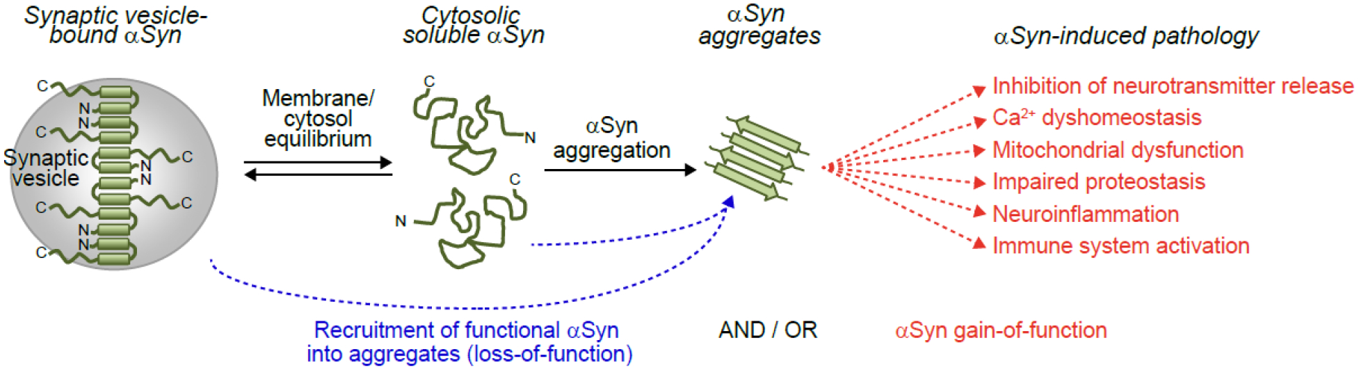
α-Synuclein is a neuronal protein that is enriched in presynaptic terminals. Under physiological conditions, it binds to synaptic vesicle membranes and functions in neurotransmitter release, although the molecular details remain unclear, and it is controversial whether α-synuclein inhibits or facilitates neurotransmitter release. Pathologically, in synucleinopathies including Parkinson's disease (PD), α-synuclein forms aggregates that recruit monomeric α-synuclein and spread throughout the brain, which triggers neuronal dysfunction at molecular, cellular, and organ levels. Here, we present an overview of the effects of α-synuclein on SNARE-complex assembly, neurotransmitter release, and synaptic vesicle pool homeostasis, and discuss how the observed divergent effects of α-synuclein on neurotransmitter release can be reconciled. We also discuss how gain-of-function versus loss-of-function of α-synuclein may contribute to pathogenesis in synucleinopathies.

Keywords: Parkinson’s disease; SNARE; neurotransmission; synapse; synaptic vesicle; synucleinopathies.


Conflict of interest statement

Declaration of interests The authors declare no competing interests.

Figures

Figure 1.
Figure 1.. α-Synuclein domain structure.
(A, B) α-Synuclein is composed of an N-terminal membrane binding region that mediates its association with synaptic vesicles through formation of an amphipathic α-helix, and a C-terminal region that binds to VAMP2 (A). The N-terminal region can be further divided into eleven imperfect KTKEGV repeats (B), and contains the aggregation-prone NAC domain. Several mutations in α-synuclein have been identified that lead to disease (purple) or with yet unknown significance (pink). (C, D) Analysis of the position of mutations within the repetitive KTKEGV regions highlights that mutations cluster in repeat position 5, 10 and 11, while other positions are spared (C). Clusters at positions 10 and 11 face towards the membrane, while cluster 5 faces towards the cytosol (D).
Figure 2.
Figure 2.. The physiological role of α-synuclein in the presynaptic nerve terminal.
Synaptic vesicles can be functionally divided into a reserve pool, recycling pool and readily releasable pool (RRP), which includes docked and fusing vesicles. Upon invasion of an action potential, RRP vesicles fuse with the plasma membrane to release neurotransmitters, and are retrieved via kiss-and-run, ultrafast, or clathrin-independent endocytosis (CIE) to reform the recycling pool of synaptic vesicles, or via clathrin-mediated endocytosis (CME), upon sorting through a recycling compartment, to reform the reserve pool of synaptic vesicles. The recycling pool is supplemented from the reserve pool under conditions of high synaptic activity, to fill the demand in synaptic vesicles. α-Synuclein maintains pool homeostasis via clustering synaptic vesicles, through interaction with the vesicle SNARE protein VAMP2/Synaptobrevin-2 and synapsins, which restricts synaptic vesicle mobility (Inset A). Separately, α-synuclein acts on the fusion pore formed between the synaptic vesicle membrane and the presynaptic plasma membrane where it stabilizes SNARE-complex assembly, which may result in an increased pore size and/or longer pore opening, and increased amount of neurotransmitter release (Inset B). Note that for simplicity, synaptic vesicle pools are depicted as spatially separated.
Figure 3.
Figure 3.. Contributions of loss-of-function of versus gain-of-function of α-synuclein to disease.
α-Synuclein exists in equilibrium between a membrane-bound α-helical pool that mediates its physiological activities (see also Figure 2), and a largely unstructured state in cytosol. At some point during disease pathogenesis, α-synuclein aggregates form from its cytosolic state. α-Synuclein aggregates sequester functional α-synuclein in the same neuron and connected neurons through spread, resulting in loss-of-function of α-synuclein and dysfunctional neurotransmitter release. In addition, or alternatively, α-synuclein aggregates directly lead to inhibition of neurotransmitter release, and result in calcium dyshomeostasis, mitochondrial dysfunction, impaired proteostasis and neuroinflammation, as well as immune system activation through a gain-of-toxic activity. The timeline and contribution of these events to disease remain unknown.

References

    1. George JM (2002) The synucleins. Genome Biol 3, REVIEWS3002 - PMC - PubMed
    1. Maroteaux L, et al. (1988) Synuclein: a neuron-specific protein localized to the nucleus and presynaptic nerve terminal. J Neurosci 8, 2804–2815 - PMC - PubMed
    1. Iwai A, et al. (1995) The precursor protein of non-A beta component of Alzheimer’s disease amyloid is a presynaptic protein of the central nervous system. Neuron 14, 467–475 - PubMed
    1. Spillantini MG, et al. (1997) Alpha-synuclein in Lewy bodies. Nature 388, 839–840 - PubMed
    1. George JM, et al. (1995) Characterization of a novel protein regulated during the critical period for song learning in the zebra finch. Neuron 15, 361–372 - PubMed

Publication types

MeSH terms