. 2022 Nov 30;25(12):105685.
doi: 10.1016/j.isci.2022.105685. eCollection 2022 Dec 22.

Neonatal exposures to sevoflurane in rhesus monkeys alter synaptic ultrastructure in later life

Affiliations

Neonatal exposures to sevoflurane in rhesus monkeys alter synaptic ultrastructure in later life

Tristan Fehr et al. iScience. .

Abstract

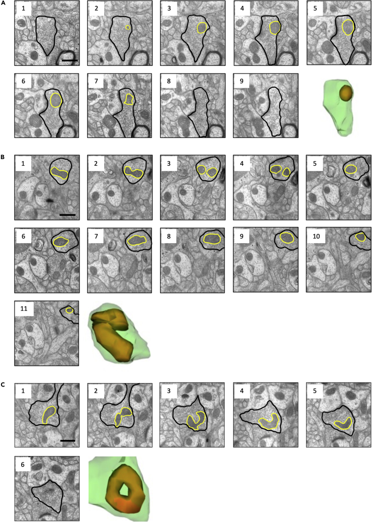
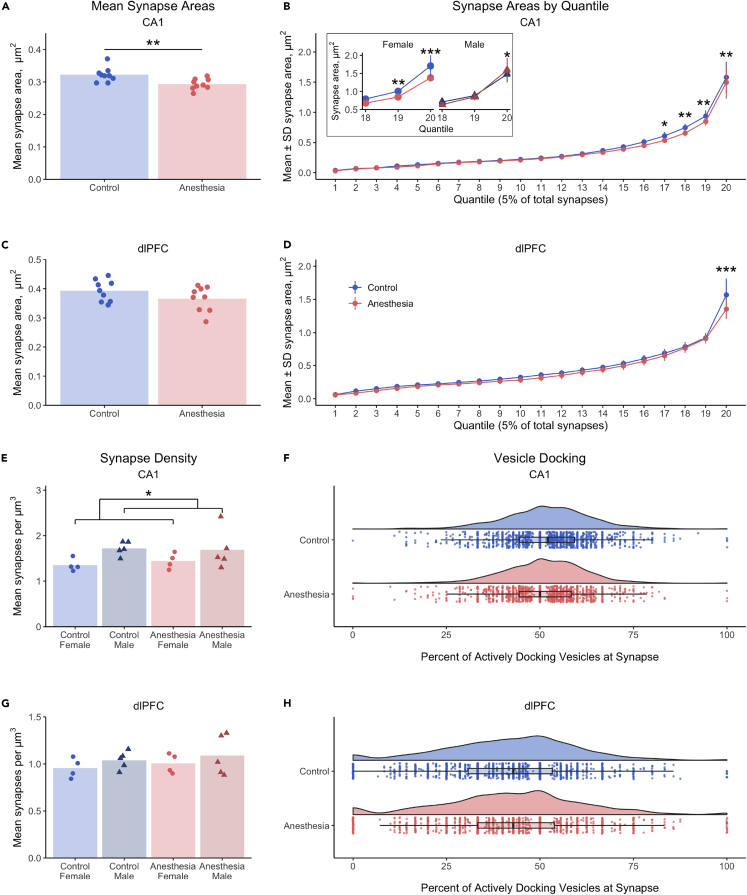
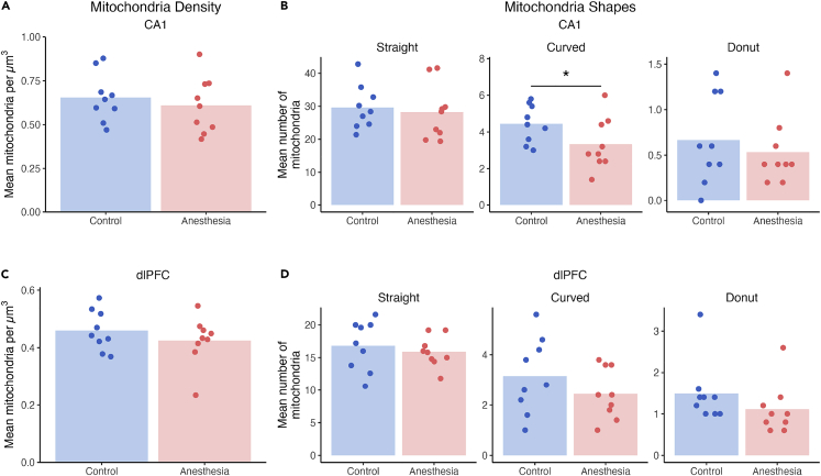
Repeated or prolonged early life exposure to anesthesia is neurotoxic in animals and associated with neurocognitive impairment in later life in humans. We used electron microscopy with unbiased stereological sampling to assess synaptic ultrastructure in dorsolateral prefrontal cortex (dlPFC) and hippocampal CA1 of female and male rhesus monkeys, four years after three 4-h exposures to sevoflurane during the first five postnatal weeks. This allowed us to ascertain long-term consequences of anesthesia exposure without confounding effects of surgery or illness. Synapse areas were reduced in the largest synapses in CA1 and dlPFC, predominantly in perforated spinous synapses in CA1 and nonperforated spinous synapses in dlPFC. Mitochondrial morphology and localization changed subtly in both areas. Synapse areas in CA1 correlated with response to a mild social stressor. Thus, exposure to anesthesia in infancy can cause long-term ultrastructural changes in primates, which may be substrates for long-term alterations in synaptic transmission and behavioral deficits.

Keywords: Cellular neuroscience; Developmental neuroscience.


Conflict of interest statement

The authors declare no competing interests.

Figures

None
Graphical abstract
Figure 1
Figure 1
Examples of synapse ultrastructure by target and complexity (A–D) Left to right, serial sections of a perforated spinous (A), perforated dendritic (B), nonperforated spinous (C), and nonperforated dendritic synapse (D). Black arrows denote the boundaries of the synapse. Serial sections are 70 nm thick. Third section overlays indicate presynaptic (green) and postsynaptic (yellow) compartments. Scale bars are 500 nm.
Figure 2
Figure 2
Examples of mitochondria ultrastructure (A–C) Serial electron microscope images and 3D reconstructions of a straight (A), curved (B), and donut (C) mitochondrion. Curved mitochondria had at least one ≤90° bend, donut mitochondria formed ring shapes, and all other mitochondria were classified as straight. In electron microscope images, presynaptic bouton is outlined in black and mitochondrion is outlined in yellow. In reconstructions, presynaptic bouton is green and mitochondrion is orange. Serial sections are 70 nm thick. Scale bars are 500 nm.
Figure 3
Figure 3
Early life anesthesia exposures reduce areas of the largest CA1 and dlPFC synapses, but do not affect synapse density or vesicle docking (A) Mean synapse areas were reduced 8.9% in CA1 of monkeys exposed to anesthesia as infants (p < 0.006). (B) In CA1, the largest 20% of synapse areas were smaller in monkeys exposed to anesthesia, shown in the 17th (p = 0.02), 18th (p < 0.004), 19th (p = 0.003), and 20th quantiles (p < 0.001). (B inset) Females had lower synapse areas associated with anesthesia exposure in the 19th (p < 0.005) and 20th quantiles (p < 0.0001), and males had higher synapse areas in the anesthesia group versus controls in the 20th quantile (p = 0.036). (C) Mean synapse area was not affected by anesthesia or sex in dlPFC. (D) A treatment by quantile interaction in the dlPFC resulted from a reduction in area of the largest 5% of synapses (20th quantile) in monkeys exposed to anesthesia as infants (p < 0.0001). (E) Synapse density in CA1 was 22% greater in males than females (p = 0.03), but was not affected by anesthesia. (F) The proportion of docking vesicles in CA1 synapses was not significantly impacted by early-life exposure to anesthesia. (G) Synapse density in dlPFC was not affected by anesthesia, sex, or their interaction). (H) The proportion of docking vesicles in dlPFC synapses was not affected by anesthesia. (A–H) Unexposed controls shown in blue, monkeys exposed to anesthesia shown in red. (A, C, E, and G) Individual points indicate individual monkey means. (B and D) Data points indicate pooled group means per quantile, ±SEM (B inset, E, and G) Females are lighter shades with circles, males are darker shades with triangles. (F and H) Kernel distribution graphs show the relative spread of the frequency of areas, box & whisker plots show the range and quartiles of areas, and individual points are individual synapses. ∗ = p < 0.05, ∗∗ = p < 0.01, ∗∗∗ = p < 0.001, LMM. Complete statistical results are presented in Table S1.
Figure 4
Figure 4
Early-life anesthesia differentially affects synapses classified by target and complexity (A) Monkeys exposed to anesthesia as infants had 180% more perforated dendritic synapses in CA1 than unexposed controls (p = 0.013). (B) Numbers of perforated dendritic synapses in dlPFC were not affected by anesthesia. (C) Monkeys exposed to anesthesia as infants had 63% more nonperforated dendritic synapses in CA1 than unexposed controls (p = 0.014). (D) Numbers of nonperforated dendritic synapses in dlPFC were not affected by anesthesia. (E) Perforated spinous synapse areas in CA1 were 7.5% smaller in monkeys exposed to anesthesia in infancy compared to unexposed controls (p = 0.042). (F) Areas of nonperforated spinous synapses in CA1 were not affected by anesthesia. (G) Perforated spinous synapse areas in dlPFC were not affected by anesthesia. (H) Areas of nonperforated spinous synapses in dlPFC were 10.4% smaller in monkeys exposed to anesthesia in infancy compared to controls (p = 0.021). (A–D) Individual points indicate individual monkey means. (E–H) Kernel distribution graphs show the relative spread of the frequency of areas, box & whisker plots show the range and quartiles of areas, and individual points are individual synapses. Colors and symbols are as in Figure 3. ∗ = p < 0.05, χ2 (A–D), LMM (E–H). Complete statistical results are presented in Table S1.
Figure 5
Figure 5
Early life exposure to anesthesia selectively affected mitochondria shapes but not overall density mitochondria (A) In CA1, the overall density of mitochondria in presynaptic boutons was not affected by exposure to early life anesthesia, sex, or their interaction. (B) Monkeys exposed to anesthesia in infancy showed a 25% reduction in curved mitochondria in CA1 compared to unexposed controls (p = 0.042), and no differences in straight or donut mitochondria. (C) In dlPFC, the density of mitochondria in presynaptic boutons was not affected by early life anesthesia, sex, or their interaction. (D) Exposure to anesthesia in early life did not alter numbers of straight, curved, or donut mitochondria. Unexposed controls shown in blue, monkeys exposed to anesthesia shown in red. Individual points indicate individual monkey means. ∗ = p < 0.05, χ2. Complete statistical results are presented in Table S2.
Figure 6
Figure 6
Early life anesthesia exposure only significantly altered the numbers of mitochondria per presynaptic bouton in the dlPFC (A) The numbers of mitochondria per presynaptic bouton in CA1 showed a trend toward a significant interaction of early life anesthesia exposure with sex (p = 0.09), and trends toward the interaction of anesthesia and sex for boutons with 0 mitochondria (p = 0.07), and sex for boutons with 3+ mitochondria (p = 0.05). (B) The numbers of mitochondria per presynaptic bouton in dlPFC showed a significant interaction of early life anesthesia exposure with sex (p = 0.013), with an interaction of early life anesthesia with sex for boutons with 0 mitochondria (p = 0.042), and a trend toward an interaction effect of anesthesia and sex for boutons with 1 mitochondrion (p = 0.088). Unexposed controls shown in blue, monkeys exposed to anesthesia shown in red. Females are lighter shades with circles, males are darker shades with triangles. Individual points indicate individual monkey means. ∗ = p < 0.05, χ2, for individual categories. Complete statistical results are presented in Table S2.

References

    1. Ing C., Jackson W.M., Zaccariello M.J., Goldberg T.E., McCann M.-E., Grobler A., Davidson A., Sun L., Li G., Warner D.O. Prospectively assessed neurodevelopmental outcomes in studies of anaesthetic neurotoxicity in children: a systematic review and meta-analysis. Br. J. Anaesth. 2021;126:433–444. doi: 10.1016/j.bja.2020.10.022. - DOI - PMC - PubMed
    1. Flick R.P., Katusic S.K., Colligan R.C., Wilder R.T., Voigt R.G., Olson M.D., Sprung J., Weaver A.L., Schroeder D.R., Warner D.O. Cognitive and behavioral outcomes after early exposure to anesthesia and surgery. Pediatrics. 2011;128:e1053–e1061. doi: 10.1542/peds.2011-0351. - DOI - PMC - PubMed
    1. Ing C., DiMaggio C., Whitehouse A., Hegarty M.K., Brady J., von Ungern-Sternberg B.S., Davidson A., Wood A.J.J., Li G., Sun L.S. Long-term differences in language and cognitive function after childhood exposure to anesthesia. Pediatrics. 2012;130:e476–e485. doi: 10.1542/peds.2011-3822. - DOI - PubMed
    1. Ing C.H., DiMaggio C.J., Malacova E., Whitehouse A.J., Hegarty M.K., Feng T., Brady J.E., von Ungern-Sternberg B.S., Davidson A.J., Wall M.M., et al. Comparative analysis of outcome measures used in examining neurodevelopmental effects of early childhood anesthesia exposure. Anesthesiology. 2014;120:1319–1332. doi: 10.1097/ALN.0000000000000248. - DOI - PubMed
    1. Wilder R.T., Flick R.P., Sprung J., Katusic S.K., Barbaresi W.J., Mickelson C., Gleich S.J., Schroeder D.R., Weaver A.L., Warner D.O. Early exposure to anesthesia and learning disabilities in a population-based birth cohort. Anesthesiology. 2009;110:796–804. doi: 10.1097/01.anes.0000344728.34332.5d. - DOI - PMC - PubMed

LinkOut - more resources