Review
. 2022;1(4-5):289-307.
doi: 10.1007/s44164-022-00028-7. Epub 2022 Aug 4.

Modelling skeletal pain harnessing tissue engineering

Affiliations
Review

Modelling skeletal pain harnessing tissue engineering

Lucia Iafrate et al. In Vitro Model. 2022.

Abstract

Bone pain typically occurs immediately following skeletal damage with mechanical distortion or rupture of nociceptive fibres. The pain mechanism is also associated with chronic pain conditions where the healing process is impaired. Any load impacting on the area of the fractured bone will stimulate the nociceptive response, necessitating rapid clinical intervention to relieve pain associated with the bone damage and appropriate mitigation of any processes involved with the loss of bone mass, muscle, and mobility and to prevent death. The following review has examined the mechanisms of pain associated with trauma or cancer-related skeletal damage focusing on new approaches for the development of innovative therapeutic interventions. In particular, the review highlights tissue engineering approaches that offer considerable promise in the application of functional biomimetic fabrication of bone and nerve tissues. The strategic combination of bone and nerve tissue engineered models provides significant potential to develop a new class of in vitro platforms, capable of replacing in vivo models and testing the safety and efficacy of novel drug treatments aimed at the resolution of bone-associated pain. To date, the field of bone pain research has centred on animal models, with a paucity of data correlating to the human physiological response. This review explores the evident gap in pain drug development research and suggests a step change in approach to harness tissue engineering technologies to recapitulate the complex pathophysiological environment of the damaged bone tissue enabling evaluation of the associated pain-mimicking mechanism with significant therapeutic potential therein for improved patient quality of life.

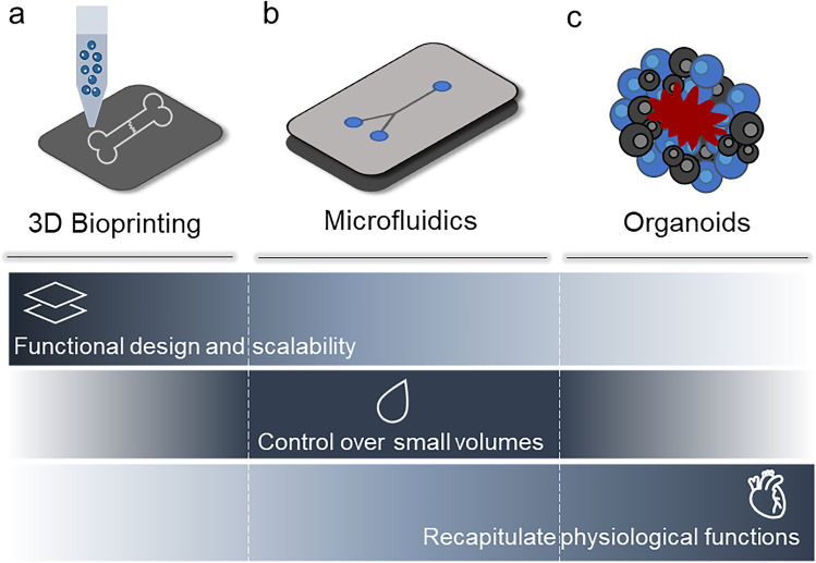
Graphical abstract: Rationale underlying novel drug testing platform development. Pain detected by the central nervous system and following bone fracture cannot be treated or exclusively alleviated using standardised methods. The pain mechanism and specificity/efficacy of pain reduction drugs remain poorly understood. In vivo and ex vivo models are not yet able to recapitulate the various pain events associated with skeletal damage. In vitro models are currently limited by their inability to fully mimic the complex physiological mechanisms at play between nervous and skeletal tissue and any disruption in pathological states. Robust innovative tissue engineering models are needed to better understand pain events and to investigate therapeutic regimes.

Keywords: Bone cancer; Bone pain; Fracture; In vitro models; Nerve.


Figures

Fig. 1
Fig. 1
Bone pain mechanism. Bone pain cycle between osteoclasts, cancer cells and bone fractures in osteolytic metastases. Bone-derived growth factors promote proliferation and stimulate epithelial–mesenchymal transition (EMT) of cancer cells and the production of bone-modifying cytokines, in bone-colonising cancer cells. These factors further stimulate osteoclastic bone resorption via activation of the receptor activator of the nuclear factor-kB (RANKL)/RANK pathway in osteoblasts and osteoclasts, increasing the release of bone-stored growth factors. Osteoclast activity induces a local acidosis increasing TRPV1 and ASIC3 ion channel activity of nociceptor fibres. The response is sent to the central nervous system (CNS) via the dorsal root ganglion (DRG) together with mechanical stimulation due to the growing tumour mass–induced bone fractures
Fig. 2
Fig. 2
Tissue engineering platforms for in vitro disease modelling. Modelling platforms for the recapitulation of bone-neuro pathological conditions include (a) 3D bioprinting, (b) microfluidics and (c) organoids. These systems hold great potential in mimicking the disease conditions present in bone and neural tissue. 3D bioprinting technologies offer the ability to pattern functional architectures and design as well as the ability to 3D print scalable and complex tissues. Microfluidics lack the above-listed abilities, but can precisely control small volumes of liquid required to create compartmentalised micro-environments for the development of in vitro models. Organoids, in contrast, offer biomaterial-free approaches with application of self-assembling properties of different types of cells to build and recapitulate physiologically functional tissue substitutes/models
Fig. 3
Fig. 3
In vivo and in vitro bone models. (a) In vivo models. (a, i) A novel osteoporotic mouse model developed using an innovative movable and non-invasive unloading device (ULD). Micro-CT scan images (yellow highlighted analysis region) of the trabecular and cortical structures of mice femurs from control, tail suspension and 3D-ULD groups; adapted from [73] Copyright © 2021. This is an open-access article distributed under the terms of the Creative Commons Attribution License (CC BY). (a, ii) The similarities between the anatomical and biomechanical characteristics of the sheep spine and the human spine allow for the study of chronic cervical spinal cord compression. A digital remote-controlled intervertebral compression device (IVCD) allowed the application of a progressive compression for 1, 5, 10, and 20 weeks, simulating a human cervical spinal cord compression [77]. This is an open-access article distributed under the terms of the Creative Commons CC BY license. (b) In vitro models. (b, i) Harnessing nanoclay-based material, a 3D mineralising micro-environment for HBMSCs to proliferate and differentiate can studies in vitro, capable of developing a functional bone model in 21 days. ALP and Von Kossa staining for HBMSC-laden 3D-bioprinted scaffolds cultured in basal and osteogenic conditioned media. Adapted from [81]; Copyright© 2020. This is an open-access article distributed under the terms of the Creative Commons Attribution License (CC BY). (b, ii) The ability of nanoclay to foster BMSC differentiation towards bone and the culturing in osteogenic media conditioning demonstrate the ability of Laponite-based constructs to generate a bone 3D model in a shorter time (D1) with significantly increased ALP deposition [82]. This is an open-access article distributed under the terms of the Creative Commons Attribution License (CC BY). (b, iii) A high shape fidelity of nanoclay-based bioprinted construct can be obtained by temporarily increasing the ink viscosity with the use of methylcellulose (i–ii), ensuring a stable cell viability over the time (iii). Furthermore, the nanoclay content can be exploited for a controlled release of biological active agents able to allow the development of bone tissue. [83]
Fig. 4
Fig. 4
In vivo and in vitro neuro models. (a) In vivo models. (a, i) A model for the study of functional treatment for the crossing of the blood–brain barrier (BBB) to inhibit central reporter gene expression and study glial signalling to alleviate chronic pain. GFP-positive reported gene expression (green) is extensively observed in the spinal cord tissue (left panels), while not localised in the sciatic nerve (right panels) at different time points. Glial fibrillary acidic protein (GFAP)–positive glial cells and eGFP expression were found to co-localise in specific regions—white arrows. Adapted from [105]. Copyright © 2022. This is an open-access article distributed under the terms of the Creative Commons Attribution License (CC BY). (b) In vitro models. (b, i) 3D-bioprinted brain-like cortical tissue with 3D compartmentalised modular and concentrical architectures. The use of a silk-fibroin/ECM scaffold with a rudimentary structure provided relevant features useful for brain neural network development. In addition, the developed brain tissue showed electrophysiological functions in response to traumatic brain injury (TBI): a change in baseline signal is shown after a weigh-drop impact, with a consequent injury-triggered Glu release that mimics observations in vivo. This modular 3D brain-like tissue is capable of real-time nondestructive assessments offering the possibility to model brain disorders such as TBI. Adapted from [107]. This is an open-access article distributed under the terms of the Creative Commons Attribution License (CC BY). (b, ii) Printing neural tissues using a lipid-bilayer-supported droplet bioprinting (i) allows for the development of a functional stimulus-responsive neural network in some weeks (ii). Neural tissue was obtained by 3D printing aqueous droplets conjoined by lipid bilayers, with a spatial pattern which not only gives a control of cell self-organisation, but also provides a neuronal network which can be obtained only after months of organoid cultures. Furthermore, diseases could also be modelled by incorporating reprogrammed patient cells with specific genetic mutations. Adapted from [108]. This is an open-access article distributed under the terms of the Creative Commons Attribution License (CC BY). (b, iii) 3D-printed nerve system on a chip. 3D-printed device comprising silicone microchannel for axonal guidance. Superior cervical ganglia (SCG) neurons with green-labelled tau protein aligned within the microchannel. Triple channels with self-assembled network of Schwann cells stained with PRV brainbow. Close-up images of above-mentioned detailed micrographs. Adapted from [109]. This article is licensed under a Creative Commons Attribution 3.0 Unported Licence
Fig. 5
Fig. 5
Bone pain models. (a) Bone pain in vitro model—TRPV1 expression. (a, i) L3 DRG section highlighting NF200 + neurons, TRPV1 + bone afferent neurons. (a, ii) The stimulation with 0.1 μM and 1 μM capsaicin increased the discharge frequency of both small (C fibre) and medium (Aδ fibre) amplitude. Adapted with permission from [143]. Copyright 2019. (b) Bone cancer pain model. Destruction of bone tissue (b, i) confirmed by X-ray imaging and histological analysis, confirming the presence of tumour cells within the marrow cavity. (b, ii) Paw mechanical withdrawal threshold (PMWT) and paw withdrawal thermal latency (PWTL) with cold and hot surfaces investigated in a bone cancer rat model to evaluate the pain response in vivo. Relative expression of mRNA and Transient receptor potential ankyrin 1 (TRPA1) was enhanced in the bone cancer pain animal model. Targeting via TRPA1 antisense oligodeoxynucleotide (AS-ODN) relieved PMWT and PWTL. Adapted from [136]. Copyright© 2021 under the terms of the Creative Commons Attribution License (CC BY). (c) Human spinal organoids in a chip (c, i) positive staining for CB1 expression, for sensory, inhibitory, and excitatory neurons (CGRP + , GAD1 + and vGlut1 + , respectively). (c, ii) Response to capsaicin and electrical stimulation of spinal organoids towards nociceptive modulation, with enhanced mean firing rate and average burst frequency for BDNF and capsaicin-stimulated group. Adapted with permission from [131]. Copyright 2022 American Chemical Society. (d) Human sensorimotor organoids based on (d, i) TUJ1 + neurons and sarcomeric α-actinin (SAA) + myocytes can include both cell types (d-ii) and observed to be functional over 4 weeks. Adapted from [149]. Creative Commons CC BY

References

    1. Alves CJ, Neto E, Sousa DM, et al. Fracture pain-traveling unknown pathways. Bone. 2016;85:107–14. 10.1016/j.bone.2016.01.026. - PubMed
    1. Lortet-Tieulent J, Georges D, Bray F, Vaccarella S. Profiling global cancer incidence and mortality by socioeconomic development. Int J Cancer. 2020;147:3029–36. 10.1002/ijc.33114. - PubMed
    1. Frost CØ, Hansen RR, Heegaard AM. Bone pain: current and future treatments. Curr Opin Pharmacol. 2016;28:31–7. 10.1016/j.coph.2016.02.007. - PubMed
    1. Ripamonti C, Fulfaro F. Malignant bone pain: pathophysiology and treatments. Curr Rev Pain. 2000;4:187–96. 10.1007/s11916-000-0078-3. - PubMed
    1. Zajączkowska R, Kocot-Kępska M, Leppert W, Wordliczek J. Bone pain in cancer patients: mechanisms and current treatment. Int J Mol Sci. 2019;20:65–75. 10.3390/ijms20236047. - PMC - PubMed

LinkOut - more resources