

Prenatal diagnosis of Lowe syndrome in a male fetus with isolated bilateral cataract
- PMID: 36568675
- PMCID: PMC9768307
- DOI: 10.1016/j.heliyon.2022.e12210
Prenatal diagnosis of Lowe syndrome in a male fetus with isolated bilateral cataract
Abstract
Background: Lowe syndrome is a rare disease characterized by the association of congenital cataract, hypotonia, followed by global psychomotor delay and intellectual disability, as well as progressive renal dysfunction, and renal failure occurring at around 20 years of age.
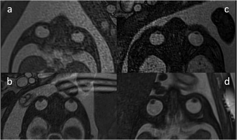
Case presentation: We discuss the case of a male fetus diagnosed with isolated bilateral cataract on the sonography performed at 21 + 5 weeks of gestation, confirmed by a fetal MRI at 23 weeks of gestation.After ruling out infectious etiologies, a genetic consult was conducted at 26 weeks of gestation, and an amniocentesis was realized to search for a chromosomal cause, Norrie's disease and Lowe syndrome by Sanger analysis. A c.1351G > A (p.Asp451Asn) hemizygous mutation in OCRL gene was identified, inherited from the mother, which led to the diagnosis of Lowe syndrome in the fetus.
Conclusions: This is the first case of Lowe syndrome diagnosed prenatally on an isolated cataract, which allows the discussion of a more extensive etiological research when a male fetus is diagnosed with isolated bilateral cataract, by including notably a systematic analysis of the OCRL gene.
Keywords: Fetus; Isolated cataract; Lowe syndrome; OCRL; X-linked.
© 2022 The Author(s).
Conflict of interest statement
The authors declare no conflict of interest.
Figures


References
-
- Zhang D.D., Du J.Z., Topolewski J., Wang X.M. Review Recent progress in identification and characterization of loci associated with sex-linked congenital cataract. Genet Mol Res. 2016;15(3) - PubMed
-
- Pichi F., Lembo A., Serafino M., Nucci P. Genetics of congenital cataract. Dev. Ophthalmol. 2016;57:1–14. - PubMed
-
- Ashwal E., Achiron A., Gilboa Y., Berkenstadt M., Rosner M., Achiron R. Prenatal ultrasonographic diagnosis of cataract: in utero manifestations of cryptic disease. Ultraschall Med. 2018;39(2):213–218. - PubMed
Publication types
LinkOut - more resources
Full Text Sources
