

Malignant atrophic papulosis: Two case reports
- PMID: 36569027
- PMCID: PMC9782946
- DOI: 10.12998/wjcc.v10.i35.12971
Malignant atrophic papulosis: Two case reports
Abstract
Background: Malignant atrophic papulosis is a rare and potentially lethal thrombo-occlusive microvasculopathy characterized by cutaneous papules and gastrointestinal perforation. The precise pathogenesis of this disease remains obscure.
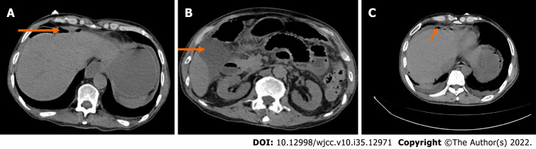
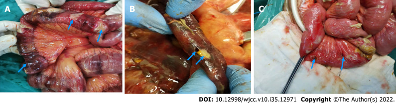
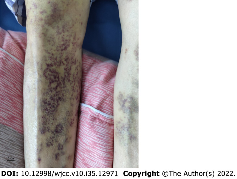
Case summary: We describe the case of a 67-year-old male patient who initially presented with cutaneous aubergine papules and dull pain in the epigastrium. One week after symptom onset, he was admitted to the hospital for worsening abdominal pain. Exploratory laparotomy showed patchy necrosis and subserosal white plaque lesions on the small intestinal wall, along with multiple perforations. Histological examination of the small intestine showed extensive hyperemia, edema, necrosis with varying degrees of inflammatory reactions in the small bowel wall, small vasculitis with fibrinoid necrosis and intraluminal thrombosis in the mesothelium. Based on the mentioned evidence, a diagnosis of malignant atrophic papulosis was made. We also present the case of a 46-year-old man with known cutaneous manifestations, abdominal pain, nausea and vomiting. His physical examination showed positive rebound tenderness. A computed tomography scan revealed free intraperitoneal air. He required surgical intervention on admission and then developed an esophageal perforation. He ultimately died of a massive hemorrhage.
Conclusion: In previously published cases of this disease, the cutaneous lesions initially appeared as small erythematous papules. Subsequently, the papules became porcelain-white atrophic depression lesions with a pink, telangiectatic peripheral rim. In one of the patients, the cutaneous lesions appeared as aubergine papules. The other patient developed multiple perforations in the gastrointestinal tract. Due to malignant atrophic papulosis affecting multiple organs, many authors speculated that it is not a specific entity. This case series serves as additional evidence for our hypothesis.
Keywords: Case report; Gastrointestinal perforation; Malignant atrophic papulosis; Papulosis; Thrombo-occlusive microvasculopathy.
©The Author(s) 2022. Published by Baishideng Publishing Group Inc. All rights reserved.
Conflict of interest statement
Conflict-of-interest statement: All authors report having no relevant conflicts of interest for this article.
Figures


References
-
- Hohwy T, Jensen MG, Tottrup A, Steiniche T, Fogh K. A fatal case of malignant atrophic papulosis (Degos' disease) in a man with factor V Leinden mutation and lupus anticoagulant. Acta Derm Venereol. 2006;86:245–247. - PubMed
-
- Kohlmeier W. Multiple Hautneknosen bei Thromboangiitis obliterans. Arch Dermatol Syphilol. 1941;181:783–784.
-
- Degos R DJ, Tricot R. Dermatite papulosquameuse atrophiante. ull Soc Fr Dermatol Syphiligr. 1942;49:148–150.
Publication types
LinkOut - more resources
Full Text Sources
