. 2022 Nov 30;5(4):e222.
doi: 10.1097/OI9.0000000000000222. eCollection 2022 Dec.

Intraoperative assessment of bone viability through improved analysis and visualization of dynamic contrast-enhanced fluorescence imaging: technique report

Affiliations

Intraoperative assessment of bone viability through improved analysis and visualization of dynamic contrast-enhanced fluorescence imaging: technique report

Jonathan Thomas Elliott et al. OTA Int. .

Abstract

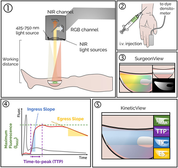
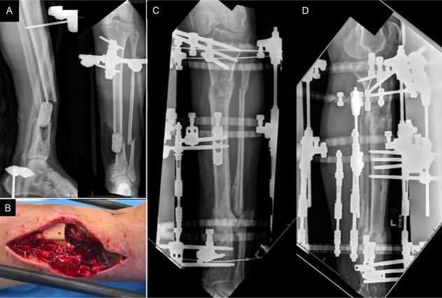
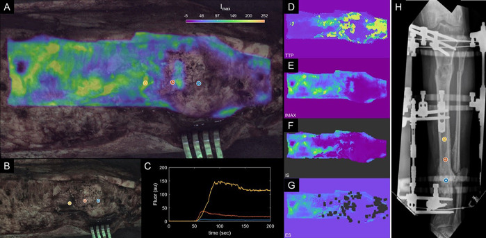
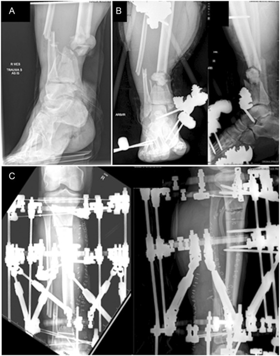
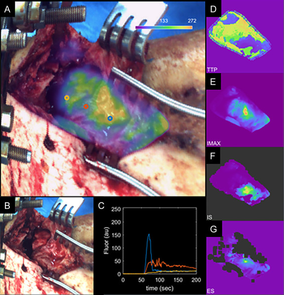
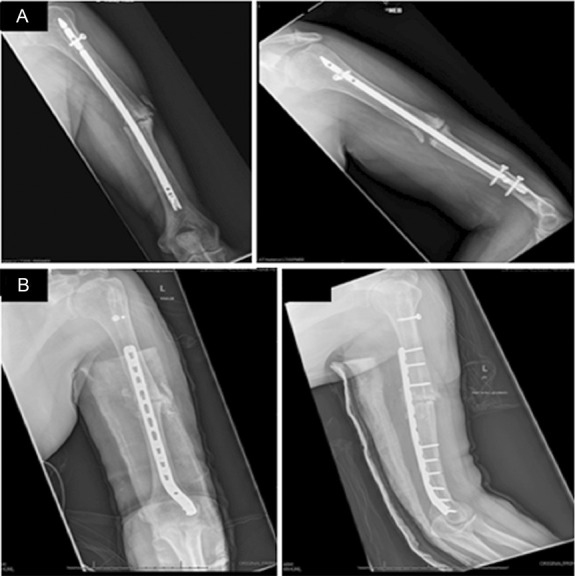
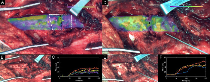
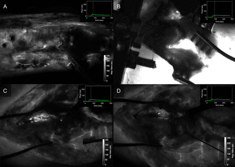
Bone devitalization is believed to be a critical determinant of complications such as infection or nonunion. However, intraoperative assessment of bone devitalization, particularly in open fractures and infections, remains highly subjective resulting in variation in treatment. Optical imaging tools, particularly dynamic contrast-enhanced fluorescence imaging, can provide real-time, intraoperative assessment of bone and soft tissue perfusion, which informs the tissues' ability to heal and fight infection. We describe a novel technique to apply indocyanine green-based fluorescence imaging, using a device that is frequently used in the operating room to assess skin or flap perfusion in plastic surgery, to assess bone and deep tissue perfusion in three pertinent cases: (1) a chronic infection/nonunion after a Gustilo type 3A tibia fracture (patient 1), (2) an acute Gustilo type 3C tibia open fracture with extensive degloving/soft tissue stripping (patient 2), and (3) an atrophic nonunion of the humerus (patient 3). In all three cases, fluorescence imaging (both time-specific fluorescence and maximum fluorescence) and derived kinetic maps of time-to-peak, ingress slope, and egress slope demonstrated clear spatial variation in perfusion that corresponded to the patient pathogenesis. The impact of this information on patient outcome will need to be evaluated in future clinical trials; however, these cases demonstrate in principle that optical imaging information has the potential to inform surgical practice, reduce the variation in treatment, and improve outcomes observed in these challenging patients.

Keywords: debridement; fluorescence-guided surgery; fracture fixation; open fracture; surgical wound infection; trauma.


Conflict of interest statement

The authors report no conflict of interest.

Figures

Figure 1.
Figure 1.
Step 1: Fluorescence imaging system is positioned over the surgical field, and fluorescence video acquisition is started. Step 2: A dye densitometer finger probe acquires the time-dependent arterial dye concentration (called arterial input function). Dye is injected intravenously. Step 3: Real-time fluorescence video, including overlay onto the color image of the surgical field, is displayed on the “SurgeonView.” Step 4: Collection of video rate recorded data allows for analysis of the inflow/outflow kinetics, which can be assessed using several parameters including maximum fluorescence intensity (Imax), time-to-peak (TTP), ingress or inflow slope (IS), and egress or outflow slope (ES). These parameters are computed for the time–concentration curve of each pixel (solid red line), corrected to account for differences in arterial input function (solid gray line), to produce kinetic maps. Step 5: The kinetic maps can then be displayed on a secondary monitor “KineticView” so that both the real-time SurgeonView information and the computed KineticView information are available intraoperatively.
Figure 2.
Figure 2.
Radiograph (A) and clinical image (B) after initial debridement and removal of intramedullary nail. Radiographs after revision debridement followed by distraction osteogenesis/bone transport (C, D).
Figure 3.
Figure 3.
A, “Enhanced DCE view” of patient 1 data showing maximum intensity heat map overlaid on the white light image. B, White light intraoperative image. C, Kinetic ICG inflow/outflow curves at regions of interest identified with blue, red, and yellow dots. D–G, Kinetic heat maps representing ICG time-to-peak (D), maximum ICG intensity (E), ICG inflow slope (F), and ICG outflow slope (G). H, AP radiographs with regions of interest (blue, red, yellow). ICG, indocyanine green.
Figure 4.
Figure 4.
Injury radiograph (A), initial postdebridement radiographs (B), and radiographs after frame placement with proximal tibial osteotomy for distraction osteogenesis/bone transport (C).
Figure 5.
Figure 5.
A, “Enhanced DCE view” of patient 2 showing maximum intensity heat map overlaid on the white light image. B, White light intraoperative image. C, Kinetic ICG inflow/outflow curves at regions of interest identified with the yellow, red, and blue dots. D–G, Kinetic heat maps representing ICG time-to-peak (D), maximum ICG intensity (E), ICG inflow slope (F), and ICG outflow slope (G). ICG, indocyanine green.
Figure 6.
Figure 6.
Preoperative radiographs of the atrophic humeral nonunion (A) and postoperative radiographs after compression plating, debridement with acute shortening, and iliac crest bone graft (B).
Figure 7.
Figure 7.
(A) Predibridement and (D) postdebridement kinetic maps of patient 3, showing Imax kinetic maps overlaid on (B and E) white light intraoperative images. Improvements in kinetic curves can be observed from (C) predebridement to (F) postdebridement, suggesting procedure was effective.
Figure 8.
Figure 8.
Fluorescence intensity maps acquired during the early wash-out period after ICG injection for (A) patient 1, (B) patient 2, and patient 3 (C) predebridement and (D) postdebridement, as seen with the SPY Elite standard visualization available in clinical units. ICG, indocyanine green.

References

    1. Christersson A, Larsson S, Sörensen J. Presurgical localization of infected avascular bone segments in chronic complicated posttraumatic osteomyelitis in the lower extremity using dual-tracer PET/CT. EJNMMI Res. 2018;8:65. - PMC - PubMed
    1. Yun HC, Murray CK, Nelson KJ, et al. Infection after orthopaedic trauma: prevention and treatment. J Orthop Trauma. 2016;30:S21–S26. - PubMed
    1. Metsemakers W-J, Morgenstern M, McNally MA, et al. Fracture-related infection: a consensus on definition from an international expert group. Injury. 2018;49:505–510. - PubMed
    1. Ricci WM, Collinge C, Streubel PN, et al. A comparison of more and less aggressive bone debridement protocols for the treatment of open supracondylar femur fractures. J Orthop Trauma. 2013;27:722. - PMC - PubMed
    1. Lowenberg DW, Buntic RF, Buncke GM, et al. Long-term results and costs of muscle flap coverage with Ilizarov bone transport in lower limb salvage. J Orthop Trauma. 2013;27:576–581. - PubMed