

Radiological and Morphometric Study of the Emissary Foramens and Canal in the Posterior Cranial Fossa of the Human Skull with Its Neurosurgical Significance
- PMID: 36570755
- PMCID: PMC9771628
- DOI: 10.1055/s-0042-1757429
Radiological and Morphometric Study of the Emissary Foramens and Canal in the Posterior Cranial Fossa of the Human Skull with Its Neurosurgical Significance
Abstract
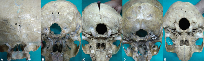
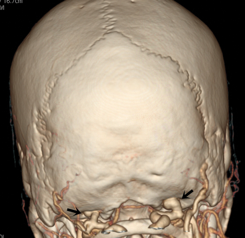
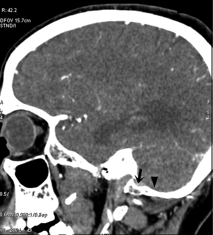
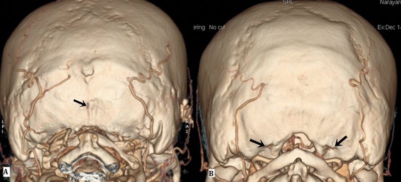
Objective The posterior condylar canals (PCCs), posterior condylar veins (PCVs), occipital foramen (OF), and occipital emissary vein (OEV) are potential anatomical landmarks for surgical approaches through the lateral foramen magnum. We performed the study to make morphometric and radiological analyses of the various emissary foramens and vein in the posterior cranial fossa. Methods Morphometric study were performed on 95 dry occipital bones and radiological analyses on computed tomography (CT) angiography images of 150 patients. The number of OFs on both sides was recorded and PCC length and mean diameters of the internal and external orifices of PCC were measured for bony specimens. Prevalence of PCV and PCV size was investigated using CT angiography. Results Mean PCC length was higher in the left side (9.85 ± 2.5). Mean diameter of the internal orifice and the external orifice diameter were almost the same. The majority of PCCs (75-79.33%) had 2 to 5 mm diameter; only 4 to 9.2% were small in size (< 2 mm). In CT angiography, PCV was not identified in 23 (15.33%) patients. PCVs were located bilaterally in 105 (70%) and unilaterally in 22 (20.5%) patients. Only 11.3% of PCVs were large in size (> 5 mm), 80% of PCVs were medium sized (2-5 mm), and 8.6% were small sized (< 2 mm). Conclusion Normal values of OF, PCC, PCV, and OEV could serve as a future reference for the understanding of the physiology of craniocervical venous drainage, which is necessary to avoid surgical complications and can also serve as a guide to surgical interventions for pathologies of the posterior cranial fossa, such as tumors and injuries.
Keywords: angiography; computed tomography; emissary vein; occipital foramen; posterior condylar canal; posterior condylar vein.
Asian Congress of Neurological Surgeons. This is an open access article published by Thieme under the terms of the Creative Commons Attribution-NonDerivative-NonCommercial License, permitting copying and reproduction so long as the original work is given appropriate credit. Contents may not be used for commercial purposes, or adapted, remixed, transformed or built upon. ( https://creativecommons.org/licenses/by-nc-nd/4.0/ ).
Conflict of interest statement
Conflict of interest None declared.
Figures

References
-
- Mortazavi M M, Tubbs R S, Riech S.Anatomy and pathology of the cranial emissary veins: a review with surgical implications Neurosurgery 201270051312–1318., discussion 1318–1319 - PubMed
-
- Coin C G, Malkasian D R.(1971). Foramen magnumIn: Newton TH, Potts DG, eds. Radiology of the Skull and Brain: The Skull.St. Louis: Mosby; 275–347.
-
- Jeevan D S, Anlsow P, Jayamohan J. Abnormal venous drainage in syndromic craniosynostosis and the role of CT venography. Childs Nerv Syst. 2008;24(12):1413–1420. - PubMed
-
- Lambert P R, Cantrell R W. Objective tinnitus in association with an abnormal posterior condylar emissary vein. Am J Otol. 1986;7(03):204–207. - PubMed
LinkOut - more resources
Full Text Sources
Research Materials