

Primary Leptomeningeal Medulloblastoma in Adults: A Diagnostic Challenge-Case Report and Systematic Review
- PMID: 36570761
- PMCID: PMC9771618
- DOI: 10.1055/s-0042-1757726
Primary Leptomeningeal Medulloblastoma in Adults: A Diagnostic Challenge-Case Report and Systematic Review
Abstract
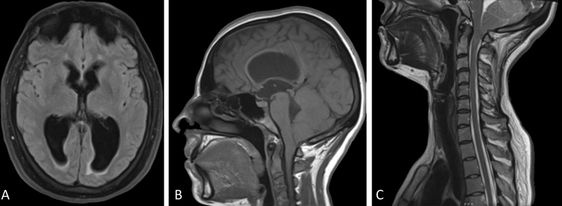
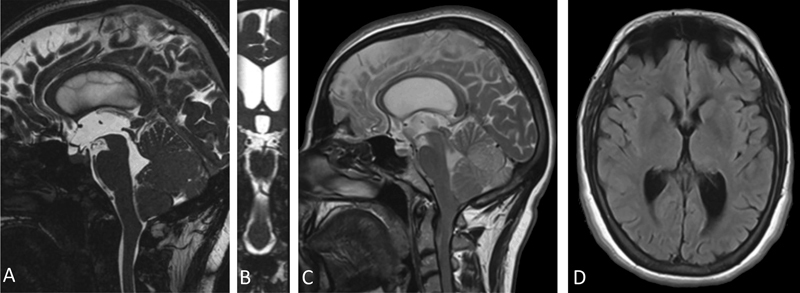
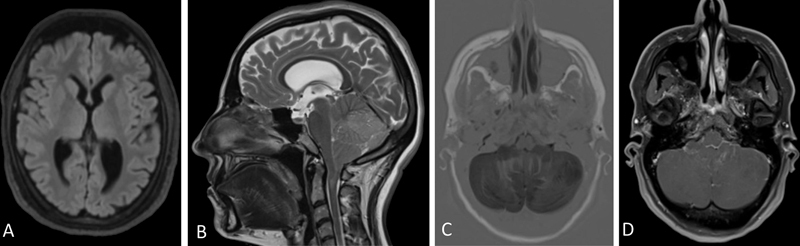
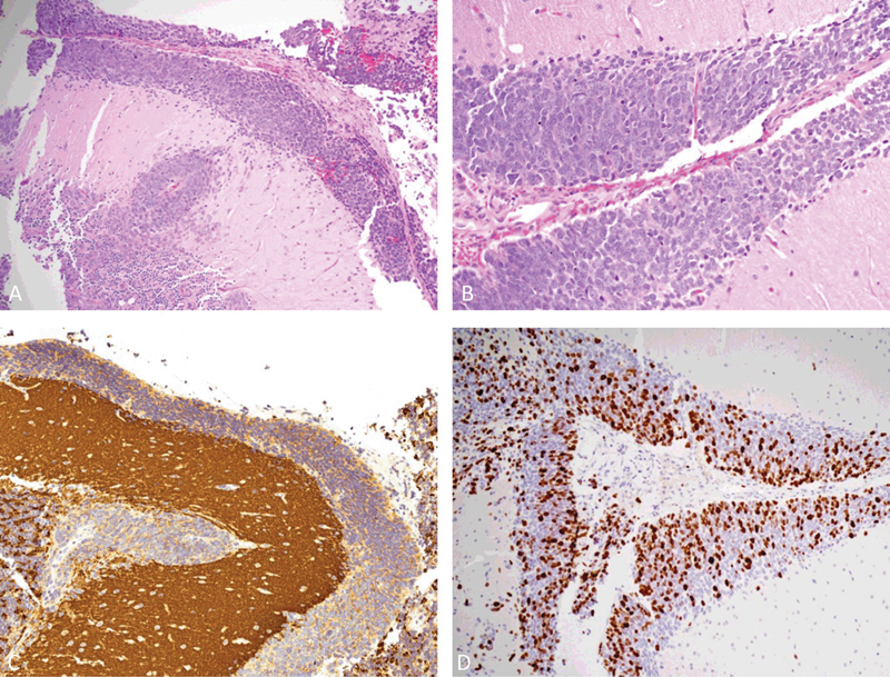
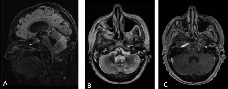
Primary leptomeningeal medulloblastoma (PL-MB) in adults is a rare disease with a severe prognosis. A 35-year-old woman presented with headaches, diplopia, and gait ataxia, with triventricular hydrocephalus and descent of the cerebellar tonsils beyond the foramen magnum. Endoscopic third ventriculostomy was performed. Six months later, headaches recurred. Dilatation of the supratentorial ventricular system and massive cerebellar swelling without contrast-enhancing nodularities were reported. Occipitocervical decompression with duraplasty was performed. A bioptic diagnosis of PL-MB was made. Craniospinal irradiation and chemotherapy were administered. After 18 months, no recurrence was observed. Few cases of PL-MB have been reported: patients die before treatment or within a few days after surgery. Our long-term survival could be ascribable to a slow clinical presentation and an early diagnosis that allowed surgical treatment and the administration of a combined chemoradiotherapy protocol. Cerebellar swelling, even without associated enhancing lesions, with or without hydrocephalus, should be a neuroradiological alarm sign, and PL-MB should be considered.
Keywords: Chiari-like malformation; endoscopic third ventriculostomy; obstructive hydrocephalus; occipitocervical decompression; primary leptomeningeal medulloblastoma.
Asian Congress of Neurological Surgeons. This is an open access article published by Thieme under the terms of the Creative Commons Attribution-NonDerivative-NonCommercial License, permitting copying and reproduction so long as the original work is given appropriate credit. Contents may not be used for commercial purposes, or adapted, remixed, transformed or built upon. ( https://creativecommons.org/licenses/by-nc-nd/4.0/ ).
Conflict of interest statement
Conflict of Interest None declared.
Figures
References
-
- Louis D N, Perry A, Reifenberger G. The 2016 World Health Organization classification of tumors of the central nervous system: a summary. Acta Neuropathol. 2016;131(06):803–820. - PubMed
-
- Roberts R O, Lynch C F, Jones M P, Hart M N. Medulloblastoma: a population-based study of 532 cases. J Neuropathol Exp Neurol. 1991;50(02):134–144. - PubMed
-
- Blessing M M, Alexandrescu S. Embryonal tumors of the central nervous system: an update. Surg Pathol Clin. 2020;13(02):235–247. - PubMed
-
- Hankey G J, Khangure M S, Spagnolo D, Quinlan M F. Adult onset medulloblastoma cerebelli with leptomeningeal dissemination and coincidental primary hyperparathyroidism. Australas Radiol. 1989;33(01):111–115. - PubMed
-
- David K M, Casey A T, Hayward R D, Harkness W F, Phipps K, Wade A M. Medulloblastoma: is the 5-year survival rate improving? A review of 80 cases from a single institution. J Neurosurg. 1997;86(01):13–21. - PubMed
Publication types
LinkOut - more resources
Full Text Sources
Miscellaneous