. 2023 Jul;18(7):1548-1552.
doi: 10.4103/1673-5374.360246.

Inhibition of autophagy rescues HT22 hippocampal neurons from erastin-induced ferroptosis

Affiliations

Inhibition of autophagy rescues HT22 hippocampal neurons from erastin-induced ferroptosis

Nora Hanke et al. Neural Regen Res. 2023 Jul.

Abstract

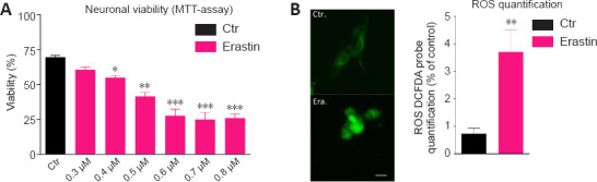
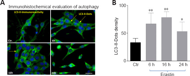
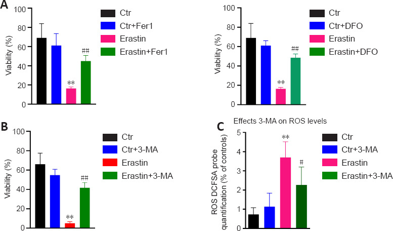
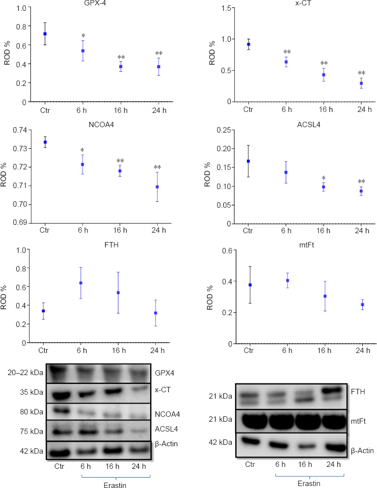
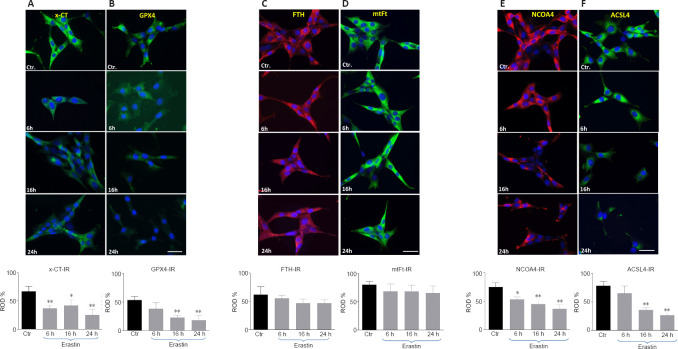
Ferroptosis is a regulated form of cell death which is considered an oxidative iron-dependent process. The lipid hydroperoxidase glutathione peroxidase 4 prevents the iron (Fe2+)-dependent formation of toxic lipid reactive oxygen species. While emerging evidence indicates that inhibition of glutathione peroxidase 4 as a hallmark of ferroptosis in many cancer cell lines, the involvement of this biochemical pathway in neuronal death remains largely unclear. Here, we investigate, first whether the ferroptosis key players are involved in the neuronal cell death induced by erastin. The second objective was to examine whether there is a cross talk between ferroptosis and autophagy. The third main was to address neuron response to erastin, with a special focus on ferritin and nuclear receptor coactivator 4-mediated ferritinophagy. To test this in neurons, erastin (0.5-8 µM) was applied to hippocampal HT22 neurons for 16 hours. In addition, cells were cultured with the autophagy inhibitor, 3-methyladenin (10 mM) and/or ferroptosis inhibitors, ferrostatin 1 (10-20 µM) or deferoxamine (10-200 µM) before exposure to erastin. In this study, we demonstrated by immunofluorescence and western blot analysis, that erastin downregulates dramatically the expression of glutathione peroxidase 4, the sodium-independent cystine-glutamate antiporter and nuclear receptor coactivator 4. The protein levels of ferritin and mitochondrial ferritin in HT22 hippocampal neurons did not remarkably change following erastin treatment. In addition, we demonstrated that not only the ferroptosis inhibitor, ferrostatin1/deferoxamine abrogated the ferroptotic cell death induced by erastin in hippocampal HT22 neurons, but also the potent autophagy inhibitor, 3-methyladenin. We conclude that (1) erastin-induced ferroptosis in hippocampal HT22 neurons, despite reduced nuclear receptor coactivator 4 levels, (2) that either nuclear receptor coactivator 4-mediated ferritinophagy does not occur or is of secondary importance in this model, (3) that ferroptosis seems to share some features of the autophagic cell death process.

Keywords: HT22 neurons; erastin; ferritin; ferritinophagy; ferroptosis; glutathione peroxidase 4; nuclear receptor coactivator 4.


Conflict of interest statement

None

Figures

Figure 1
Figure 1
Erastin induces cell death (A) and ROS production (B) in hippocampal HT22 neurons. (A) HT22 neurons were treated with different concentrations of erastin (0.3, 0.4, 0.5, 0.6, 0.7, and 0.8 µM) for 12 hours and examined by light microscopy. Control neurons have been treated with vehicle (DMSO). Neurons were severely damaged after treatment with erastin at 0.5, 0.6, 0.7, and 0.8 µM. 3-(4,5-Dimethylthiazol-2-yl)-2,5-diphenyltetrazolium bromide (MTT) assay for the viability of HT22 neurons after treatment with erastin at 0.5, 0.6, 0.7, and 0.8 µM shows a significant decrease in viability. (B) ROS expression was quantified by the 2′,7′-dichlorodihydrofluorescein diacetate (CM-H2DCFDA) probe. Fluorescence semi-quantitative analysis highlighting the role of erastin in increasing ROS production. The intracellular ROS-derived fluorescence is expressed as the percentage of fluorescence compared to control, untreated cells, taken as 100%. Each experiment point was performed in triplicate. Five different microscopic fields for each experimental point were analyzed (scale bar: 25 µm). Data are expressed as the mean + SD (n = 6).*P < 0.05, **P < 0.01, ***P < 0.001, vs. control group (one-way analysis of variance with Tukey’s post hoc test). ROS: Reactive oxygen species.
Figure 2
Figure 2
Erastin induced changes in the levels of the autophagy related protein LC3-II. (A) LC3-II staining (green) exhibited punctuate dots in erastin-treated hippocampal HT22 cells. Arrows show LC3-immunoreactive dots in A. Blue indicates 4′,6-diamidino-2-phenylindole-stained nuclei. Scale bar: 50 µm. (B) Quantification of autophagosomes (LC3-II-immunoreactive dots). *P < 0.05, ** P < 0.01, vs. control group (paired Student’s t test). LC3: Light chain 3.
Figure 3
Figure 3
Effects of DFO/Fer-1 and 3-MA on ersatin-induced ferroptosis. (A) DFO/Fer-1 inhibits erastin-induced cell death in HT22 neurons. (B) 3-MA protects HT22 cells from erastin-induced cell death. (C) 3-MA attenuated ROS production in erastin-treated HT22 neurons. Fer-1 (20 µM), DFO (100 μM), and 3-MA (10 mM) were dissolved in dimethyl sulfoxide and added to wells immediately after erastin treatment. Each experiment was performed in triplicate. **P < 0.01, vs. control (Ctr) group; #P < 0.05, ##P < 0.01, vs. erastin group (one-way analysis of variance with Tukey’s post hoc test). DFO: Deferoxamine; Fer-1: ferrostatin 1; 3-MA: 3-methyladenin.
Figure 4
Figure 4
Western blot assay and densitometric analysis of immunoblots of GPX4, x-CT protein, NCOA4, ACSL4, FTH and, mtFt 6, 16 and 24 hours after incubation with 0.5 μM erastin in HT22 neurons. Each experiment was performed in triplicate. *P < 0.05, **P < 0.01, vs. control (Ctr) group (one-way analysis of variance with Tukey’s post hoc test). ACSL4: Acyl-CoA synthetase long-chain family member 4; FTH: ferritin; GPX4: glutathione peroxidase 4; mtFT: mitochondrial ferritin; NCOA4: nuclear receptor coactivator 4; x-CT: sodium-independent cystine-glutamate antiporter.
Figure 5
Figure 5
The x-CT- (A), GPX4 (B), FTH (C), mtFt (D), NCOA4 (E) and ACSL4 (F) immunoreactivities (IR) in HT 22 neurons collected from controls and at 6, 16, and 24 hours after incubation with erastin. Red and green colors represent the immunoreactivities of the corresponding antibodies. Blue (DAPI) corresponds to nuclei. Scale bar: 50 µm. Each experiment was performed in triplicate. *P < 0.05 and **P < 0.01, vs. control (Ctr) group (one-way analysis of variance with Tukey’s post hoc test). ACSL4: Acyl-CoA synthetase long-chain family member 4; FTH: ferritin; GPX4: glutathione peroxidase 4; mtFT: mitochondrial ferritin; NCOA4: nuclear receptor coactivator 4; x-CT: sodium-independent cystine-glutamate antiporter.

References

    1. Andrews NC, Schmidt PJ. Iron homeostasis. Annu Rev Physiol. 2007;69:69–85. - PubMed
    1. Friedmann Angeli JP, Schneider M, Proneth B, Tyurina YY, Tyurin VA, Hammond VJ, Herbach N, Aichler M, Walch A, Eggenhofer E, Basavarajappa D, Rådmark O, Kobayashi S, Seibt T, Beck H, Neff F, Esposito I, Wanke R, Förster H, Yefremova O, et al. Inactivation of the ferroptosis regulator Gpx4 triggers acute renal failure in mice. Nat Cell Biol. 2014;16:1180–1191. - PMC - PubMed
    1. Angeli JPF, Shah R, Pratt DA, Conrad M. Ferroptosis Inhibition:Mechanisms and Opportunities. Trends Pharmacol Sci. 2017;38:489–498. - PubMed
    1. Arosio P, Levi S. Ferritin, iron homeostasis, and oxidative damage. Free Radic Biol Med. 2002;33:457–463. - PubMed
    1. Arosio P, Elia L, Poli M. Ferritin, cellular iron storage and regulation. IUBMB Life. 2017;69:414–422. - PubMed