. 2023 Jul;18(7):1578-1583.
doi: 10.4103/1673-5374.357904.

Inhibiting phosphatase and actin regulator 1 expression is neuroprotective in the context of traumatic brain injury

Affiliations

Inhibiting phosphatase and actin regulator 1 expression is neuroprotective in the context of traumatic brain injury

Yao Jing et al. Neural Regen Res. 2023 Jul.

Abstract

Studies have found that the phosphatase actin regulatory factor 1 expression can be related to stroke, but it remains unclear whether changes in phosphatase actin regulatory factor 1 expression also play a role in traumatic brain injury. In this study we found that, in a mouse model of traumatic brain injury induced by controlled cortical impact, phosphatase actin regulatory factor 1 expression is increased in endothelial cells, neurons, astrocytes, and microglia. When we overexpressed phosphatase actin regulatory factor 1 by injection an adeno-associated virus vector into the contused area in the traumatic brain injury mice, the water content of the brain tissue increased. However, when phosphatase actin regulatory factor 1 was knocked down, the water content decreased. We also found that inhibiting phosphatase actin regulatory factor 1 expression regulated the nuclear factor kappa B signaling pathway, decreased blood-brain barrier permeability, reduced aquaporin 4 and intercellular adhesion molecule 1 expression, inhibited neuroinflammation, and neuronal apoptosis, thereby improving neurological function. The findings from this study indicate that phosphatase actin regulatory factor 1 may be a potential therapeutic target for traumatic brain injury.

Keywords: apoptosis; aquaporin 4; blood brain barrier; intercellular adhesion molecule 1; neuroinflammation; nuclear factor kappa B; occludin; phosphatase and actin regulator-1; traumatic brain injury; zonula occludens 1.


Conflict of interest statement

None

Figures

Figure 1
Figure 1
PHACTR-1 expression in the brain after TBI. (A, B) PHACTR-1 protein expression was significantly increased in the TBI group compared with the sham group, and the increased expression was maintained for at least 7 days. Values were normalized to the sham group and are expressed as mean ± SD (n = 3/group). *P < 0.05, **P < 0.01, vs. sham group (one-way analysis of variance followed by Tukey’s honestly significant difference post hoc test). (C) Immunofluorescence staining for PHACTR-1 (Alexa Fluor 488, green) in endothelial cells (CD31-positive cells, Alexa Fluor 555, red), astrocytes (GFAP-positive cells, Alexa Fluor 594, red), microglial cells (Iba1-positive cells, Alexa Fluor 647, red), and neurons (NeuN-positive cells, Alexa Fluor 594, red). PHACTR-1 expression was significantly increased in these cells 3 days after TBI. White arrows indicate co-staining. Scale bar: 25 μm. GFAP: Glial fibrillary acidic protein; Iba: ionized calcium-binding adapter molecule; PHACTR: phosphatase and actin regulator; TBI: traumatic brain injury.
Figure 2
Figure 2
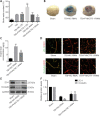
Effect of AAV targeting PHACTR-1 on the contusion point after TBI. (A) Contusion point (blue area) and AAV injection sites (red arrows) in the peri-contusion area. The fluorescence image shows transfected AAV (green) in the peri-contusion area. Scale bar: 50 μm. (B, C) Western blot analysis of PHACTR-1 expression in the different groups 3 days after TBI. Values were normalized to the sham group and are expressed as mean ± SD (n = 3/group). **P < 0.01, vs. TBI group (one-way analysis of variance followed by Tukey’s honestly significant difference post hoc test). AAV: Adeno-associated virus; PHACTR: phosphatase and actin regulator; TBI: traumatic brain injury.
Figure 3
Figure 3
Effect of PHACTR-1 on brain edema and BBB integrity 3 days after TBI. (A) Brain water content in the different groups (*P < 0.05, **P < 0.01, vs. TBI) (one-way analysis of variance followed by Tukey’s Honestly Significant Difference post hoc test). (B, C) The EB concentration was significantly decreased in the TBI + PHACTR-1-RNAi group compared with the TBI + NC-RNAi group. (D–F) Expression of the TJ proteins ZO-1 and occludin (both Alexa Fluor 488, green) in endothelial cells (CD31) (Alexa Fluor 555, red) was significantly increased in the TBI + PHACTR-1-RNAi group compared with the TBI + NC-RNAi group, and measurement of quantification by western blotting (normalized to the sham group). Scale bar: 25 μm. Data are expressed as mean ± SD (n = 5/group). *P < 0.05, **P < 0.01, vs. TBI + NC-RNAi group (Student’s t-test). BBB: Blood-brain barrier; EB: Evans Blue; PHACTR: phosphatase and actin regulator; TBI: traumatic brain injury; TJ: tight junction; ZO: zonula occludens.
Figure 4
Figure 4
Effect of PHACTR-1 on AQP-4 and ICAM-1 expression in injured brain tissues 3 days after TBI. (A–C) AQP-4 (Alexa Fluor 647, red) expression levels were significantly decreased after PHACTR-1 knockdown. (D–F) ICAM-1 (Alexa Fluor 647, red) expression levels were significantly decreased after PHACTR-1 knockdown. Scale bar: 35 μm in A, 60 μm in D. Data are expressed as mean ± SD (n = 5/group). **P < 0.01, vs. TBI + NC-RNAi group (Student’s t-test). AQP: Aquaporin; ICAM: intercellular cell adhesion molecule; PHACTR: phosphatase and actin regulator; TBI: traumatic brain injury.
Figure 5
Figure 5
Effect of PHACTR-1 on neuroinflammation in injured brain tissues 3 days after TBI. (A) Immunofluorescence staining showed that astrocyte (GFAP-positive cells, Alexa Fluor 594, red) and M1 microglial cell (CD16/32 (Alexa Fluor 488, green)/Iba1 (Alexa Fluor 647, red)-positive cells) activation was increased after TBI but reduced by PHACTR-1 knockdown. The opposite effect was seen for M2 microglial cells (CD206 (Alexa Fluor 488, green)/Iba1 (Alexa Fluor 647, red)-positive cells). Scale bars: 60 μm. (B, C) After TBI, expression of the inflammatory cytokines TNF-α, IL-1β, and IL-6 was decreased by PHACTR-1 knockdown, while expression of the anti-inflammatory cytokines IL-4 and IL-10 was increased by PHACTR-1 knockdown, as detected by real-time polymerase chain reaction. Data are expressed as mean ± SD (n = 5/group). *P < 0.05, **P < 0.01, vs. TBI + NC-RNAi group (Student’s t-test). GFAP: Glial fibrillary acidic protein; Iba: ionized calcium-binding adapter molecule; IL: interleukin; PHACTR: phosphatase and actin regulator; TBI: traumatic brain injury, TNF: tumor necrosis factor alpha.
Figure 6
Figure 6
Effect of PHACTR-1 on neuronal apoptosis and brain volume loss 7 days after TBI. (A–C) The number of apoptotic nerve cells and neurons (NeuN positive cell, Alexa Fluor 488, green) observed after TBI was significantly reduced by PHACTR-1 knockdown. Scale bars: 25 μm. (D, E) Nissl staining showed that the brain volume loss induced by TBI was attenuated by PHACTR-1 knockdown. Data are expressed as mean ± SD (n = 5/group). *P < 0.05, **P < 0.01, vs. TBI + NC-RNAi group (Student’s t-test). DAPI: 4′,6-Diamidino-2-phenylindole; PHACTR: phosphatase and actin regulator; TBI: traumatic brain injury; TUNEL: TdT-mediated dUTP nick-end labeling.
Figure 7
Figure 7
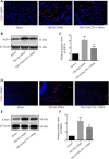
Effect of PHACTR-1 on NF-κB/p65 expression in injured brain tissue 3 days after TBI. (A–C) Expression of NF-κB/p65 (Alexa Fluor 594, red) in both the cytoplasm and nucleus, as well as translocation of p65 to the nucleus, induced by TBI were significantly decreased by PHACTR-1 knockdown. Scale bar: 25 μm. Data are expressed as mean ± SD (n = 5/group). *P < 0.05, **P < 0.01, vs. TBI + NC-RNAi group (Student’s t-test). DAPI: 4′,6-Diamidino-2-phenylindole; GAPDH: glyceraldehyde-3-phosphate dehydrogenase; NF-κB: nuclear factor kappa B; PHACTR: phosphatase and actin regulator; TBI: traumatic brain injury.
Figure 8
Figure 8
Effect of PHACTR-1 on neurological function after TBI. (A, B) After PHACTR-1 knockdown, the mNSS decreased significantly from day 3 after TBI, and rotarod latency increased from day 7 after TBI. Data are expressed as mean ± SD (n = 12/group). *P < 0.05, **P < 0.01, vs. TBI + NC-RNAi group (Student’s t-test). mNSS: Modified neurological severity score; PHACTR: phosphatase and actin regulator; TBI: traumatic brain injury.

References

    1. Abbott NJ, Patabendige AA, Dolman DE, Yusof SR, Begley DJ. Structure and function of the blood-brain barrier. Neurobiol Dis. 2010;37:13–25. - PubMed
    1. Allain B, Jarray R, Borriello L, Leforban B, Dufour S, Liu WQ, Pamonsinlapatham P, Bianco S, Larghero J, Hadj-Slimane R, Garbay C, Raynaud F, Lepelletier Y. Neuropilin-1 regulates a new VEGF-induced gene, Phactr-1 , which controls tubulogenesis and modulates lamellipodial dynamics in human endothelial cells. Cell Signal. 2012;24:214–223. - PubMed
    1. Allen PB, Greenfield AT, Svenningsson P, Haspeslagh DC, Greengard P. Phactrs 1-4:a family of protein phosphatase 1 and actin regulatory proteins. Proc Natl Acad Sci U S A. 2004;101:7187–7192. - PMC - PubMed
    1. Alluri H, Grimsley M, Anasooya Shaji C, Varghese KP, Zhang SL, Peddaboina C, Robinson B, Beeram MR, Huang JH, Tharakan B. Attenuation of blood-brain barrier breakdown and hyperpermeability by calpain inhibition. J Biol Chem. 2016;291:26958–26969. - PMC - PubMed
    1. Bieber M, Gronewold J, Scharf AC, Schuhmann MK, Langhauser F, Hopp S, Mencl S, Geuss E, Leinweber J, Guthmann J, Doeppner TR, Kleinschnitz C, Stoll G, Kraft P, Hermann DM. Validity and reliability of neurological scores in mice exposed to middle cerebral artery occlusion. Stroke. 2019;50:2875–2882. - PubMed