

Advances in Optical Tools to Study Taste Sensation
- PMID: 36572557
- PMCID: PMC9794552
- DOI: 10.14348/molcells.2022.0116
Advances in Optical Tools to Study Taste Sensation
Abstract
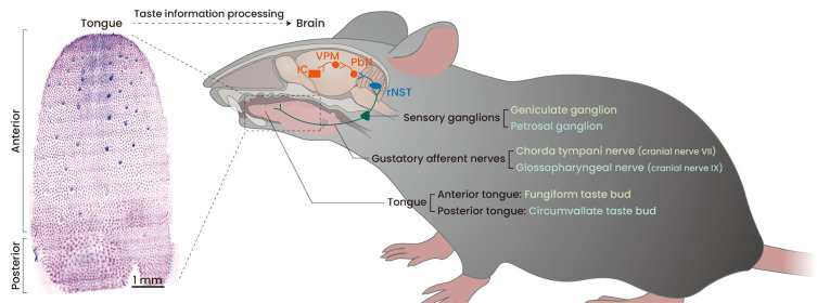
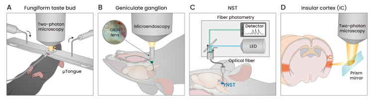
Taste sensation is the process of converting chemical identities in food into a neural code of the brain. Taste information is initially formed in the taste buds on the tongue, travels through the afferent gustatory nerves to the sensory ganglion neurons, and finally reaches the multiple taste centers of the brain. In the taste field, optical tools to observe cellular-level functions play a pivotal role in understanding how taste information is processed along a pathway. In this review, we introduce recent advances in the optical tools used to study the taste transduction pathways.
Keywords: geniculate ganglion; gustation; imaging; insular cortex; optical tools; taste bud.
Conflict of interest statement
The authors have no potential conflicts of interest to disclose.
Figures
Similar articles
-
Types of taste circuits synaptically linked to a few geniculate ganglion neurons.J Comp Neurol. 2008 Dec 20;511(6):753-72. doi: 10.1002/cne.21869. J Comp Neurol. 2008. PMID: 18925565 Free PMC article.
-
Oral Sensory Neurons of the Geniculate Ganglion That Express Tyrosine Hydroxylase Comprise a Subpopulation That Contacts Type II and Type III Taste Bud Cells.eNeuro. 2022 Oct 13;9(5):ENEURO.0523-21.2022. doi: 10.1523/ENEURO.0523-21.2022. Print 2022 Sep-Oct. eNeuro. 2022. PMID: 36216506 Free PMC article.
-
Hedgehog Signaling Regulates Taste Organs and Oral Sensation: Distinctive Roles in the Epithelium, Stroma, and Innervation.Int J Mol Sci. 2019 Mar 16;20(6):1341. doi: 10.3390/ijms20061341. Int J Mol Sci. 2019. PMID: 30884865 Free PMC article. Review.
-
Organization of geniculate and trigeminal ganglion cells innervating single fungiform taste papillae: a study with tetramethylrhodamine dextran amine labeling.Neuroscience. 1999;93(3):931-41. doi: 10.1016/s0306-4522(99)00115-3. Neuroscience. 1999. PMID: 10473258
-
[Progress in the effects of injury and regeneration of gustatory nerves on the taste functions in animals].Sheng Li Xue Bao. 2014 Oct 25;66(5):519-27. Sheng Li Xue Bao. 2014. PMID: 25331997 Review. Chinese.
Cited by
-
c-Kit signaling confers damage-resistance to sweet taste cells upon nerve injury.Int J Oral Sci. 2025 Jul 29;17(1):57. doi: 10.1038/s41368-025-00387-3. Int J Oral Sci. 2025. PMID: 40730572 Free PMC article.
-
Cerebral hemodynamics and functional connectivity changes in stroke patients with dysphagia under acidic taste stimulation: a preliminary study.Front Neurol. 2025 Jun 5;16:1533099. doi: 10.3389/fneur.2025.1533099. eCollection 2025. Front Neurol. 2025. PMID: 40538661 Free PMC article.
References
Publication types
MeSH terms
LinkOut - more resources
Full Text Sources
