. 2022 Dec 26;13(1):7953.
doi: 10.1038/s41467-022-35661-z.

Diverse monogenic subforms of human spermatogenic failure

Affiliations

Diverse monogenic subforms of human spermatogenic failure

Liina Nagirnaja et al. Nat Commun. .

Abstract

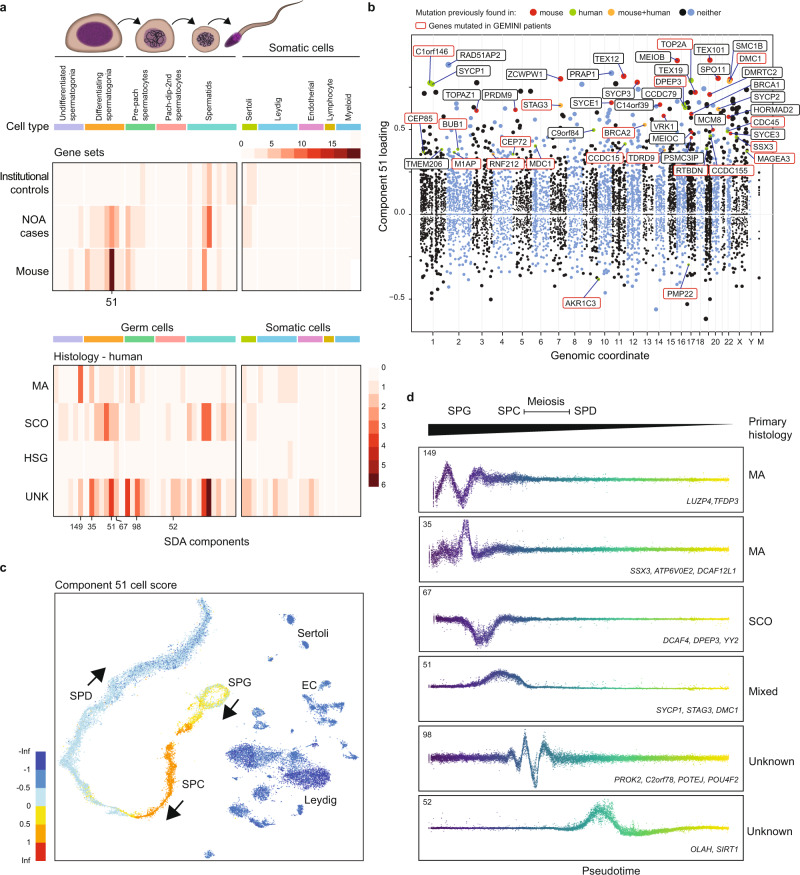
Non-obstructive azoospermia (NOA) is the most severe form of male infertility and typically incurable. Defining the genetic basis of NOA has proven challenging, and the most advanced classification of NOA subforms is not based on genetics, but simple description of testis histology. In this study, we exome-sequenced over 1000 clinically diagnosed NOA cases and identified a plausible recessive Mendelian cause in 20%. We find further support for 21 genes in a 2-stage burden test with 2072 cases and 11,587 fertile controls. The disrupted genes are primarily on the autosomes, enriched for undescribed human "knockouts", and, for the most part, have yet to be linked to a Mendelian trait. Integration with single-cell RNA sequencing data shows that azoospermia genes can be grouped into molecular subforms with synchronized expression patterns, and analogs of these subforms exist in mice. This analysis framework identifies groups of genes with known roles in spermatogenesis but also reveals unrecognized subforms, such as a set of genes expressed across mitotic divisions of differentiating spermatogonia. Our findings highlight NOA as an understudied Mendelian disorder and provide a conceptual structure for organizing the complex genetics of male infertility, which may provide a rational basis for disease classification.


Conflict of interest statement

The authors declare no competing interests.

Figures

Fig. 1
Fig. 1. Overview of the study.
DNA samples from NOA cases recruited in 11 centers across the world underwent WES to detect rare recessive variants underlying the disease. The identified Mendelian causes of NOA were additionally screened for in independent male infertility cohorts to map recurrent causes of NOA. An integrated analysis with scRNAseq atlas of adult human testis enabled to cluster the molecular NOA causes into ‘subforms’ that go beyond the visual classification of the disease based on histological findings. Map image created by Serhii Brovko and used under license from iStock.
Fig. 2
Fig. 2. Variant prioritization and burden testing in NOA cases.
a Distribution of the prioritized variation types across inheritance modes. AR autosomal recessive, Chet Compound heterozygous, XL X-linked, YL Y-linked. b Predicted hemizygous loss-of-function genotypes appear to be enriched on sex chromosomes compared to biallelic loss-of-function genotypes on autosomes (A) (Fisher’s exact p = 0.018 for chrX and p = 0.032 for chrY vs A). *p < 0.05. c Summary of all genes with multiple case findings in the GEMINI cohort. d PSAP p values of all variants detected in patient GEMINI-295, the carrier of the biallelic STRA8 deletion. STRA8 CNV (the smallest p value) was prioritized as the most likely cause of NOA in GEMINI-295. The gray area represents the 95% confidence interval. e Z-scores of normalized read depth of exome sequencing data around the STRA8 locus, plotted against the STRA8 gene model. NLS nuclear localization; NES, nuclear export signal. Green arrows, PCR primers. f PCR spanning the predicted deletion region yielded a short ~5 kb product in GEMINI-295 (P) validating the homozygous STRA8 deletion. The experiment repeated twice with the same result. C, control; M, ladder. g In control testis, clear staining of STRA8 was observed in spermatogonia (arrows), together with background staining in peritubular and interstitial cells (asterisk). In GEMINI-295, STRA8 protein staining in spermatogonia is much lower (arrows), presumably due to nonsense-mediated decay of the transcript. Three different sections of the controls (n = 8) and the case (n = 1) showed consistent staining patterns. The bar represents 10 µm. h Burden test results from comparison of a combined cohort of 2072 NOA cases with 11,587 fertile controls. Thirty-four genes with prioritized variation in the GEMINI cohort were selected for burden testing; of these 21 were nominally associated with NOA (“Methods”). * indicates genes with at least “limited” clinical validity defined by ref. .
Fig. 3
Fig. 3. Characteristics of NOA genes in the context of male infertility and the broader Mendelian disease landscape.
a Estimation of the total number of azoospermia genes. To detect 9.4% of genes recurrently in non-consanguineous cases with predominantly monogenic NOA, as seen in GEMINI, a gene pool of roughly 625 azoospermia genes would be expected. Blue area represents standard deviation around the mean of 1000 iterations. b Projected number of samples required for observing recurrent gene hits in NOA cohorts considering the estimated pool of 625 azoospermia genes and discovery rates of 76% for consanguineous cases, 20% for GEMINI overall (8% of consanguineous cases) and 15% for non-consanguineous cases. c High confidence protein-protein interactions between GEMINI genes and known male infertility disease genes from Oud et al. 2019 (Supplementary Data 5). d Expression patterns of NOA genes are distinct from that of known recessive Mendelian disease genes (n = 615) in the GTEx atlas of 52 human tissues. NOA genes are more likely to have maximum expression in testis (top panel) and greater testis-specific expression (lower panel). Center line, median; box limits, upper and lower quartiles; whiskers, 1.5× interquartile range; points, outliers.
Fig. 4
Fig. 4. Decomposition of testis gene expression patterns with sparse decomposition of arrays (SDA).
a We applied sparse decomposition analysis (SDA) to testis scRNA-seq data from 12 human donors to identify latent factors (‘components’) representing gene modules. These components are defined by two vectors – one that indicates the loading of each cell on the component, and one that indicates the loading of each gene on the component. b The same scRNA-seq data was summarized using a conventional tSNE analysis, and testicular cell types labels assigned to all cells. c By plotting the cell scores for three representative germ cell components on the tSNE, and as a function of pseudotime, it is apparent that transcription during spermatogenesis can be modeled as series of components overlapping in time, coming on and off gradually on different timescales. Shown are components with activity that start in spermatogonia (35), spermatocytes (59), and spermatids (92).
Fig. 5
Fig. 5. Using SDA components to define molecular subforms of genetic infertility.
a Heatmaps summarizing the count of genes loading on each SDA component. Top: The distribution of NOA candidate genes across components is more similar to the distribution of mouse infertility genes, compared to genes with rare damaging genotypes in population controls (p = 3 × 10−4, Pearson and Filon’s Z, two-sided). Bottom: Distribution of NOA candidate genes by testicular histology across SDA components. Genes found in patients with MA show a different pattern of expression compared to genes in SCO patients. SPG spermatogonia, SPC spermatocytes, SPD spermatids, UNK undetermined histology. b Expression of SDA component 51 visualized on a t-SNE representation of the human testis scRNA-seq dataset. This component is expressed in spermatogonia, and more highly in pre-leptotene spermatocytes. c Gene loadings for SDA component 51. Gene loadings reflect which genes are active in a component, with stronger positive or negative loadings indicating greater expression changes in the component compared to the total dataset. d Germ cell expression of components with multiple gene loadings ordered by pseudotime, shown with representative genes and patient histologies.
Fig. 6
Fig. 6. Disruption of piRNA biogenesis causes spermatogenic failure in men.
a A schematic of piRNA biogenesis with the components affected among NOA cases in this study highlighted in red. piRNAs are produced by two biogenesis pathways. The primary pathway involves transcription of long precursor-transcripts from genomic clusters, which are then processed in the cytoplasm. b Cell scores for component 59, which encodes both piRNA processing genes and target pre-pre-piRNAs, indicate that it is expressed primarily in pre-leptotene spermatocytes. c Gene loadings for component 59. Note TDRD10 (black box), a protein-coding gene that has yet to be characterized for a role in mammalian piRNA processing. d H&E stain of TDRD9 patient biopsy showing spermatogenic arrest. The most mature germ cell observed were early round spermatids, which often appeared multinucleated (arrows). In addition, many pyknotic cells were observed (arrowheads). Scale bar = 50 µm. e Size distribution of piRNAs detected in the NOA case with a biallelic missense variant in TDRD12 and matching control, derived from small RNA-sequencing of testis tissue. f Significantly decreased fraction of mature (<32 bases) piRNAs was detected in testis of patients with piRNA pathway mutations, indicative of faulty processing of the immature piRNA transcripts.

References

    1. Jarow JP, Espeland MA, Lipshultz LI. Evaluation of the Azoospermic patient. J. Urol. 1989;142:62–65. doi: 10.1016/S0022-5347(17)38662-7. - DOI - PubMed
    1. Willott GM. Frequency of azoospermia. Forensic Sci. Int. 1982;20:9–10. doi: 10.1016/0379-0738(82)90099-8. - DOI - PubMed
    1. Itoh N, Kayama F, Tatsuki TJ, Tsukamoto T. Have sperm counts deteriorated over the past 20 years in healthy, young Japanese men? Results from the Sapporo area. J. Androl. 2001;22:40–44. - PubMed
    1. Tüttelmann F, et al. Clinical experience with azoospermia: aetiology and chances for spermatozoa detection upon biopsy. Int. J. Androl. 2011;34:291–298. doi: 10.1111/j.1365-2605.2010.01087.x. - DOI - PubMed
    1. Giudice FD, et al. Clinical correlation among male infertility and overall male health: a systematic review of the literature. Investig. Clin. Urol. 2020;61:355. doi: 10.4111/icu.2020.61.4.355. - DOI - PMC - PubMed

Publication types

Supplementary concepts