Review
. 2022 Dec 21;2(6):517-541.
doi: 10.1021/acsmeasuresciau.2c00042. Epub 2022 Aug 29.

Enzymatic and Microbial Electrochemistry: Approaches and Methods

Affiliations
Review

Enzymatic and Microbial Electrochemistry: Approaches and Methods

Giada Bedendi et al. ACS Meas Sci Au. .

Abstract

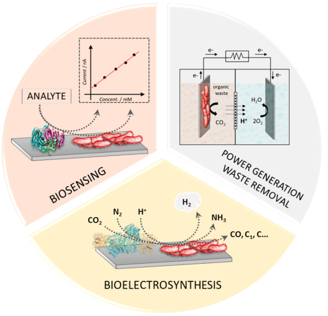
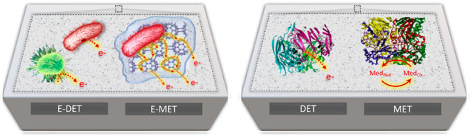
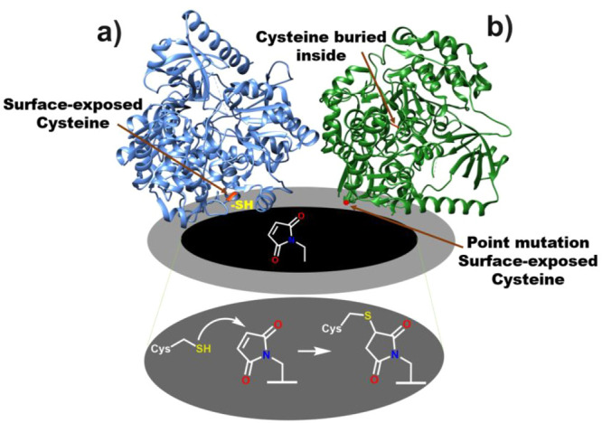
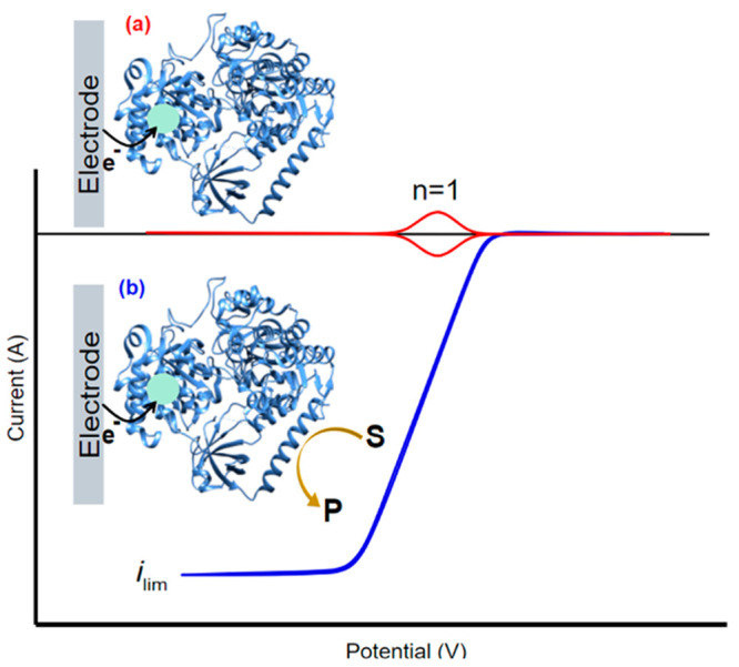
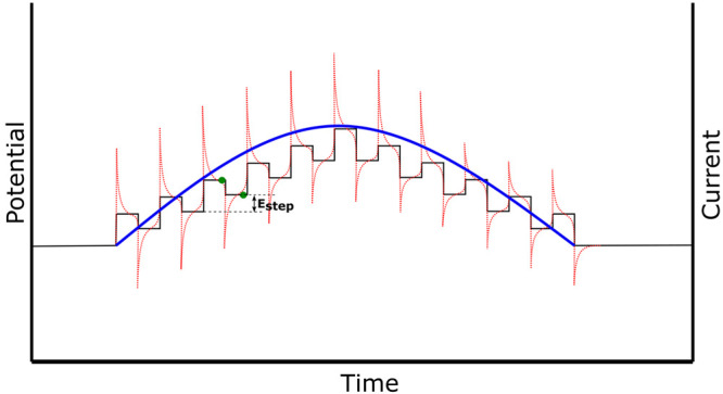
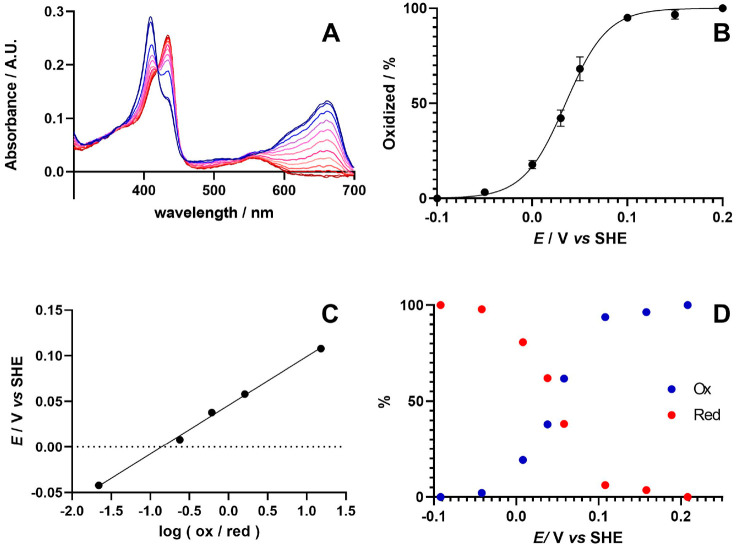
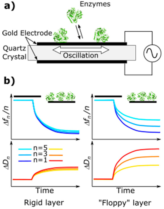
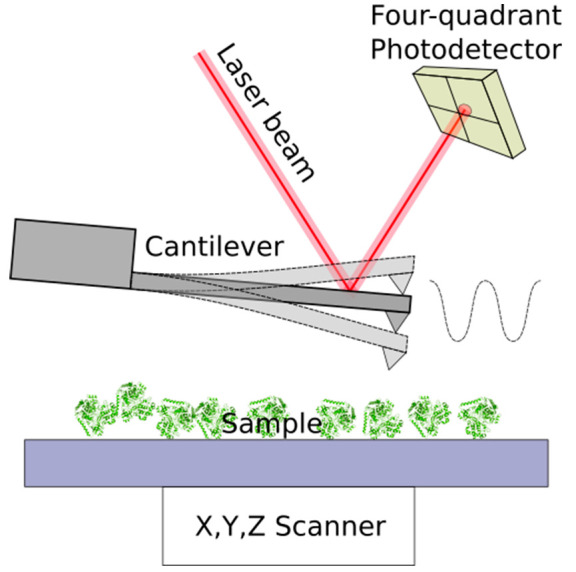
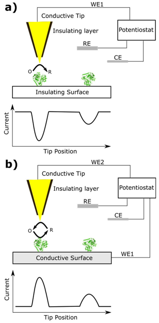
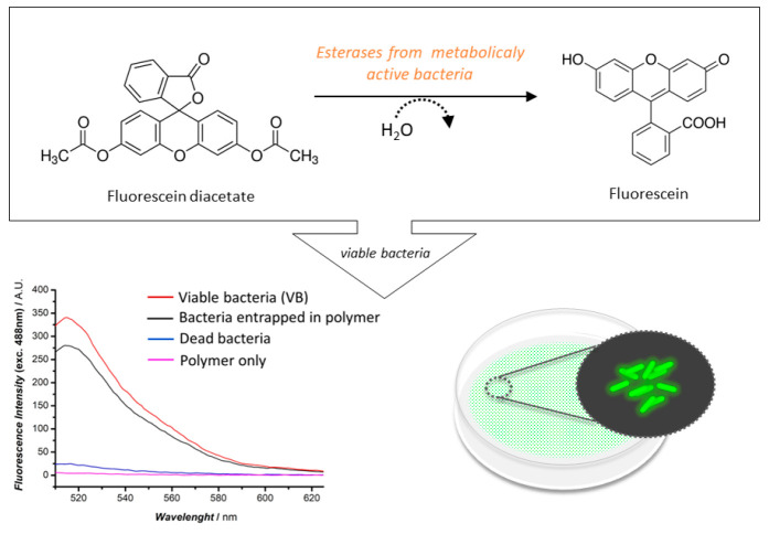
The coupling of enzymes and/or intact bacteria with electrodes has been vastly investigated due to the wide range of existing applications. These span from biomedical and biosensing to energy production purposes and bioelectrosynthesis, whether for theoretical research or pure applied industrial processes. Both enzymes and bacteria offer a potential biotechnological alternative to noble/rare metal-dependent catalytic processes. However, when developing these biohybrid electrochemical systems, it is of the utmost importance to investigate how the approaches utilized to couple biocatalysts and electrodes influence the resulting bioelectrocatalytic response. Accordingly, this tutorial review starts by recalling some basic principles and applications of bioelectrochemistry, presenting the electrode and/or biocatalyst modifications that facilitate the interaction between the biotic and abiotic components of bioelectrochemical systems. Focus is then directed toward the methods used to evaluate the effectiveness of enzyme/bacteria-electrode interaction and the insights that they provide. The basic concepts of electrochemical methods widely employed in enzymatic and microbial electrochemistry, such as amperometry and voltammetry, are initially presented to later focus on various complementary methods such as spectroelectrochemistry, fluorescence spectroscopy and microscopy, and surface analytical/characterization techniques such as quartz crystal microbalance and atomic force microscopy. The tutorial review is thus aimed at students and graduate students approaching the field of enzymatic and microbial electrochemistry, while also providing a critical and up-to-date reference for senior researchers working in the field.


Conflict of interest statement

The authors declare no competing financial interest.

Figures

Figure 1
Figure 1
Scheme of different applications of enzymatic and microbial electrochemical systems, including the development of biosensors for the monitoring of medically relevant analytes or toxic compounds, the local generation of micropower with simultaneous waste removal, and the development of bioelectrosynthetic factories.
Scheme 1
Scheme 1. Electron Transfer Pathways for Microbial (Left) and Enzymatic Electrodes (Right)
E-DET refers to the EET of electrons through microbial nanowires or membrane-bound proteins. E-MET refers to the EET by means of diffusible or polymer bound-redox mediators. DET refers to the direct transfer from the redox-active sites of an enzyme to the electrode surface, while MET refers to the electron transfer through artificial redox mediators.
Figure 2
Figure 2
(a) Metalloprotein with a surface-exposed cysteine can readily react with a scaffold incorporated on the electrode, such as maleimide. (b) In case of a protein having a cysteine residue not exposed on the surface, a point mutation can help to introduce a cysteine residue at the suitable position.
Figure 3
Figure 3
Idealized cyclic voltammetry showing the redox response of an enzyme under reductive conditions with one active redox center (n = 1) under (a) non-turnover conditions where there is only an electron transfer between the redox center of the enzyme and the electrode, and (b) turnover conditions in the presence of a substrate and steady-state conditions. The current is a direct measure of enzyme activity.
Figure 4
Figure 4
SWV trace where the potential (black) is applied as a staircase potential, the absolute current (red) is sampled twice at the end of each potential step (green dots), and the final current is reported as an average of both values (blue line).
Figure 5
Figure 5
(A) Spectroelectrochemical oxidation (blue) and reduction (red) of myoglobin in the presence of methylene blue as an electron mediator. (B) Nonlinear regression and (C) linear fit of data obtained in (A) to the Nernst equation to determine the formal redox potential (E0′) and the number of electrons transferred (n). (D) Changes in the oxidized and reduced features of myoglobin’s heme cofactor at different applied potentials. Adapted with permission under a Creative Commons Attribution License from ref (186). Copyright 2022 The Authors.
Figure 6
Figure 6
(a) Working principle of the QCM-D. The quartz disk is oscillated at its resonance frequencies with an alternating electric field. (b) During the adsorption of proteins on the surface, the resonance frequencies decrease (Δf) and the dissipations increase (ΔD). Typical responses for a rigid and “floppy” add-layer are shown on the left and right, respectively.
Figure 7
Figure 7
Typical setup of an AFM system. Sample is moved with a nanopositioning scanner. Tip–sample interaction deflects the cantilever, whose deformations are recorded using a laser beam reflected from the back of the cantilever onto a four-quadrant photodetector.
Figure 8
Figure 8
AFM-SECM working sketch. (a) Tip functions as primary working electrode (WE1). The electrochemical reaction takes place when the tip interacts with the adsorbed enzyme. (b) Tip functions as secondary working electrode (WE2). The electrochemical reaction products are detected when the tip is situated in the vicinity of the adsorbed molecule.
Figure 9
Figure 9
(Top) Reaction of fluorescein diacetate with esterases present intracellularly or bound to the external membrane of bacterial cells. (Bottom left) FS result obtained for free R. capsulatus cells (red), R. capsulatus cells entrapped in PDA (black), heat-treated R. capsulatus cells at 120 °C for 4 h (blue), and PDA only (purple), adapted with permission under a Creative Commons Attribution License from ref (81). Copyright 2022 The Authors. (Bottom right) Scheme of a microscopy slide covered in bacteria for WFM analysis.

Similar articles

Cited by

References

    1. Unden G.; Bongaerts J. Alternative respiratory pathways of Escherichia coli: energetics and transcriptional regulation in response to electron acceptors. Biochim. Biophys. Acta - Bioenerg. 1997, 1320, 217–234. 10.1016/S0005-2728(97)00034-0. - DOI - PubMed
    1. Kano K.; et al. Voltammetric determination of acid dissociation constants of pyrroloquinoline quinone and its reduced form under acidic conditions. Bioelectrochemistry Bioenerg. 1990, 24, 193–201. 10.1016/0302-4598(90)85021-9. - DOI
    1. Wagner T.; Koch J.; Ermler U.; Shima S. Methanogenic heterodisulfide reductase (HdrABC-MvhAGD) uses two noncubane [4Fe-4S] clusters for reduction. Science (80-.). 2017, 357, 699–703. 10.1126/science.aan0425. - DOI - PubMed
    1. Buckel W.; Thauer R. K. Flavin-Based Electron Bifurcation, A New Mechanism of Biological Energy Coupling. Chem. Rev. 2018, 118, 3862–3886. 10.1021/acs.chemrev.7b00707. - DOI - PubMed
    1. Blankenship R. E. Early Evolution of Photosynthesis. Plant Physiol. 2010, 154, 434–438. 10.1104/pp.110.161687. - DOI - PMC - PubMed