

Association of daily physical activity with brain volumes and cervical spinal cord areas in multiple sclerosis
- PMID: 36573559
- PMCID: PMC9972237
- DOI: 10.1177/13524585221143726
Association of daily physical activity with brain volumes and cervical spinal cord areas in multiple sclerosis
Abstract
Background: Remote activity monitoring has the potential to evaluate real-world, motor function, and disability at home. The relationships of daily physical activity with spinal cord white matter and gray matter (GM) areas, multiple sclerosis (MS) disability and leg function, are unknown.
Objective: Evaluate the association of structural central nervous system pathology with ambulatory disability.
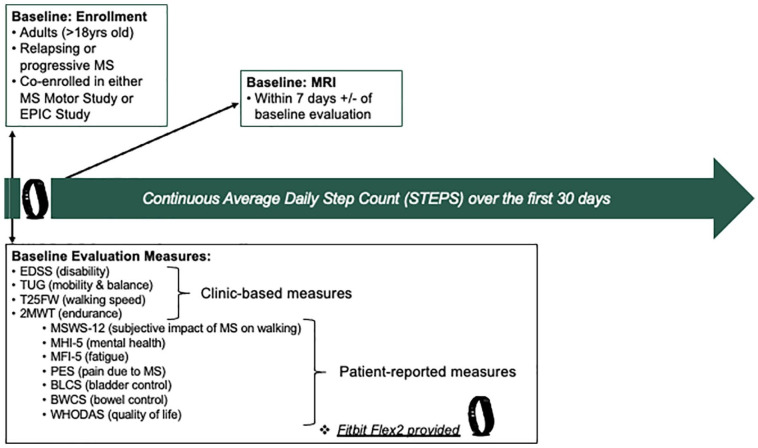
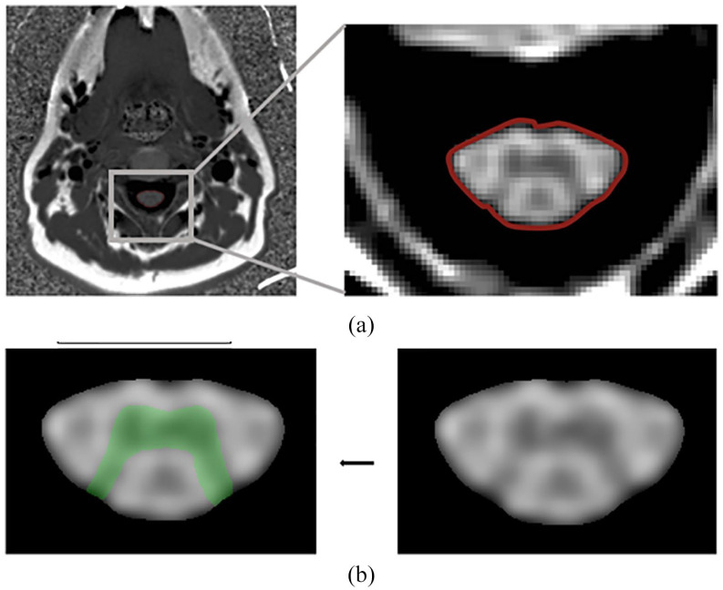
Methods: Fifty adults with progressive or relapsing MS with motor disability who could walk >2 minutes were assessed using clinician-evaluated, patient-reported outcomes, and quantitative brain and spinal cord magnetic resonance imaging (MRI) measures. Fitbit Flex2, worn on the non-dominant wrist, remotely assessed activity over 30 days. Univariate and multivariate analyses were performed to assess correlations between physical activity and other disability metrics.
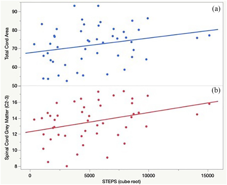
Results: Mean age was 53.3 years and median Expanded Disability Status Scale (EDSS) was 4.0. Average daily step counts (STEPS) were highly correlated with EDSS and walking measures. Greater STEPS were significantly correlated with greater C2-C3 spinal cord GM areas (ρ = 0.39, p = 0.04), total cord area (TCA; ρ = 0.35, p = 0.04), and cortical GM volume (ρ = 0.32, p = 0.04).
Conclusion: These results provide preliminary evidence that spinal cord GM area is a neuroanatomical substrate associated with STEPS. STEPS could serve as a proxy to alert clinicians and researchers to possible changes in structural nervous system pathology.
Keywords: Fitbit; Multiple sclerosis; activity level; brain MRI; cervical MRI; remote monitoring; spinal cord gray matter area.
Conflict of interest statement
The author(s) declared the following potential conflicts of interest with respect to the research, authorship, and/or publication of this article: Dr V.J.B. is funded by the National MS Society Career Transition Award. Ms S.C., Mr J.J., Ms G.K., Mrs A.M.A., Mr M.K., Mr A.A., and Mr W.A.S. have no relevant disclosures. Drs R.C., E.C., M.J.P., J.E.O., and G.M.M. have no relevant disclosures. Dr N.P. has received research support to UCSF from the Race to Erase MS Foundation and from the National Center for Advancing Translational Sciences, National Institutes of Health, through a UCSF-CTSI Grant. Dr S.L.H. currently serves on the scientific advisory board of Accure, Annexon, Alector, board of directors of Neurona, and has previously consulted for NGM Bio and Moderna. Dr S.L.H. also has received travel reimbursement and writing assistance from Roche and Novartis for CD20-related meetings and presentations. Dr J.M.G. has received research support to UCSF from Genentech/Roche and Vigil Neuroscience, and consulting fees from Biogen. Dr R.B. is funded by the NMSS Harry Weaver Award, NIH, DOD, NSF as well as Biogen, Novartis, and Roche Genentech. She has received personal fees for consulting from Alexion, EMD Serono, Horizon, Jansen, Genzyme Sanofi, and TG Therapeutics. Dr B.A.C.C. has received personal compensation for consulting from Alexion, Atara, Biogen, EMD Serono, Novartis, Sanofi, and TG Therapeutics. Dr R.G.H. has received consulting fees from Roche/Genentech, Sanofi/Genzyme, Novartis, Celgene, Atara Bio, QIA Consulting, Boston Pharma, and Neurona Therapeutic. Grants from Roche/Genentech and Atara Bio.
Figures
References
-
- Kearney H, Miller DH, Ciccarelli O. Spinal cord MRI in multiple sclerosis—Diagnostic, prognostic and clinical value. Nat Rev Neurol 2015; 11(6): 327–338. - PubMed
-
- Kremenchutzky M, Rice GP, Baskerville J, et al.. The natural history of multiple sclerosis: A geographically based study 9: Observations on the progressive phase of the disease. Brain 2006; 129(Pt 3): 584–594. - PubMed
Publication types
MeSH terms
Grants and funding
LinkOut - more resources
Full Text Sources
Medical
Miscellaneous
