. 2023 Feb 1;324(2):L154-L168.
doi: 10.1152/ajplung.00248.2022. Epub 2022 Dec 27.

The matricellular protein CCN3 supports lung endothelial homeostasis and function

Affiliations

The matricellular protein CCN3 supports lung endothelial homeostasis and function

Kalpana R Betageri et al. Am J Physiol Lung Cell Mol Physiol. .

Abstract

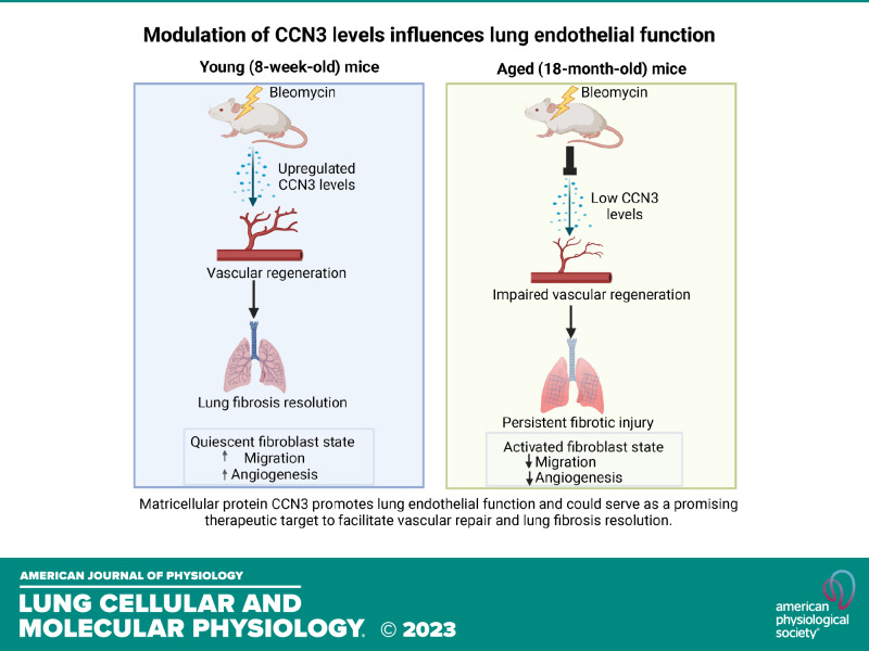
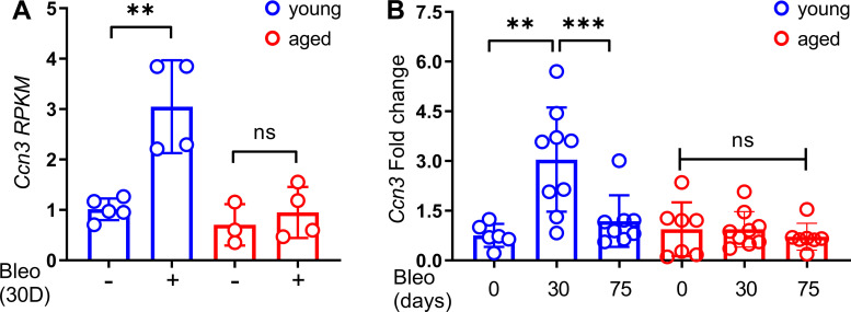
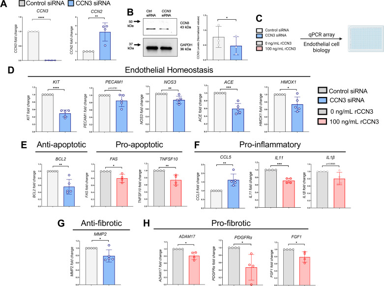
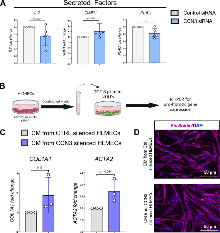
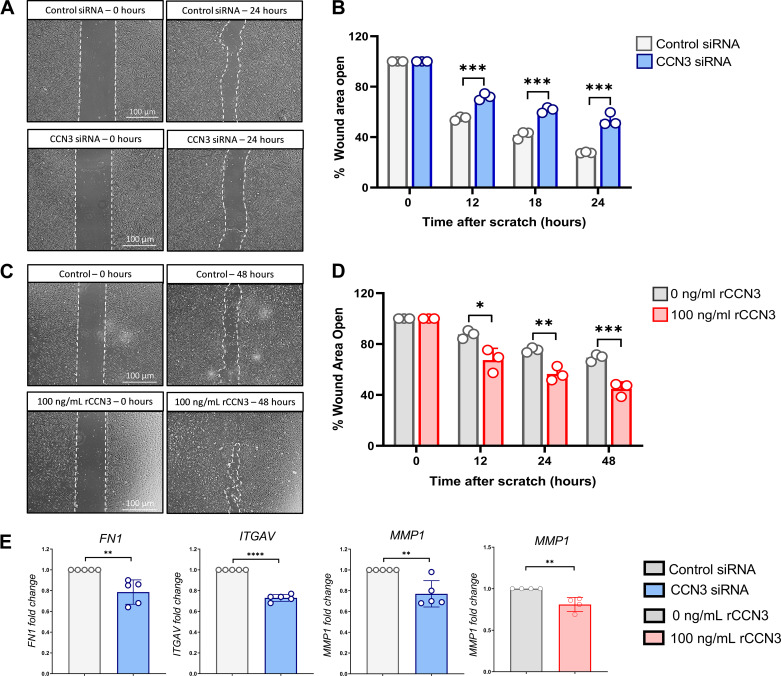
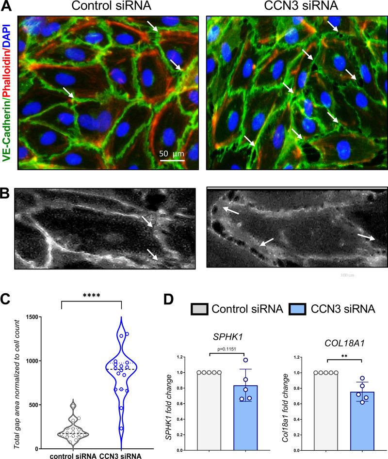
Aberrant vascular remodeling contributes to the progression of many aging-associated diseases, including idiopathic pulmonary fibrosis (IPF), where heterogeneous capillary density, endothelial transcriptional alterations, and increased vascular permeability correlate with poor disease outcomes. Thus, identifying disease-driving mechanisms in the pulmonary vasculature may be a promising strategy to limit IPF progression. Here, we identified Ccn3 as an endothelial-derived factor that is upregulated in resolving but not in persistent lung fibrosis in mice, and whose function is critical for vascular homeostasis and repair. Loss and gain of function experiments were carried out to test the role of CCN3 in lung microvascular endothelial function in vitro through RNAi and the addition of recombinant human CCN3 protein, respectively. Endothelial migration, permeability, proliferation, and in vitro angiogenesis were tested in cultured human lung microvascular endothelial cells (ECs). Loss of CCN3 in lung ECs resulted in transcriptional alterations along with impaired wound-healing responses, in vitro angiogenesis, barrier integrity as well as an increased profibrotic activity through paracrine signals, whereas the addition of recombinant CCN3 augmented endothelial function. Altogether, our results demonstrate that the matricellular protein CCN3 plays an important role in lung endothelial function and could serve as a promising therapeutic target to facilitate vascular repair and promote lung fibrosis resolution.

Keywords: CCN3; idiopathic pulmonary fibrosis; lung homeostasis; pulmonary fibrosis; pulmonary vasculature.


Conflict of interest statement

No conflicts of interest, financial or otherwise, are declared by the authors.

Figures

None
Graphical abstract
Figure 1.
Figure 1.
CCN3 is transiently upregulated in lung ECs from young but not aged mice after bleomycin injury. A: histograms of RNA expression RPKM values showing upregulation of Ccn3 in lung ECs isolated from young mice 30 days after bleomycin but no elevation in lung ECs isolated from aged mice at the same time point (young sham 2-mo-old, n = 5; young 2-mo-old day 30 after bleomycin, n = 4; aged sham 18-mo-old, n = 3; aged 18-mo-old after bleomycin, n = 4). Values are summarized as means and SD. B: qPCR analysis of Ccn3 transcripts in FACS-isolated lung endothelial cells (CD45−/CD326−/GFP−/CD31+ population) isolated from young and aged mice either sham or challenged with bleomycin (young sham 2-mo-old, n = 6; young 2-mo-old, day 30 after bleomycin, n = 9; young 2-mo-old, day 75 after bleomycin, n = 8; aged sham 18-mo-old, day 0, n = 7; aged 18-mo-old, day 30 after bleomycin, n = 9; aged 18-mo-old, day 75 after bleomycin, n = 7). Data are expressed as means ± SD and analyzed using one-way analysis of variance (followed by Tukey’s post hoc test). Aged lung ECs failed to upregulate Ccn3 at any of the time points analyzed. EC, endothelial cell; RPKM, reads per kilobase of exon per million reads mapped. **P ≤ 0.01; ***P ≤ 0.001; ns, not significant.
Figure 2.
Figure 2.
CCN3 loss and gain of function leads to transcriptional changes. A: gene expression analysis by qPCR demonstrates significant increase of CCN2 transcript in CCN3-silenced HLMECs. Data are expressed as means ± SD from n = 4 independent experiments and analyzed using Student’s t test (****P ≤ 0.0001). B: Western blot analysis and relative quantification confirm reduction of CCN3 protein expression in CCN3-silenced HLMECs. Data are expressed as means ± SD from n = 3 independent experiments and analyzed using Student’s t test (*P ≤ 0.05). C: schematic of the experimental workflow. DH: transcriptional changes in control- and CCN3-silenced HLMECs for 72 h (n = 5 independent experiments) or control- and HLMECs treated with 100 ng/mL recombinant CCN3 for 9 h (n = 4 independent experiments) were analyzed by using an endothelial cell biology profiler PCR array. Data are expressed as means ± SD and analyzed using Student’s t test (*P ≤ 0.05, **P ≤ 0.01, ***P ≤ 0.001, ****P ≤ 0.0001). HLMEC, human lung microvascular endothelial cell; PCR, polymerase chain reaction. [Image created with BioRender.com and published with permission.]
Figure 3.
Figure 3.
Loss of CCN3 in human lung ECs enhances lung fibroblast activation via paracrine signals. A: loss of CCN3 in HLMECs leads to alterations in genes encoding for secreted factors IL7, TIMP1, and PLAU. Data are expressed as means ± SD and analyzed using Student’s t test (P = 0.08, P = 0.12, *P ≤ 0.05). B: schematic of the experimental workflow. Conditioned media was collected and concentrated from HLMECs transfected with either nontargeting or CCN3 siRNA (72 h). The conditioned media was then applied to NHLFs primed with 2 ng/mL TGF-β and RT-PCR was performed to evaluate profibrotic gene expression on recipient fibroblasts. C: primed fibroblasts cultured in conditioned medium collected from CCN3-silenced HLMECs displayed elevated levels of profibrotic markers COL1A1 and ACTA2 compared with fibroblasts exposed to conditioned medium generated from control silenced HLMECs. Data are expressed as means ± SD from n = 3 independent experiments and analyzed using Student’s t test (P = 0.1 and P = 0.054, respectively). D: fibroblasts were primed with TGFβ and then exposed for 3 days to conditioned media collected from control or CCN3-silenced HLMECs. At the end of the incubation, cells were stained with phalloidin to visualize cell morphology and counterstained with DAPI. Cells exposed to conditioned medium generated from CCN3-silenced HLMECs exhibit a more elongated phenotype. EC, endothelial cell; HLMEC, human lung microvascular endothelial cell; NHLF, normal human lung fibroblast. [Image created with BioRender.com and published with permission.]
Figure 4.
Figure 4.
Loss of CCN3 impairs endothelial migration while exogeneous CCN3 restores this function. HLMECs were transfected with control- or CCN3 siRNA or treated with vehicle or rCCN3 before a scratch was performed to study their migratory ability. A, B: cell migration in HLMECs transfected with CCN3 siRNA after a 24-h time course was significantly impaired compared with cells transfected with control siRNA as quantified using FIJI and normalized to wound area. C, D: cell migration in HLMECs treated with 100 ng/mL recombinant CCN3 after a 48-h time course was significantly improved compared with untreated cells as quantified using FIJI and normalized to wound area. Data are expressed as means ± SD from n = 3 independent experiments and analyzed using one-way analysis of variance (followed by Tukey’s post hoc test). (**P ≤ 0.01, ***P ≤ 0.001, ****P ≤ 0.0001). E: loss and gain of function of CCN3 in HLMECs leads to changes in genes associated with endothelial migration. Data are expressed as means ± SD and analyzed using Student’s t test (**P ≤ 0.01, ***P ≤ 0.001, ****P ≤ 0.0001). HLMEC, human lung microvascular endothelial cell.
Figure 5.
Figure 5.
Loss of CCN3 impairs endothelial barrier integrity. A: representative immunofluorescence images (×10 objective magnification) of HLMECs transfected with control or CCN3 siRNA (72 h), stained for VE-cadherin (green) and Phalloidin (red) (F-actin). Nuclei were counterstained for DAPI. Arrows highlight the inter-cellular gaps. B: arrows highlight the intercellular gaps. C: three frames per well were analyzed (n ≥ 40 cells) and total gap area was quantified via ImageJ and normalized for cell count (obtained through quantification of nuclei visualized by DAPI). Data distribution is visualized via violin plot from control siRNA (n = 15 images) and CCN3 siRNA (n = 16 images) expressing the median as the dotted line. Data are analyzed using Student’s t test, n = 3 independent experiments (****P ≤ 0.0001). D: loss of CCN3 in HLMECs leads to reduction in genes associated with endothelial barrier integrity. Data are expressed as means ± SD and analyzed using Student’s t test (P = 0.11, **P ≤ 0.01). DAPI, 4′,6-diamidino-2-phenylindole; HLMEC, human lung microvascular endothelial cell.
Figure 6.
Figure 6.
Loss of CCN3 impairs in vitro angiogenic function while exogeneous CCN3 recovers this function. A: schematic depicting the experimental workflow. Control- and CCN3-silenced HLMECs were plated on polymerized Matrigel basement membrane for 6 h and analyzed for the ability to form tube-like structures. B: representative images (×10 objective magnification) of HLMECs transfected with control or CCN3 siRNA (72 h) indicate that loss of CCN3 resulted in a more disordered, less continuous network. C: quantification of total number of junctions and total vessel length, parameters of in vitro angiogenesis, via AngioTool 0.6 software shows reduction of both parameters in CCN3-silenced cells compared with control cells. D: schematic depicting the experimental workflow. Control HLMECs cells or those treated with 100 ng/mL recombinant CCN3 were plated on Matrigel basement membrane for 6 h and analyzed for the ability to form tube-like structures. E: representative images (×10 objective magnification) of HLMECs treated with 0 ng/mL or 100 ng/mL human recombinant CCN3 protein indicate that exogeneous CCN3 promoted a more continuous, ordered network. F: quantification via AngioTool 0.6 software shows increases in total number of junctions and total vessels length in HLMECs treated with recombinant CCN3 in comparison with untreated control cells deficient in S1P. Data are expressed as means ± SD from n = 3 independent experiments and analyzed using Student’s t test (*P ≤ 0.05, **P ≤ 0.01). G: loss and gain of function of CCN3 in HLMECs leads to changes in genes associated with angiogenesis. Data are expressed as means ± SD and analyzed using Student’s t test (P = 0.057, *P ≤ 0.05, **P ≤ 0.01, ****P ≤ 0.0001). Representative immunofluorescence images (×10 objective magnification) of HLMECs transfected with control or CCN3 siRNA (72 h) (H) and HLMECs treated with 0 ng/mL or 100 ng/mL human recombinant CCN3 protein (I), stained for Ki67 (green). Nuclei were counterstained with DAPI. Quantification of percent of Ki67 positive, DAPI positive cells, via Bio-Tek Gen5 Image software shows no significant change in CCN3-silenced cells (H) and recombinant CCN3 protein treated cells compared with respective controls (I). Data are expressed as means ± SD from n = 3 independent experiments and analyzed using Student’s t test (*P ≤ 0.05, **P ≤ 0.01). DAPI, 4′,6-diamidino-2-phenylindole; HLMEC, human lung microvascular endothelial cells. [Image created with BioRender.com and published with permission.]

Similar articles

Cited by

References

    1. Evans CE, Iruela-Arispe ML, Zhao YY. Mechanisms of endothelial regeneration and vascular repair and their application to regenerative medicine. Am J Pathol 191: 52–65, 2021. doi:10.1016/j.ajpath.2020.10.001. - DOI - PMC - PubMed
    1. Caporarello N, Lee J, Pham TX, Jones DL, Guan J, Link PA, Meridew JA, Marden G, Yamashita T, Osborne CA, Bhagwate AV, Huang SK, Nicosia RF, Tschumperlin DJ, Trojanowska M, Ligresti G. Dysfunctional ERG signaling drives pulmonary vascular aging and persistent fibrosis. Nat Commun 13: 4170, 2022. doi:10.1038/s41467-022-33197-w. - DOI - PMC - PubMed
    1. Caporarello N, Meridew JA, Aravamudhan A, Jones DL, Austin SA, Pham TX, Haak AJ, Moo Choi K, Tan Q, Haresi A, Huang SK, Katusic ZS, Tschumperlin DJ, Ligresti G. Vascular dysfunction in aged mice contributes to persistent lung fibrosis. Aging Cell 19: e13196, 2020. doi:10.1111/acel.13196. - DOI - PMC - PubMed
    1. Ungvari Z, Tarantini S, Kiss T, Wren JD, Giles CB, Griffin CT, Murfee WL, Pacher P, Csiszar A. Endothelial dysfunction and angiogenesis impairment in the ageing vasculature. Nat Rev Cardiol 15: 555–565, 2018. doi:10.1038/s41569-018-0030-z. - DOI - PMC - PubMed
    1. Herrera MD, Mingorance C, Rodríguez-Rodríguez R, Alvarez de Sotomayor M. Endothelial dysfunction and aging: an update. Ageing Res Rev 9: 142–152, 2010. doi:10.1016/j.arr.2009.07.002. - DOI - PubMed

Publication types

Substances