. 2023 Apr 1;14(4):e00560.
doi: 10.14309/ctg.0000000000000560.

ABX464 (Obefazimod) Upregulates miR-124 to Reduce Proinflammatory Markers in Inflammatory Bowel Diseases

Affiliations

ABX464 (Obefazimod) Upregulates miR-124 to Reduce Proinflammatory Markers in Inflammatory Bowel Diseases

Cécile Apolit et al. Clin Transl Gastroenterol. .

Abstract

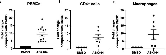
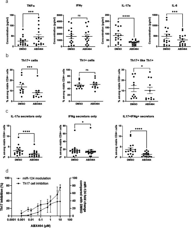
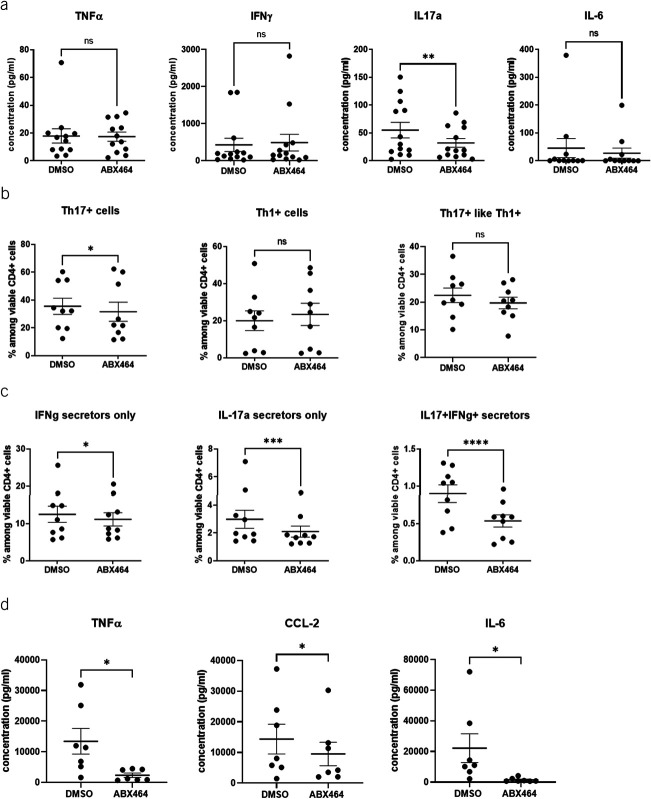
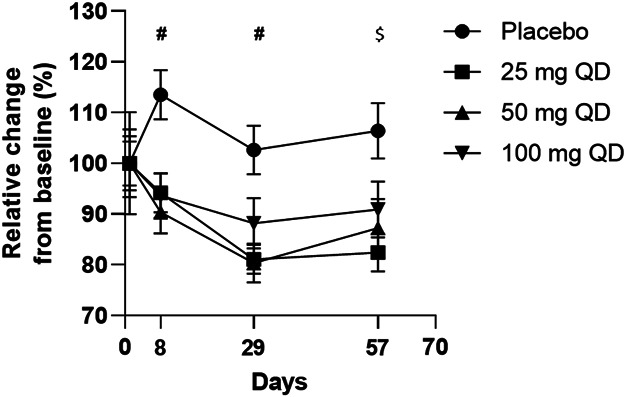
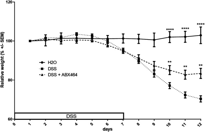
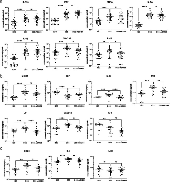
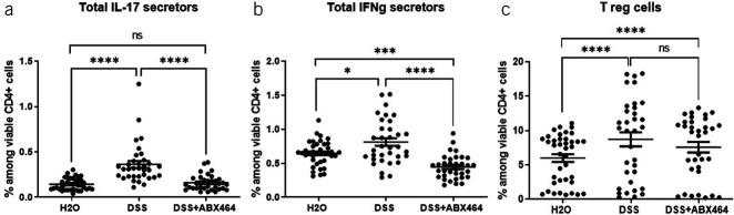
Advanced therapies have transformed the treatment of inflammatory bowel disease; however, many patients fail to respond, highlighting the need for therapies tailored to the underlying cell and molecular disease drivers. The first-in-class oral molecule ABX464 (obefazimod), which selectively upregulates miR-124, has demonstrated its ability to be a well-tolerated treatment with rapid and sustained efficacy in patients with ulcerative colitis (UC). Here, we provide evidence that ABX464 affects the immune system in vitro , in the murine model of inflammatory bowel disease, and in patients with UC. In vitro , ABX464 treatment upregulated miR-124 and led to decreases in proinflammatory cytokines including interleukin (IL) 17 and IL6, and in the chemokine CCL2. Consistently, miR-124 expression was upregulated in the rectal biopsies and blood samples of patients with UC, and a parallel reduction in Th17 cells and IL17a levels was observed in serum samples. In a mouse model of induced intestinal inflammation with dextran sulfate sodium, ABX464 reversed the increases in multiple proinflammatory cytokines in the colon and the upregulation of IL17a secretion in the mesenteric lymph nodes. By upregulating miR-124, ABX464 acts as "a physiological brake" of inflammation, which may explain the efficacy of ABX464 with a favorable tolerability and safety profile in patients with UC.


Conflict of interest statement

Guarantor of the article: Jamal Tazi, PhD.

Specific author contributions: C.A. and N.C. contributed equally to the experiments and should be considered as co-first authors. A.V., C.B.-P., L.L., A.G., and J.S. contributed to the experiments; A.G., J.S., and J.T. contributed to conducting experiments, acquiring data, analyzing data; A.G., J.S., D.S., P.G., H.E., and J.T. contributed to the conception of the manuscript; A.G., J.S., and J.T. contributed to the writing of the manuscript and literature review. The final version was approved by all authors. The authors meet criteria for authorship as recommended by the International Committee of Medical Journal Editors (ICMJE).

Financial support: This study was supported by Abivax. The authors received no direct compensation related to the development of this manuscript.

Potential competing interests: C.A., N.C., A.V., L.L., D.S., P.G., A.G., J.S., H.E., and J.T. are employees at Abivax.

Figures

Figure 1.
Figure 1.
Upregulation of miR-124 expression. Changes in miR-124 levels after 6 days of ABX464 5 μM treatment of (a) peripheral blood mononuclear cells (PBMCs), (b) CD4+ cells, and (c) macrophages. Fold change compared with DMSO-treated cells as a negative control. Dots represent blood samples from different donors, and mean ± SEM are reported.
Figure 2.
Figure 2.
Effects of ABX464 on peripheral blood mononuclear cells (PBMCs). Cells were treated for 6 days with 5 μM ABX464. (a) Cytokine concentrations in the supernatant of PBMCs. (b) Th1/Th17 cells (CD196/CD183 expression) among viable CD4+ cells. (c) Intracellular IFNγ and IL17a expression among viable CD4+ cells. (d) Correlation between miR-124 modulation and Th17-cell inhibition in PBMCs related to ABX464 concentration. Cells were treated for 6 days with different concentrations of ABX464 (not above 10 μM to avoid a cytotoxic effect). Th17-cell inhibition and fold changes in miR-124 were determined in comparison with DMSO-treated cells as a negative control. Mean ± SD are reported. Results were obtained on PBMC isolated from 15 or 16 healthy blood donors and 5 different experiments (panel a), from 11 donors and 3 experiments (panel b), and from 16 donors and 5 experiments (panel c). Dots represent blood samples from different, and mean ± SEM are reported. Dunnett or Dunn test (ns = P > 0.05, *P < 0.05, ***P < 0.001, ****P < 0.0001).
Figure 3.
Figure 3.
Effects of ABX464 on CD4+ cells and macrophages. Cells were treated for 6 days with 5 μM ABX464. (a) Cytokine concentrations in the supernatant of CD4+ cells. (b) Th1/Th17 cells (CD196/CD183 expression) among viable CD4+ cells. (c) Intracellular interferon (IFN) γ and interleukin (IL) 17a expression among viable CD4+ cells. Dots represent blood samples from different donors, and mean ± SEM are reported. (d) Effects of ABX464 on macrophages. Cells were treated for 7 days with 5 μM ABX464. Results were obtained from 15 or 16 healthy blood donors and 5 different experiments (panel a), from 11 donors and 3 experiments (panel b), from 16 donors and 5 experiments (panel c), and from 7 donors and 3 experiments (panel d). Dots represent blood samples from different donors, and mean ± SEM are reported. Dunnett or Dunn test (ns = P > 0.05, *P < 0.05, **P < 0.01, ***P < 0.001, ****P < 0.0001).
Figure 4.
Figure 4.
MiR-124 modulation in rectal tissue (ac) and blood (df) in patients with UC. The number of miR-124 copies was assessed by droplet digital PCR at baseline (a, d) and week 8 (b, e), and fold increase at week 8 compared with baseline (c, f). A total of 576 rectal biopsies from 139 patients and 1,104 blood samples from 229 patients who received ABX464 (25, 50, or 100 mg) or placebo were analyzed. Kruskal-Wallis test (***P < 0.001 vs placebo; ****P < 0.0001 vs placebo).
Figure 5.
Figure 5.
IL17a levels in serum from patients with UC treated with 25, 50, or 100 mg ABX464 daily or placebo for 8 weeks. A total of 1,185 serum samples (500 μL of serum per sample) from 254 patients who received either ABX464 (25, 50, or 100 mg) or placebo were assessed during 5 visits (day 1, weeks 1, 4, 8, and 16) of the phase 2b trial (10). ****P < 0.0001 vs placebo. #P < 0.01 for 25, 50, and 100 mg daily; $P < 0.01 for 25 and 50 mg daily. QD, every day (quaque die); UC, ulcerative colitis.
Figure 6.
Figure 6.
ABX464 treatment reduces disease severity in dextran sodium sulfate (DSS)-induced colitis. C57BL6 mice subjected to the DSS colitis protocol received orally once a day ABX464 (40 mg/kg) in methylcellulose (MC) or MC only through gavage. The weight profiles are representative of the curves obtained in all DSS experiments. They were obtained with 10 mice per group. **P < 0.01, ****P < 0.0001, 1-way ANOVA parametric test.
Figure 7.
Figure 7.
Effects of ABX464 on cytokine secretion by colons of dextran sodium sulfate (DSS)-induced mice. The colons of untreated healthy mice (H2O group) or DSS-induced colitis mice treated by ABX464 (DSS + ABX464) or untreated (DSS) were cultured ex vivo at the end of an acute DSS experiment and assessed for cytokine secretions with (a) LegendPlex mu inflammation panel or (b) a LegendPlex mu HSC panel or (c) ELISA. Dots represent colons, and mean ± SEM are reported. The statistics are based on the fold change compared with DSS. Kruskal-Wallis with the Dunn multiple comparison test (*P < 0.05, **P < 0.01, ***P < 0.001, ****P < 0.0001). N = 26 mice per group.
Figure 8.
Figure 8.
Effects of ABX464 on CD4+ subsets in the mesenteric lymph nodes (MLNs) of dextran sodium sulfate (DSS)-induced mice. Cells in the MLNs of untreated healthy mice (H2O group), untreated mice with DSS-induced colitis (DSS group), or treated mice with DSS-induced colitis (DSS + ABX464) were collected and stained to determine proportions of (a) interleukin (IL) 17 secretors, (b) interferon (IFN) γ secretors, and (c) Treg cells. Dots represent samples from individual mice, and mean ± SEM are reported. Kruskal-Wallis tests with the Dunn multiple comparison test (*P < 0.05, **P < 0.01, ***P < 0.001, ****P < 0.0001).

References

    1. Lee SH, Kwon Je, Cho ML. Immunological pathogenesis of inflammatory bowel disease. Intest Res 2018;16(1):26–42. - PMC - PubMed
    1. Abraham C, Cho JH. Inflammatory bowel disease. N Engl J Med 2009;361(21):2066–78. - PMC - PubMed
    1. Torres J, Mehandru S, Colombel JF, et al. . Crohn's disease. Lancet 2017;389(10080):1741–55. - PubMed
    1. Ungaro R, Mehandru S, Allen PB, et al. . Ulcerative colitis. Lancet 2017;389(10080):1756–70. - PMC - PubMed
    1. Mowat C, Cole A, Windsor A, et al. . Guidelines for the management of inflammatory bowel disease in adults. Gut 2011;60(5):571–607. - PubMed