. 2022 Oct;43(10):1539-1543.
doi: 10.3174/ajnr.A7628. Epub 2022 Sep 8.

Utility of Dual-Energy CT to Improve Diagnosis of CSF Leaks on CT Myelography following Lateral Decubitus Digital Subtraction Myelography with Negative Findings

Affiliations

Utility of Dual-Energy CT to Improve Diagnosis of CSF Leaks on CT Myelography following Lateral Decubitus Digital Subtraction Myelography with Negative Findings

S J Huls et al. AJNR Am J Neuroradiol. 2022 Oct.

Abstract

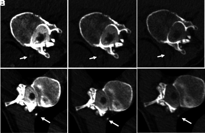
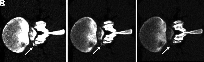
CSF leaks, including CSF-venous fistulas, which cause spontaneous intracranial hypotension, remain difficult to diagnose, even on digital subtraction myelography and CT myelography. Dual-energy CT technology has been used to improve diagnostic utility within multiple organ systems. The capability of dual-energy CT to create virtual monoenergetic images can be leveraged to increase conspicuity of contrast in CSF-venous fistulas and direct epidural CSF leakage to improve the diagnostic utility of CT myelography. Six cases (in 5 patients) are shown in which virtual monoenergetic images demonstrate a leak location that was either occult or poorly visible on high- or low-kilovolt series. This clinical report describes the novel application of dual-energy CT for the detection of subtle CSF leaks including CSF-venous fistulas.


Figures

FIG 1.
FIG 1.
A, 50-keV VMI. B, 100 kV. C, 140 kV. Case 1: Left-side-down CTM with linear contrast at the left T12–L1 neural foramen, thought to reflect a CVF (arrows). Contrast is also noted in the renal collecting system. ROI Hounsfield units: A, 50-keV VMI Hounsfield unit maximum (max): 283 HU; mean, 76 HU. B, 100 -kV Hounsfield unit max: 147 HU; mean, 68 HU. C, 140-kV Hounsfield unit max: 116 HU; mean, 46 HU. D, 50-keV VMI. E, 100 kV. F, 140 kV. Case 1: Right-side-down CTM with paraspinal contrast at the level of T11–12, representing a second CVF (arrows). ROI Hounsfield units: D, 50-keV VMI Hounsfield unit max: 981 HU; mean, 693 HU. E, 100-kV Hounsfield unit max: 542 HU; mean, 369 HU. F, 140-kV Hounsfield unit max: 233 HU; mean, 107 HU.
FIG 2.
FIG 2.
A, 50-keV VMI. B, 100 kV. C, 140 kV. Case 2: Left-side-down CTM with a suspected distal nerve root sleeve tear at the left L2–3 (arrows). The patient also had contrast in the renal collecting system (not shown). ROI Hounsfield units: A, 50-keV VMI Hounsfield unit maximum (max): 806 HU; mean, 271 HU. B, 100-kV Hounsfield unit max: 435 HU; mean, 133 HU. C, 140-kV Hounsfield unit max: 210 HU; mean, 68 HU.
FIG 3.
FIG 3.
A, 50 -keV VMI. B, 100 kV. C, 150 kV. Case 3: Right-side-down CTM with linear contrast at the right L2–3 neuroforamen (arrows), extending into the paraspinal soft tissues, thought to reflect a vessel associated with contrast leakage at a higher right-T10 distal nerve root sleeve tear (not shown). ROI Hounsfield units: A, 50-keV VMI Hounsfield unit maximum (max): 485 HU; mean, 118 HU. B, 100-kV Hounsfield unit max: 271 HU; mean, 51 HU. C, 150-kV Hounsfield unit max: 151 HU; mean, 16 HU.
FIG 4.
FIG 4.
A, 50-keV VMI. B, 100 kV. C, 140 kV. Case 4: Right-side-down CTM with a small focus of extradural contrast at the right aspect of the thecal sac at T7–8 (arrows). ROI Hounsfield units: A, 50-keV VMI Hounsfield unit maximum (max): 441 HU; mean, 275 HU). B, 100-kV Hounsfield unit max: 277 HU; mean, 165 HU. C, 140-kV Hounsfield unit max: 163 HU; mean, 102 HU.
FIG 5.
FIG 5.
A, 50-keV VMI. B, 100 kV. C, 140 kV. Case 5: CTM right-side-down at the level of T11–12 shows linear focus most consistent with a CSF venous fistula (arrows). ROI Hounsfield units: A, 50-keV Hounsfield unit maximum (max): 436 HU; mean, 154 HU. B, 100-kV Hounsfield unit max: 251 HU; mean, 88 HU. C, 140-kV Hounsfield unit max: 125 HU; mean, 33 HU.

Comment in

  • Myelographic Timing Matters.
    Mamlouk MD, Shen PY. Mamlouk MD, et al. AJNR Am J Neuroradiol. 2023 Mar;44(3):E16. doi: 10.3174/ajnr.A7726. Epub 2023 Feb 23. AJNR Am J Neuroradiol. 2023. PMID: 36822824 Free PMC article. No abstract available.

References

    1. Schievink WI, Meyer FB, Atkinson JL, et al. . Spontaneous spinal cerebrospinal fluid leaks and intracranial hypotension. J Neurosurg 1996;84:598–605 10.3171/jns.1996.84.4.0598 - DOI - PubMed
    1. Farb RI, Nicholson PJ, Peng PW, et al. . Spontaneous intracranial hypotension: a systematic imaging approach for CSF leak localization and management based on MRI and digital subtraction myelography. AJNR Am J Neuroradiol 2019;40:745–53 10.3174/ajnr.A6016 - DOI - PMC - PubMed
    1. Mamlouk MD, Ochi RP, Jun P, et al. . Decubitus CT myelography for CSF-venous fistulas: a procedural approach. AJNR Am J Neuroradiol 2021;42:32–36 10.3174/ajnr.A6844 - DOI - PMC - PubMed
    1. Kim DK, Carr CM, Benson JC, et al. . Diagnostic yield of lateral decubitus digital subtraction myelogram stratified by brain MRI findings. Neurology 2021;96:e1312–18 10.1212/WNL.0000000000011522 - DOI - PubMed
    1. Kranz PG, Gray L, Malinzak MD, et al. . CSF-venous fistulas: anatomy and diagnostic imaging. AJR Am J Roentgenol 2021;217:1418–29 10.2214/AJR.21.26182 - DOI - PubMed

MeSH terms