Review
. 2023 Jan;90(1):3-13.
doi: 10.1002/mrd.23665. Epub 2022 Dec 27.

Optical coherence tomography for dynamic investigation of mammalian reproductive processes

Affiliations
Review

Optical coherence tomography for dynamic investigation of mammalian reproductive processes

Kohei Umezu et al. Mol Reprod Dev. 2023 Jan.

Abstract

The biological events associated with mammalian reproductive processes are highly dynamic and tightly regulated by molecular, genetic, and biomechanical factors. Implementation of live imaging in reproductive research is vital for the advancement of our understanding of normal reproductive physiology and for improving the management of reproductive disorders. Optical coherence tomography (OCT) is emerging as a promising tool for dynamic volumetric imaging of various reproductive processes in mice and other animal models. In this review, we summarize recent studies employing OCT-based approaches toward the investigation of reproductive processes in both, males and females. We describe how OCT can be applied to study structural features of the male reproductive system and sperm transport through the male reproductive tract. We review OCT applications for in vitro and dynamic in vivo imaging of the female reproductive system, staging and tracking of oocytes and embryos, and investigations of the oocyte/embryo transport through the oviduct. We describe how the functional OCT approach can be applied to the analysis of cilia dynamics within the male and female reproductive systems. We also discuss the areas of research, where OCT could find potential applications to progress our understanding of normal reproductive physiology and reproductive disorders.

Keywords: cilia; in vivo imaging; mouse; oocyte; optical coherence tomography; oviduct; spermatozoa.


Conflict of interest statement

Conflict of Interests

The authors declare no conflicts of interest.

Figures

Figure 1
Figure 1
Volumetric OCT imaging of mouse male reproductive tract. (A-D) Three-dimensional OCT reconstruction of testis (A), epididymis (B), efferent ducts (C), and vas deferens (D). (A’-D’) Cross-sectional OCT image of testis (A’), epididymis (B’), efferent ducts (C’), and vas deferens (D’). Yellow, cyan, magenta and green arrowheads indicate seminiferous tubules inside the testis, lumen of epididymal duct, muscle layers and epithelium of vas deferens, respectively. Scale bars in (A), (A’), and (B) correspond to 500 μm, and the other scale bars correspond to 200 μm. Reproduced from Umezu et al., 2022.
Figure 2
Figure 2
In vivo 3D sperm tracking in the mouse oviduct using OCT. (A) The representative trajectory shows sperm swimming along the mucosa folds in the ampulla. (B) Sperm trajectory captured next to the cumulus-oocyte complexes in the ampulla. Scale bars correspond to 50 μm. Reproduced from Wang & Larina, 2018.
Figure 3
Figure 3
In vivo 3D imaging of oocytes and embryos in the mouse oviduct. (A) The circular movement of cumulus-oocyte complexes in the upper ampulla. (B) Embryo movement and separation in the lower ampulla. Red arrows indicate the large embryo group, and purple arrowheads indicate the movement of a single embryo. Scale bars in (A) and (B) correspond to 300 μm and 200 μm, respectively. Reproduced from Wang & Larina, 2021.
Figure 4
Figure 4
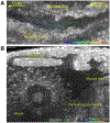
CBF mapping of female and male reproductive tracts with functional OCT. (A) Mapping of CBF in the mouse oviduct. (B) Cross-sectional image with OCT mapping of the CBF in the mouse efferent ducts. Reproduced from Wang et al., 2015 and Umezu et al., 2022.

References

    1. Aprea I, Nothe-Menchen T, Dougherty GW, Raidt J, Loges NT, Kaiser T, Wallmeier J, Olbrich H, Strunker T, Kliesch S, Pennekamp P, & Omran H (2021). Motility of efferent duct cilia aids passage of sperm cells through the male reproductive system. Mol Hum Reprod, 27(3). 10.1093/molehr/gaab009 - DOI - PMC - PubMed
    1. Ardon F, Markello RD, Hu L, Deutsch ZI, Tung CK, Wu M, & Suarez SS (2016). Dynamics of Bovine Sperm Interaction with Epithelium Differ Between Oviductal Isthmus and Ampulla. Biol Reprod, 95(4), 90. 10.1095/biolreprod.116.140632 - DOI - PMC - PubMed
    1. Besenfelder U, Havlicek V, & Brem G (2012). Role of the oviduct in early embryo development. Reprod Domest Anim, 47 Suppl 4, 156–163. 10.1111/j.1439-0531.2012.02070.x - DOI - PubMed
    1. Bezerra HG, Costa MA, Guagliumi G, Rollins AM, & Simon DI (2009). Intracoronary optical coherence tomography: a comprehensive review clinical and research applications. JACC Cardiovasc Interv, 2(11), 1035–1046. 10.1016/j.jcin.2009.06.019 - DOI - PMC - PubMed
    1. Bochner F, Fellus-Alyagor L, Kalchenko V, Shinar S, & Neeman M (2015). A Novel Intravital Imaging Window for Longitudinal Microscopy of the Mouse Ovary. Sci Rep, 5, 12446. 10.1038/srep12446 - DOI - PMC - PubMed

Publication types

LinkOut - more resources