. 2023 Jan 3;120(1):e2210967120.
doi: 10.1073/pnas.2210967120. Epub 2022 Dec 27.

Role of intracortical neuropil growth in the gyrification of the primate cerebral cortex

Affiliations

Role of intracortical neuropil growth in the gyrification of the primate cerebral cortex

Brian G Rash et al. Proc Natl Acad Sci U S A. .

Abstract

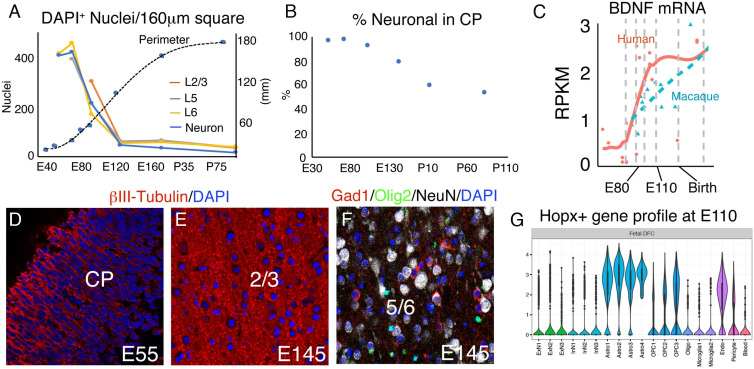
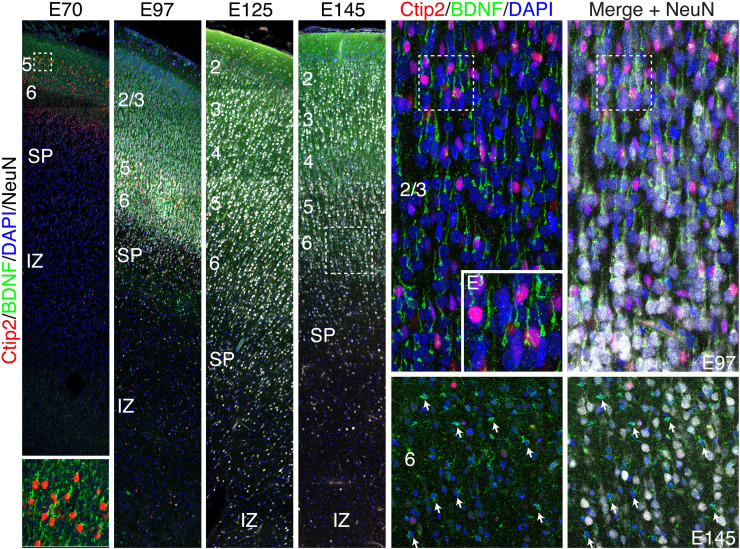
The convolutions of the mammalian cerebral cortex allow the enlargement of its surface and addition of novel functional areas during evolution while minimizing expansion of the cranium. Cognitive neurodevelopmental disorders in humans, including microcephaly and lissencephaly, are often associated with impaired gyrification. In the classical model of gyrification, surface area is initially set by the number of radial units, and the forces driving cortical folding include neuronal growth, formation of neuropil, glial cell intercalation, and the patterned growth of subcortical white matter. An alternative model proposes that specified neurogenic hotspots in the outer subventricular zone (oSVZ) produce larger numbers of neurons that generate convexities in the cortex. This directly contradicts reports showing that cortical neurogenesis and settling of neurons into the cortical plate in primates, including humans, are completed well prior to the formation of secondary and tertiary gyri and indeed most primary gyri. In addition, during the main period of gyrification, the oSVZ produces mainly astrocytes and oligodendrocytes. Here we describe how rapid growth of intracortical neuropil, addition of glial cells, and enlargement of subcortical white matter in primates are the primary forces responsible for the post-neurogenic expansion of the cortical surface and formation of gyri during fetal development. Using immunohistochemistry for markers of proliferation and glial and neuronal progenitors combined with transcriptomic analysis, we show that neurogenesis in the ventricular zone and oSVZ is phased out and transitions to gliogenesis prior to gyral development. In summary, our data support the classical model of gyrification and provide insight into the pathogenesis of congenital cortical malformations.

Keywords: cortex; gyrification; neurogenesis.


Conflict of interest statement

The authors declare no competing interest.

Figures

Fig. 1.
Fig. 1.
Rapid cortical surface area growth and gyrification after the completion of excitatory neurogenesis. (A and B) Coronal sections of macaque frontal cerebrum at E90 and E138 stained for Nissl. Abbreviations: CTX, cerebral cortex; IZ, intermediate zone; WM, white matter; CGS, cingulate sulcus; SAR, superior arcuate sulcus; STR, striatum; PS, principal sulcus; IAR, inferior arcuate sulcus; LORB, lateral orbital sulcus; OLFS, olfactory sulcus. (Scale bar, 2 mm.)
Fig. 2.
Fig. 2.
Development of the cortical plate in macaque from E55 to E125. (A) Whole brain (Inset) and panoramic view of E97 caudo-frontal cortex in coronal section stained by immunohistochemistry for the cortical layer markers, Brn2 (red; layer 2/3), Ctip2 (white; layer 5), and Tbr1 (green; layer 6/SP). (B–E) Time series of high magnification images of dorsal caudo-frontal cortex at the indicated ages; (D) corresponds to the boxed region in (A). (F and G) High magnification views of boxed regions in (D) and (E), respectively. The excitatory neurons of the cortex are almost finished migrating by E97, when a few remaining Brn2+ cells were observed still migrating through the IZ/SP (D, F, arrows in F’’’’). However, such cells were not found at E125 (E and G). Position of future SF and superior temporal sulcus demarcating prospective cerebral lobes are indicated (A, Inset). Abbreviations: ARC, arcuate sulcus; SF, Sylvian fissure; STS, superior temporal sulcus; IP, intraparietal sulcus; LU, lunate sulcus; TL, temporal lobe; CP, cortical plate; WM, white matter; IFL, inner fiber layer. A–E represent confocal montages.
Fig. 3.
Fig. 3.
Cessation of neurogenesis and transition to gliogenesis by E97. (A) Transcriptome analysis using the online Psychencode database (34) (http://evolution.psychencode.org/#) indicates loss of Tbr2 expression in dorsolateral prefrontal cortex (DFC) prior to E110 in macaque (blue line) and prior to the equivalent developmental stage in humans (red line). Y-axis represents normalized gene expression as log2(RPKM+1) where RPKM is number of reads per million kilobases. The vertical gray lines separate developmental stages: 3, early fetal; 4 to 6, mid fetal; 7, late fetal; 8 to 9, infancy; 10 to 11, childhood; 12, adolescence; 13 to 15, adulthood. The vertical gray line between 7 and 8 represents birth in both species as described (34). (BE) Immunohistochemistry (IHC) for Tbr2 and EGFR in coronal macaque dorsal parietal brain sections at the indicated ages. By IHC, robust Tbr2 expression was found in the iSVZ and oSVZ at E70 (B), but only a few cells were found at E92 (C). Tbr2 expression was not detected at E125 or E145 (D and E). In contrast, EGFR expression continues in the iSVZ and oSVZ through E145. (F) Acute BrdU (1 h) labeling shows the position of cells in S phase in a whole cerebral hemisphere; boxed region magnified in (G). (H) E97 IHC showing triple labeling of Ki67, Tbr2, and EGFR and quantified in (I). Images are confocal montages. (Scale bar, 200 μm in (BE), (H), 800 μm in (F).) Error bars represent SEM. FL, frontal lobe; IC, internal capsule; LV, lateral ventricle; SF, Sylvian fissure; STR, striatum; TL, temporal lobe.
Fig. 4.
Fig. 4.
Cortical plate surface expansion and gyrification due to neuropil production. The decrease in cellular density in different cortical laminae is indicated by DAPI staining in a series of coronal sections of dorsal parietal macaque cerebrum from E55-P91 (A). The decrease in cellular density becomes evident between E70 and E92, but dramatically accelerates between E92 and E125, just prior to initial cortical gyral development (A–C). (B) Coronal Nissl-stained macaque sections at the indicated ages showing dramatic cortical surface area growth, particularly after E90, at the completion of neurogenesis, which is explained largely by the massive decrease in neuronal density. (C) Coronal cortical perimeter measured from the corpus callosum to the lateroventral edge of the cortical plate at the level of the anterior commissure. Error bars represent SEM. Abbreviations: SPCD, superior precentral dimple; SF, Sylvian fissure; IF, interhemispheric fissure; CgS, cingulate sulcus; RF, rhinal fissure; ARSP, arcuate sulcus spur; ASD, anterior subcentral dimple; AMT, anterior middle temporal sulcus; CirS, circular sulcus; STS, superior temporal sulcus.(Scale bar in A, 40 μm; scale bar in B, 4 mm.)
Fig. 5.
Fig. 5.
Neuropil growth, regulation, and cell-type diversification in the developing macaque cortical plate. (A) DAPI nuclear density in 160 μm square regions of single confocal images, by cortical layer as shown in Fig. 4A, with neuronal density plotted in blue. (B) Percentage of total DAPI-labeled nuclei that stain for neuronal markers; Tbr1 at E55, Tbr1/Ctip2/Brn2 at E70-E97, and NeuN at E145, P7, and P91. Neuronal density declines faster than cellular density after E70. (C) Single-cell transcriptome data via Psychencode showing that humans express higher levels of BDNF in dorsal frontal cortex than do macaques during gyrification. (D and E) Neuropil labeled by βIII-tubulin IHC. (F) Neurons, OPCs, and interneurons revealed by NeuN, Olig2, and Gad1, respectively. (G) Psychencode data showing that Hopx+ cells express gene clusters principally associated with astrocyte and OPC lineages at E110. [Scale bar: 40 μm in (D–F).]
Fig. 6.
Fig. 6.
Enriched BDNF expression in macaque cortical neurons and glia during gyrification. (A–D) Series of coronal sections of macaque dorsal frontal cortex from E70-E145 stained for Ctip2, BDNF, NeuN, and DAPI. At all ages examined, BDNF was highly expressed in the apical dendrite of excitatory cortical neurons and enriched in Ctip2+ layer five neurons. (A–F, A’). (E and F) High magnification confocal Z-stack of cortical neurons at E97. (E’) Magnified image of the boxed region in (E). (F) NeuN labels almost all cortical cells at E97. (G and H) However, by E145 many cells are NeuN-negative putative glia, displaying diminutive somal size, and multipolar or stellate morphology, most of which express high levels of BDNF.

References

    1. Rakic P., Specification of cerebral cortical areas. Science 241, 170–176 (1988). - PubMed
    1. Rakic P., Evolution of the neocortex: A perspective from developmental biology. Nat. Rev. Neurosci. 10, 724–735 (2009). - PMC - PubMed
    1. Rakic P., A small step for the cell, a giant leap for mankind: A hypothesis of neocortical expansion during evolution. Trends Neurosci. 18, 383–388 (1995). - PubMed
    1. Dehay C., Kennedy H., Cell-cycle control and cortical development. Nat. Rev. Neurosci. 8, 438–450 (2007). - PubMed
    1. Noctor S. C., Flint A. C., Weissman T. A., Dammerman R. S., Kriegstein A. R., Neurons derived from radial glial cells establish radial units in neocortex. Nature 409, 714–720 (2001). - PubMed

Publication types

LinkOut - more resources