. 2023 Jan 3;120(1):e2211927120.
doi: 10.1073/pnas.2211927120. Epub 2022 Dec 27.

ERK and c-Myc signaling in host-derived tumor endothelial cells is essential for solid tumor growth

Affiliations

ERK and c-Myc signaling in host-derived tumor endothelial cells is essential for solid tumor growth

Zehua Zuo et al. Proc Natl Acad Sci U S A. .

Abstract

The limited efficacy of the current antitumor microenvironment strategies is due in part to the poor understanding of the roles and relative contributions of the various tumor stromal cells to tumor development. Here, we describe a versatile in vivo anthrax toxin protein delivery system allowing for the unambiguous genetic evaluation of individual tumor stromal elements in cancer. Our reengineered tumor-selective anthrax toxin exhibits potent antiproliferative activity by disrupting ERK signaling in sensitive cells. Since this activity requires the surface expression of the capillary morphogenesis protein-2 (CMG2) toxin receptor, genetic manipulation of CMG2 expression using our cell-type-specific CMG2 transgenic mice allows us to specifically define the role of individual tumor stromal cell types in tumor development. Here, we established mice with CMG2 only expressed in tumor endothelial cells (ECs) and determined the specific contribution of tumor stromal ECs to the toxin's antitumor activity. Our results demonstrate that disruption of ERK signaling only within tumor ECs is sufficient to halt tumor growth. We discovered that c-Myc is a downstream effector of ERK signaling and that the MEK-ERK-c-Myc central metabolic axis in tumor ECs is essential for tumor progression. As such, disruption of ERK-c-Myc signaling in host-derived tumor ECs by our tumor-selective anthrax toxins explains their high efficacy in solid tumor therapy.

Keywords: ERK signaling; anthrax lethal toxin; c-Myc; endothelial cells; tumor microenvironment.


Conflict of interest statement

The authors declare no competing interest.

Figures
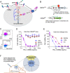
Fig. 1.
Fig. 1.
Anthrax toxin receptor CMG2-based tumor–host genetic platform for assessing tumor ECs. (A) Anthrax toxin protein delivery system as a unique platform for cancer therapy with high specificity. Tumor specificity of PA-L1-I656Q is achieved by engineering the delivering vehicle PA to bind to the CMG2 receptor and rely on tumor-associated proteases (MMPs) for activation. Thus, LF (or LF fusions) can be selectively delivered into tumor cells and tumor stromal cells to inactivate MEK1/2, disrupting the ERK signaling. (B) Generation of endothelial cell–specific CMG2 receptor–expressing mice. Breeding of CMG2LSL and Cdh5-cre mice allowed specific removal of the LoxP-Stop-LoxP cassette and subsequent activation of the CMG2 transgene only in ECs. Subsequent breeding with whole-body CMG2−/− mice eliminated expression of the endogenous CMG2 gene, resulting in a mouse where only ECs express CMG2 (CMG2EC). Similarly, we can generate other tumor stromal cell-type–specific CMG2-expressing mice by using the corresponding cell-type–specific Cre transgenic mouse. (C) The identity of primary ECs isolated from CMG2EC mice was verified by the second endothelial marker CD31-positive staining. Flow cytometry analyses of ECs (ICAM2 positive) and nonECs (ICAM2 negative) bound with a CD31 antibody. (D and E) Primary cells from CMG2EC (D) and wild-type control (E) mice were treated with various concentrations of PA-L1-I656Q in the presence of FP59 (100 ng/mL) for 48 h, followed by an MTT assay evaluating cell viability. Of note, only ECs from CMG2EC mice were sensitive to PA-L1/FP59, whereas non-ECs (ICAM2 negative), VSMs, and BMDMs were resistant to the toxin. (F) A tumor–host model with tumor ECs as an only possible target. Tumors contain tumor-initiating malignant cells and a variety of tumor stromal cells, which cooperate to promote tumor growth. In this example, only tumor ECs express the toxin receptor CMG2, thereby allowing unambiguous assessment of the role of this tumor stromal cell type in the targeted therapy using the engineered toxin. The same approach could be adapted to evaluate the roles of other tumor stromal cell types in tumor development.
Fig. 2.
Fig. 2.
Disruption of the MEK–ERK signaling in tumor ECs is sufficient to inhibit tumor growth. (A) B16-F10 (CMG2-KO) cells are resistant to the CMG-specific PA-L1-I656Q/FP59. B16-F10 cells (WT) and B16-F10 (CMG2-KO) cells were incubated with various concentrations of PA-L1-I656Q in the presence of 100 ng/mL FP59 for 48 h, followed by an MTT assay for assessing cell viability. (B) MEK cleavage and ERK phosphorylation do not occur in B16(CMG2-KO) cells treated with PA-L1-I656Q/LF. B16-F10 cells (WT) and B16-F10 (CMG2-KO) cells were incubated with various concentrations of PA-L1-I656Q/LF for 3 h. Then, cell lysates were prepared and analyzed by western blotting using anti-MEK1, anti-MEK2, and anti-phospho-ERK antibodies. Of note, while LF could be efficiently delivered into WT B16-F10 cells, resulting in MEK cleavage and disruption of the ERK signaling, this could not occur in B16-F10 (CMG2-KO) cells. (C) B16(CMG2-KO) melanomas grown in CMG2−/− mice were completely resistant to the PA-L1-I656Q/LF [4 × (30 µg/10 µg)] treatments. Toxin treatments indicated by red arrows. Tumors, mean ± SE. No significant difference. (D) B16(CMG2-KO) melanomas grown in CMG2EC (endothelial cell–specific CMG2-expressing) mice are sensitive to the PA-L1-I656Q/LF [4 × (30 µg/10 µg)] treatments. Tumors (mean ± SE) and body weight (mean ± SD) (SI Appendix, Fig. S3) were monitored. *P < 0.05 and **P < 0.01. (E) Generation of endothelial cell–specific CMG2-null (EC-CMG2−/−) mice. (F) Primary ECs and ICAM2-negative nonECs from EC-CMG2−/− mice were treated with various concentrations of PA-I656Q in the presence of FP59 (100 ng/mL) for 48 h, followed by an MTT assay evaluating cell viability. (G) B16(CMG2-KO) melanoma-bearing EC-CMG2−/ mice and their littermate control (EC-CMG2+/−) mice were treated with PA-L1-I656Q/LF (30 µg/10 µg) as indicated by arrows. Of note, B16(CMG2-KO) melanomas are much less sensitive to the toxin when grown in EC-CMG2−/− mice. EC-CMG2+/− (PA-L1-I656Q/LF) vs. other groups, P < 0.01, and no statistically significant difference among other groups. Tumors (mean ± SE) and body weight (mean ± SD) (SI Appendix, Fig. S3) were monitored. **P < 0.01.
Fig. 3.
Fig. 3.
Effects of the engineered LT on metabolism of tumor ECs. (A) Flow cytometry analyses of tumor ECs (ICAM2-positive) and B16-F10 cells bound with a CD31 antibody. (B) Tumor ECs were incubated with or without PA-L1/LF (1 µg/mL each) for 72 h or with PA-L1/FP59 (0.1 µg/mL each) for 24 h. Upper: microscopic views of the cells after the treatments. Note, the cells were still viable 72 h after PA-L1/LF treatments. In contrast, the cells treated with PA-L1/FP59 for 24 h were mostly dead, with only a small number of swollen damaged cells remaining attached. Lower: flow cytometry analyses of the cells stained with propidium iodine (PI) and annexin V. Note, 95% of the cells treated with PA-L1/LF were still alive (vs. 97% alive for the untreated cells). (C) Tumor ECs cultured in 96-well plates were incubated with various concentrations of PA-L1 in the presence of LF (500 ng/mL) for 72 h; MTT assays were followed to evaluate cell numbers relative to the nontoxin-treated wells. Data are shown as mean ± SD. Inset, microscopic views of tumor ECs treated with or without PA-L1/LF (1 µg/mL each) for 72 h. (D) Tumor ECs were treated with or without PA-L1/LF (1 µg/mL each) or Trametinib (0.2 µM) for 24 h; then, the ECARs were measured using the Seahorse XF24 analyzer under basal conditions and following sequential addition of ATP synthase inhibitor oligomycin (0.5 µg/mL), uncoupler FCCP (1 µM), and complex III inhibitor antimycin A. The ECAR readings were normalized to amounts of cells having 50 µg total protein, mean ± SD. Paired Student’s t test, P < 0.0001. (E) The OCRs of tumor ECs treated under the same conditions as in D were measured using the Seahorse XF24 analyzer. The OCR readings were normalized to 50 µg of total protein, mean ± SD. Paired Student’s t test, P = 0.0019. (F) The relative cellular ATP levels of tumor ECs treated with PA-L1/LF (0.5 µg/mL or 2 µg/mL each) for 24 h vs. untreated cells. Data are shown as mean ± SD. (G) Real-time PCR analyses of selected key genes in central metabolism of tumor ECs treated with or without PA-L1/LF (1 µg/mL each) for 4 h or 24 h. Eukaryotic translation initiation factor Eif3s5 was used as an internal normalization control. The full names of the genes can be found in SI Appendix, Table S1. Note, Cpt1a, Cpt2, Mpc1, Mpc2, and Eif3s5 are among the genes not affected by the toxin. Student’s t test: *P < 0.01. (H) Downregulation of c-Myc at the protein level following the ERK signaling disruption. Tumor ECs were incubated with various concentrations of PA-L1/LF or Trametinib for 3 h. Then, cell lysates were prepared and analyzed by western blotting using antibodies as indicated. The relative protein band densities estimated using ImageJ were shown below each lane.
Fig. 4.
Fig. 4.
Expression of the c-Myc transgene in primary ECs partially rescued the metabolic stress caused by the engineered toxin. (A) Primary ECs isolated from c-MycEC or their littermate control (WT) mice were treated with PA-L1/LF (1 µg/mL) or Trametinib (0.2 µM) for 3 h. Then, cell lysates were prepared and analyzed by western blotting. Both PA-L1/LF and Trametinib could efficiently disrupt the ERK signaling in both WT and c-MycEC ECs. However, c-Myc was only partially affected by PA-L1/LF and Trametinib in c-MycEC ECs. (B) The metabolic crisis caused by PA-L1/LF was partially rescued in ECs from the c-MycEC mice. ECs were treated with or without PA-L1/LF (1 µg/mL each) for 24 h; then, the ECARs (Left) and OCRs (Right) were analyzed as described in Fig. 3. The ECAR and OCR readings were normalized to 50 µg of total protein, mean ± SD. EC (c-Myc) (PA-L1/LF) vs. EC (WT) (PA-L1/LF), P < 0.01, paired Student’s t test.

Similar articles

Cited by

References

    1. Bejarano L., Jordao M. J. C., Joyce J. A., Therapeutic targeting of the tumor microenvironment. Cancer Discov. 11, 933–959 (2021). - PubMed
    1. Baghban R., et al. , Tumor microenvironment complexity and therapeutic implications at a glance. Cell Commun. Signal 18, 59 (2020). - PMC - PubMed
    1. Hurwitz H., Integrating the anti-VEGF-A humanized monoclonal antibody bevacizumab with chemotherapy in advanced colorectal cancer. Clin. Colorectal. Cancer 4, S62–68 (2004). - PubMed
    1. Hurwitz H., et al. , Bevacizumab plus irinotecan, fluorouracil, and leucovorin for metastatic colorectal cancer. N. Engl. J. Med. 350, 2335–2342 (2004). - PubMed
    1. Schmittnaegel M., et al. , Dual angiopoietin-2 and VEGFA inhibition elicits antitumor immunity that is enhanced by PD-1 checkpoint blockade. Sci. Transl. Med. 9, eaak9670 (2017). - PubMed

Publication types