. 2023 Jan 3;120(1):e2213703120.
doi: 10.1073/pnas.2213703120. Epub 2022 Dec 27.

SUMO enhances unfolding of SUMO-polyubiquitin-modified substrates by the Ufd1/Npl4/Cdc48 complex

Affiliations

SUMO enhances unfolding of SUMO-polyubiquitin-modified substrates by the Ufd1/Npl4/Cdc48 complex

Hyein G Lee et al. Proc Natl Acad Sci U S A. .

Abstract

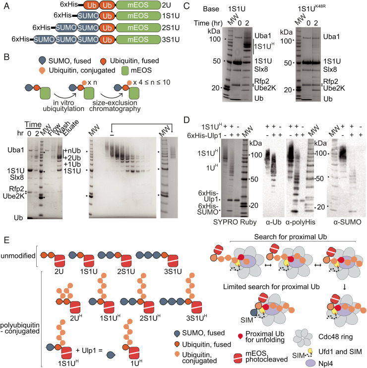
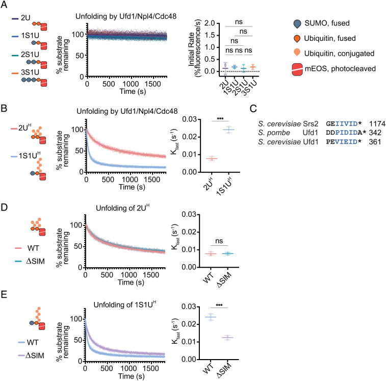
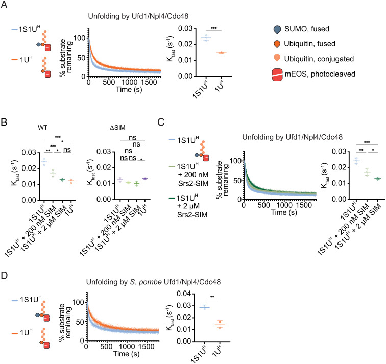
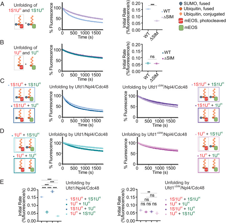
The Ufd1/Npl4/Cdc48 complex is a universal protein segregase that plays key roles in eukaryotic cellular processes. Its functions orchestrating the clearance or removal of polyubiquitylated targets are established; however, prior studies suggest that the complex also targets substrates modified by the ubiquitin-like protein SUMO. Here, we show that interactions between Ufd1 and SUMO enhance unfolding of substrates modified by SUMO-polyubiquitin hybrid chains by the budding yeast Ufd1/Npl4/Cdc48 complex compared to substrates modified by polyubiquitin chains, a difference that is accentuated when the complex has a choice between these substrates. Incubating Ufd1/Npl4/Cdc48 with a substrate modified by a SUMO-polyubiquitin hybrid chain produced a series of single-particle cryo-EM structures that reveal features of interactions between Ufd1/Npl4/Cdc48 and ubiquitin prior to and during unfolding of ubiquitin. These results are consistent with cellular functions for SUMO and ubiquitin modifications and support a physical model wherein Ufd1/Npl4/Cdc48, SUMO, and ubiquitin conjugation pathways converge to promote clearance of proteins modified with SUMO and polyubiquitin.

Keywords: SUMO; quality control; segregase; stress; ubiquitin.


Conflict of interest statement

The authors declare no competing interest.

Figures

Fig. 1.
Fig. 1.
Synthesis and characterization of SUMO/polyubiquitin dual-modified substrates. (A) Schematics of substrates. Two ubiquitin molecules (2U), one SUMO and one ubiquitin (1S1U), two SUMO and one ubiquitin (2S1U), or three SUMO and one ubiquitin (3S1U) were fused end to end with the N-terminus of mEOS (B) Top: schematic of ubiquitylation and purification of substrates. Bottom Left: Substrates were ubiquitylated with E1 Uba1, E2 Ube2K, and STUbL E3 ubiquitin ligase Slx8-Rfp2. Ubiquitylation of 1S1U shown with ubiquitylated species (+nUb) labeled. Molecular weight marker (MW) is Invitrogen BenchMark Protein Ladder, markers for 100, 50, and 20 kDa are noted with text and/or asterisk (*) in panels B–D. Bottom Right: Substrates were purified by size exclusion chromatography to select for substrates with chains estimated to include 4 to 10 ubiquitin molecules. Inset: substrate pool after purification. (C) Lysine 48 of the substrate ubiquitin is required for efficient chain elongation. Left: Ubiquitylation of 1S1U substrate as in B. Right: Substrate containing a K48R mutation was subjected to ubiquitin conjugation. (D) SUMO is not the primary site for ubiquitin conjugation. 1S1UH substrate was digested with Ulp1 to liberate SUMO, generating 1UH. SYPRO Ruby staining and western blotting for the polyhistidine tag (α-polyHis), SUMO (α-SUMO), and ubiquitin (α-Ub) in the presence or absence of Ulp1 and substrate. (E) Left: Composition of substrates is denoted by S for SUMO or U for ubiquitin and numbering of fused molecules. Superscript H denotes substrates containing ubiquitin chains estimated to be 4 to 10 ubiquitin molecules long. Right: Model of search for proximal ubiquitin and the proposed limitations on the search for proximal ubiquitin when SUMO is present in substrate.
Fig. 2.
Fig. 2.
Unfolding depends on polyubiquitin, SUMO, and SIM. (A) Non-polyubiquitylated substrates are not readily unfolded. Loss of fluorescence was measured for non-ubiquitylated substrates 2U, 1S1U, 2S1U, and 3S1U by WT Ufd1/Npl4/Cdc48. (B) Unfolding increases in the presence of SUMO. Unfolding of either 2UH or 1S1UH by WT Ufd1/Npl4/Cdc48. (C) Alignment of C-terminal residues of Srs2 from S. cerevisiae, and Ufd1 from S. pombe and S. cerevisiae. Blue lettering indicates SIM. Asterisk (*) denotes the C-terminus of the protein. Unfolding of 2UH (D) or 1S1UH (E) by WT Ufd1/Npl4/Cdc48 (WT) or Ufd1ΔSIM/Npl4/Cdc48 where the SIM was deleted from Ufd1 (ΔSIM). Values were normalized to background fluorescence in the absence of ATP. Plot of three replicates with fit of linear regression (A) or two-phase nonlinear regression (B, D, and E). Initial rate determined using the first 30 s of linear fit or Kfast (s−1) determined using two-phase exponential decay fit. Error bars represent SD. P values calculated by one-way ANOVA with Tukey’s test (A) or unpaired two-tailed t test (B, D, and E); *< 0.05, **P < 0.01, ***P < 0.001, ns (not significant).
Fig. 3.
Fig. 3.
Rate of Ufd1/Npl4/Cdc48 unfolding is enhanced in a SUMO- and SIM-dependent manner. (A) Unfolding of 1S1UH or 1UH (Ulp1-treated 1S1UH) by WT Ufd1/Npl4/Cdc48. (B) Comparison of Kfast values for WT or ΔSIM complexes for 1S1UH, 1S1UH + 200 nM SIM peptide, 1S1UH + 2 µM SIM peptide, or 1UH. (C) Unfolding of 1S1UH by WT Ufd1/Npl4/Cdc48 in presence of a competing SIM peptide. 200 nM or 2 µM of SIM peptide derived from the C-terminus of Srs2 (residues 1,107 to 1,174) was added to the unfolding reaction of 1S1UH. (D) Unfolding by S. pombe WT Ufd1/Npl4/Cdc48 of 1S1UH or 1UH. Values were normalized to background fluorescence in the absence of ATP. Plot of three replicates with fit of two-phase nonlinear regression. Kfast (s−1) determined using two-phase exponential decay fit. Error bars represent SD. P values calculated by unpaired two-tailed t test (A and D) or one-way ANOVA with Tukey’s test (B and C); *P < 0.05, **P < 0.01, ***P < 0.001, ns (not significant).
Fig. 4.
Fig. 4.
Ufd1/Npl4/Cdc48 preferentially unfolds SUMO–polyubiquitin substrates in a mixed substrate pool. Pooled substrates contained activated mEOS (denoted by red cartoon and text) or native mEOS (denoted by green cartoon and text). Unfolding of (A) 1S1UH-mEOSR and 1S1UH-mEOSG or (B) 1UH-mEOSR and 1UH-mEOSG by WT and ΔSIM Ufd1/Npl4/Cdc48. (C) Unfolding of 1S1UH-mEOSR and 1S1UH-mEOSG or 1UH-mEOSG by WT (Left) and ΔSIM (Right) Ufd1/Npl4/Cdc48. (D) Unfolding of 1UH-mEOSR and 1S1UH-mEOSG or 1UH-mEOSG by WT (Left) and ΔSIM (Right) Ufd1/Npl4/Cdc48. Values were normalized to background fluorescence in the absence of ATP. Plot of three replicates. (E) Initial rates of unfolding for WT (Left) and ΔSIM mutant (Right) Ufd1/Npl4/Cdc48 in AD. Initial rate determined using linear fit of the first 30 s of unfolding. Error bars represent SD. P values calculated by unpaired two-tailed t test (A and B) or one-way ANOVA with Tukey’s test (E); *P < 0.05, **P < 0.01, ***P < 0.001, ns (not significant).
Fig. 5.
Fig. 5.
Cryo-EM analysis of Ufd1/Npl4/Cdc48 in process of unfolding a SUMO–polyubiquitin substrate. (A) Top: cartoon of components in sample. 1S1UH was generated as in Fig. 1 and added prior to photocleavage to allow multiple rounds of unfolding. Components were preincubated with ATP prior to vitrification. Bottom: cartoon showing the three main classes of particles observed as described by their relation to the substrate—substrate-unbound, substrate-interacting, and ubiquitin-unfolded. (B) EM density and models of sub-states in substrate-interacting (denoted with the prefix “int”) and ubiquitin-unfolded (denoted with the prefix “u”) classes. Subclassification revealed at least eight states within the sample. Classes differ in the net number of folded ubiquitin molecules, their position atop Ufd1/Npl4, and the presence or absence of unfolded ubiquitin—these differences are represented by the “polyUb” cartoon. In five states that could be modeled, a cartoon representation of the model (colored; Npl4 in light blue, ubiquitin in orange, and Ufd1 in yellow) is shown overlayed with EM density (in grey). (C) EM density and model of the Npl4 loop (Top) and sequence alignment of the Npl4 loop (Bottom) with conserved residues in dark purple. Conserved Npl4 loop adopts discrete positions: closed, occluding the central pore of the Cdc48 hexamer (as seen in substrate-unbound and substrate-interacting classes) or open, allowing access to the central pore (as seen in ubiquitin-unfolded class).
Fig. 6.
Fig. 6.
Sites of ubiquitin interaction in Ufd1/Npl4/Cdc48. (A) Ufd1/Npl4 domain organization. Solid lines indicate regions modeled in cryo-EM reconstructions. The four sites of ubiquitin association are colored and residues of the sites are shown as spheres in the cartoon representation below. Ufd1 is in yellow and Npl4 is in light blue. (B) Top: Close-up of ubiquitin in UBS1 in EM density maps and models of states uA and uC. uA represents states where ubiquitin occupies UBS1-A (also observed in states intA and intB) while uC represents states where ubiquitin occupies UBS1-B (also observed in uD). Pink boxes highlight sites of ubiquitin interaction shown in Lower panels. Middle Left: Interactions between ubiquitin in UBS1-A and Npl4. Side chains of residues in ubiquitin and Npl4 that are positioned to mediate the association are shown and labeled. Middle Right: Interactions between ubiquitin in UBS1-B and Ufd1/Npl4. Side chains of residues in ubiquitin, Npl4 and Ufd1 that mediate contacts are shown and labeled. Bottom: The Npl4 pivot groove. Positioning of the β1–β2 loop of ubiquitin at the Npl4 pivot groove in UBS1-A (Left) and UBS1-B (Right) are shown. (C) EM density map and models representing UBS3 and UBS2 as derived from state uD, and their sequence alignment. UBS3 and UBS2 include two helices separated by a short linker that interact with tandem ubiquitin molecules in the Lys48 chain.
Fig. 7.
Fig. 7.
Unfolding by Ufd1/Npl4/Cdc48. (A) Ubiquitin in UBS1 in states A and B are superposed in ribbon representation color coded for each indicated structure. Amino and carboxy-terminal residues labeled N and C, respectively, with a bracket indicating locations of the β1–β2 loop. (B) Close-up of the model of ubiquitin at the C-terminal face of the ubiquitin fold with side chains in stick representation with a black dotted line indicating distance between α-carbons of Leu8 in intB (pink) and uC (orange). (C) EM densities and model with side chains in stick representation of ubiquitin from states intB (Left) and uC (Right). Select side chains are labeled. (D) Schematic of Ufd1/Npl4 and substrate in state intB. Throughout the figure, Ufd1 is represented in yellow, ubiquitin in orange, Npl4 in light blue, and Cdc48 in grey. Two views of the Ufd1/Npl4 in association with the substrate are shown—rotated 90° (E) and from above (F). A surface representation of Ufd1/Npl4 and a cartoon representation of ubiquitin (Left), a cartoon scheme of each view (Middle), and cartoon scheme with substrate (Right) are shown to illustrate positioning of the Ufd1/Npl4/Cdc48 complex relative to the substrate. Ufd1 residues 310 to 361 are indicated to approximate scale for an extended chain by a dotted black line with the “SIM” labeled at the C-terminal end. Red asterisk (*) denotes the last modeled residue of Ufd1. (G) Model for Ufd1/Npl4/Cdc48 unfolding a 1S1UH-mEOS substrate suggesting order to events left to right. Ubiquitin shown in different colors (purple, red, orange) to indicate proximity to the substrate.

Similar articles

Cited by

References

    1. Cappadocia L., Lima C. D., Ubiquitin-like protein conjugation: Structures, chemistry, and mechanism. Chem. Rev. 118, 889–918 (2018), 10.1021/acs.chemrev.6b00737. - DOI - PMC - PubMed
    1. Gareau J. R., Lima C. D., The SUMO pathway: Emerging mechanisms that shape specificity, conjugation and recognition. Nat. Rev. Mol. Cell Boil. 11, 861–871 (2010), 10.1038/nrm3011. - DOI - PMC - PubMed
    1. Flotho A., Melchior F., Sumoylation: A regulatory protein modification in health and disease. Annu. Rev. Biochem. 82, 357–385 (2013), 10.1146/annurev-biochem-061909-093311. - DOI - PubMed
    1. Komander D., Rape M., The ubiquitin code. Annu. Rev. Biochem. 81, 203–229 (2012), 10.1146/annurev-biochem-060310-170328. - DOI - PubMed
    1. Danielsen J. R., et al. , DNA damage–inducible SUMOylation of HERC2 promotes RNF8 binding via a novel SUMO-binding Zinc finger. J. Cell Biol. 197, 179–187 (2012), 10.1083/jcb.201106152. - DOI - PMC - PubMed

Publication types

MeSH terms

LinkOut - more resources