. 2023 Jan 3;120(1):e2213537120.
doi: 10.1073/pnas.2213537120. Epub 2022 Dec 27.

A RGS7-CaMKII complex drives myocyte-intrinsic and myocyte-extrinsic mechanisms of chemotherapy-induced cardiotoxicity

Affiliations

A RGS7-CaMKII complex drives myocyte-intrinsic and myocyte-extrinsic mechanisms of chemotherapy-induced cardiotoxicity

Madhuri Basak et al. Proc Natl Acad Sci U S A. .

Abstract

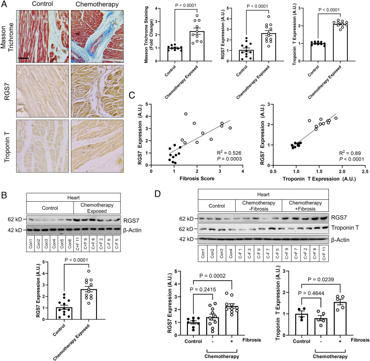
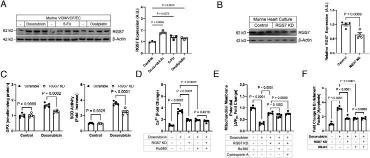
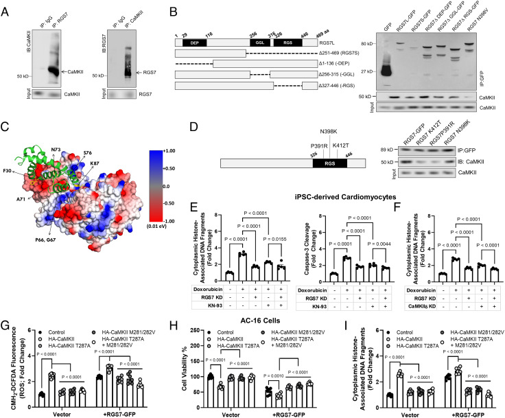
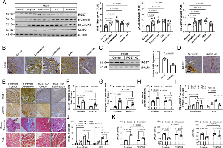
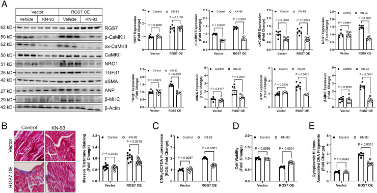
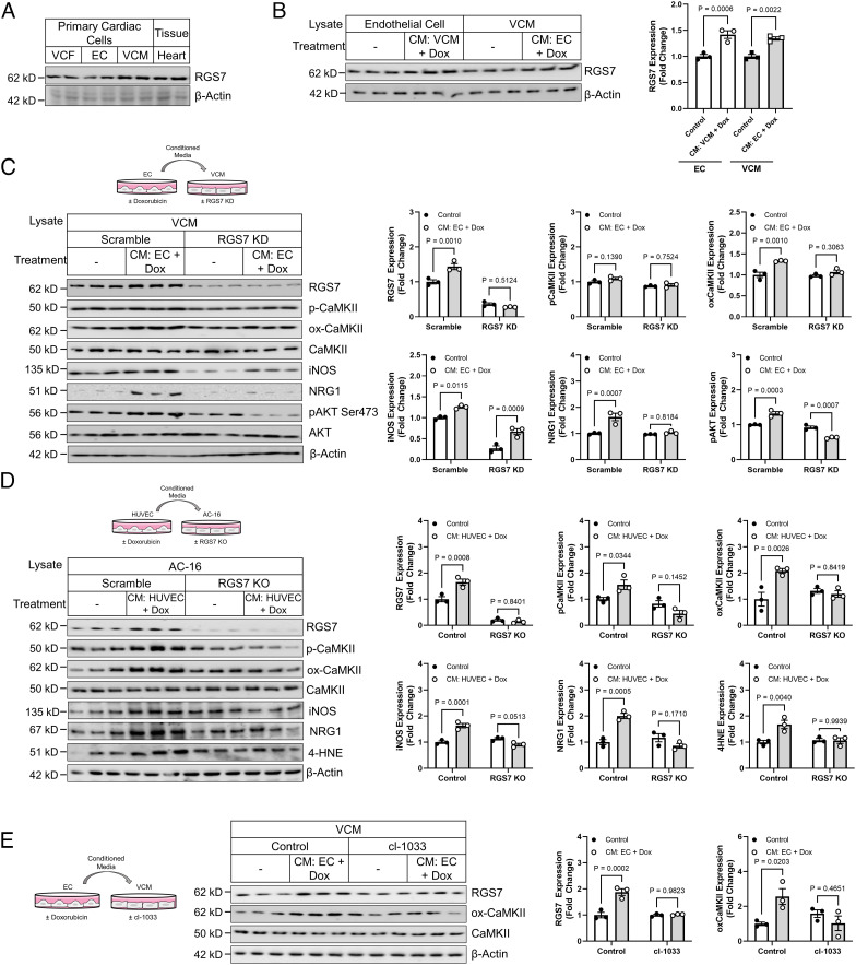
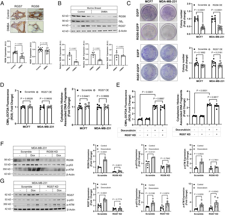
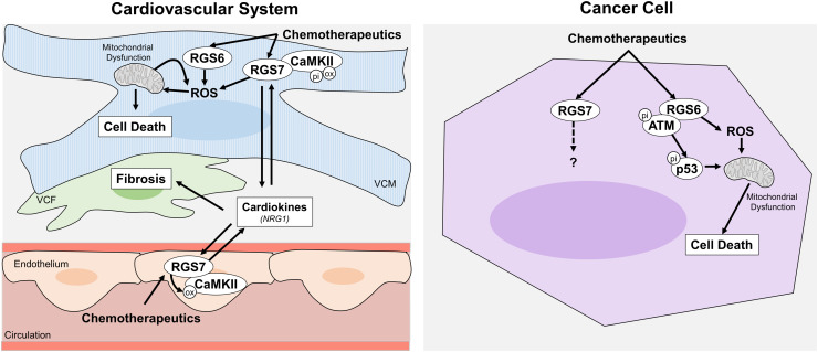
Dose-limiting cardiotoxicity remains a major limitation in the clinical use of cancer chemotherapeutics. Here, we describe a role for Regulator of G protein Signaling 7 (RGS7) in chemotherapy-dependent heart damage, the demonstration for a functional role of RGS7 outside of the nervous system and retina. Though expressed at low levels basally, we observed robust up-regulation of RGS7 in the human and murine myocardium following chemotherapy exposure. In ventricular cardiomyocytes (VCM), RGS7 forms a complex with Ca2+/calmodulin-dependent protein kinase (CaMKII) supported by key residues (K412 and P391) in the RGS domain of RGS7. In VCM treated with chemotherapeutic drugs, RGS7 facilitates CaMKII oxidation and phosphorylation and CaMKII-dependent oxidative stress, mitochondrial dysfunction, and apoptosis. Cardiac-specific RGS7 knockdown protected the heart against chemotherapy-dependent oxidative stress, fibrosis, and myocyte loss and improved left ventricular function in mice treated with doxorubicin. Conversely, RGS7 overexpression induced fibrosis, reactive oxygen species generation, and cell death in the murine myocardium that were mitigated following CaMKII inhibition. RGS7 also drives production and release of the cardiokine neuregulin-1, which facilitates paracrine communication between VCM and neighboring vascular endothelial cells (EC), a maladaptive mechanism contributing to VCM dysfunction in the failing heart. Importantly, while RGS7 was both necessary and sufficient to facilitate chemotherapy-dependent cytotoxicity in VCM, RGS7 is dispensable for the cancer-killing actions of these same drugs. These selective myocyte-intrinsic and myocyte-extrinsic actions of RGS7 in heart identify RGS7 as an attractive therapeutic target in the mitigation of chemotherapy-driven cardiotoxicity.

Keywords: RGS protein; cardiotoxicity; cell death; chemotherapy; oxidative stress.


Conflict of interest statement

The authors declare no competing interest.

Figures

Fig. 1.
Fig. 1.
RGS7 is up-regulated in the chemotherapy-exposed myocardium. (A) Cardiac staining (Scale bar, 100 μm.) and (B) correlation analysis for RGS7, Troponin T, and Masson trichrome in control or chemotherapy-exposed patients (n = 10). (C) RGS7 expression in heart tissue from chemotherapy-exposed patients or controls (n = 12). (D) RGS7 (n = 8 to 10) and Troponin T (n = 4 to 5) immunoreactivity in control and chemotherapy patients with/without detectable fibrosis. Codes corresponding to specific chemotherapy patients and controls (SI Appendix, Table S2) are provided. β-Actin serves as a loading control for immunoblots. Exact P values are provided on graphs. Data are presented as mean ± SEM.
Fig. 2.
Fig. 2.
RGS7 mediates doxorubicin-induced mitochondrial dysfunction, oxidative stress, and cell death. (A) RGS7 immunoreactivity in total heart cultures treated with doxorubicin (n = 3; 3 μM, 16 h), 5-FU (n = 3; 500 mM, 16 h), or oxaliplatin (n = 3; 0.06 mM, 16 h). (B) Verification of RGS7 knockdown in total heart cultures with scramble or RGS7-shRNA (n = 4). (CF) Control or RGS7 knockdown (KD) VCM were treated with doxorubicin ± pre-treatment of Ru360 (50 μM, 1 h) or cyclosporin A (0.2 mM, 45 min). (C) GPX activity (n = 5) and SOD activity (n = 5). (D) Mitochondrial Ca2+ flux (n = 5). (E) ΔψM (n = 5). (F) Apoptosis (cytoplasmic histone-associated DNA fragments; n = 5).
Fig. 3.
Fig. 3.
RGS7 forms a complex with CaMKII and drives CaMKII-dependent cellular dysfunction. (A) Reciprocal co-IP of RGS7 and CaMKII in human AC-16 cardiomyocytes. (B) Co-IP of CaMKII with RGS7 deletion constructs. (C) In silico modeling of the RGS7-CaMKII complex revealed key RGS7 residues (K412, P391, N398) in the RGS domain of RGS7 predicted to support a direct RGS7/CaMKII interaction. (D) Mutation of specific residues abolishes RGS7-CaMKII binding. (E and F) Apoptosis was measure in control, or RGS7 KD human iPSC-derived cardiomyocytes (n = 5) were treated with doxorubicin (3 μM, 16 h) ± pre-treatment with CaMKII inhibitor KN-93 (E, 50 μM, 1 h) or δ-CaMKII shRNA (F). (GI) AC-16 cells transfected with RGS7-GFP or control plasmid ± phosphorylation (T287A) or oxidation (M281/282V)-deficient CaMKII where indicated. (G) CM-H2-DCFDA fluorescence (ROS; n = 7). (H) Cell viability (n = 7). (I) Apoptosis (n = 7).
Fig. 4.
Fig. 4.
Cardiac-specific RGS7 knockdown ameliorates chemotherapy-dependent cardiotoxicity in mice. (A–H, K) Following administration of scramble or RGS7-shRNA via intracardiac injection, mice were treated with doxorubicin (cumulative dose of 45 mg/kg i.p.), 5-FU (200 mg/kg i.p.), oxaliplatin (45 mg/kg i.p.), irinotecan (175 mg/kg i.p.), or saline control. After 8 wk, cardiac phenotyping was performed, and tissues collected 1 wk later for biochemical and histological analyses. (A) Immunoblotting for RGS7, p-CaMKII, and ox-CaMKII in heart (n = 6). (B) RGS7 immunohistochemistry in heart. (Scale bar, 100 μm.) RGS7 knockdown was verified in heart via (C) immunoblotting and (D) immunohistochemistry. (Scale bar, 100 μm.) (E) Representative images depicting RGS7, CaMKII, Masson trichrome (fibrosis), and H&E staining (myofibrillar architecture) in mice. (Scale bar, 100 μm.) Quantification of (F) fibrotic area (blue stain) from Masson trichrome (n = 10) and (G) myocyte area from H&E images (n = 10). (H) Heart weight (n = 10). (I and J) Mice were treated with single acute dose of doxorubicin (20mg/kg, i.p.), 5-FU (150 mg/kg, i.p.), oxaliplatin (30 mg/kg, i.p.), or saline control. (I) CM-H2-DCFDA fluorescence (ROS; n = 5). (J) Quantification of TUNEL+ nuclei (apoptosis; n = 5). (K) Cardiac phenotyping (n = 6); left ventricular end diastolic and systolic pressure and LVEF.
Fig. 5.
Fig. 5.
RGS7 OE in heart is sufficient to drive CaMKII-dependent cardiotoxicity. A RGS7-containing viral construct or control was introduced into the myocardium. After 15 d, animals were given saline or KN-93 (8 mg/kg i.p. two doses, 4 d apart) and tissues collected 2 d after the last injection for downstream analyses. (A) Immunoblotting for RGS7, p-CaMKII, ox-CaMKII, NRG1, TGFβ1, αSMA, ANP, and β-MHC in cardiac tissue (n = 6). (B) Representative images and quantification of cardiac fibrosis (Masson trichrome staining; n = 6). (C) CM-H2-DCFDA fluorescence (total ROS; n = 6). (D) Cell viability (n = 6). (E) Apoptosis (n = 6).
Fig. 6.
Fig. 6.
RGS7 knockdown protects VCM from EC-driven myocyte dysfunction. (A) RGS7 expression in heart, VCM, VCF, and EC. (B) VCM and EC were treated with conditioned media from doxorubicin-treated (3 μM, 24 h) EC or VCM, respectively. Lysates were probed for RGS7 expression (n = 3). (C) Control or RGS7 KD VCM cultures (n = 3) were treated with conditioned media from doxorubicin-treated (3 μM, 24 h) EC and the resultant lysates were probed for RGS7, p-CaMKII, ox-CaMKII, iNOS, NRG1, and p-AKT. (D) Control or RGS7 KO AC-16 cardiomyocytes (n = 3) were treated with conditioned media from doxorubicin-treated (3 μM, 24 h) HUVEC cells, and immunoblotting was performed to detect RGS7, p-CaMKII, ox-CaMKII, iNOS, NRG1, and 4-HNE. (E) Murine ECs (n = 3) were treated with doxorubicin (3 μM, 24 h), and the culture media transplanted onto VCM cultures in the presence or absence of the tyrosine kinase inhibitor cl-1033 (2 μM, 1 h). RGS7, ox-CaMKII, and CaMKII expression were determined.
Fig. 7.
Fig. 7.
RGS6, not RGS7, drives chemotherapy-dependent death of breast cancer cells. (A and B) Female mice (n = 10) were treated with the carcinogen DMBA (1 mg/20 g body weight), and protein expression detected in breast tissue via (A) immunohistochemistry (n = 10) and (B) immunoblot (n = 4 to 5). (C) Colony formation in MCF7 or MDA-MB-231 cells following transfection of control (GFP), RGS6-GFP or RGS7-GFP (n = 8). (D) RGS7-GFP was transfected into MCF7 or MDA-MB-231 breast cancer cells and CM-H2-DCFDA fluorescence (total ROS; n = 6) and apoptosis (n = 6) measured. (E) MDA-MB-231 cells were treated with doxorubicin (2 µM, 12 h) following introduction of scramble or RGS7-shRNA and total ROS (n = 6); and apoptosis (n = 6) measured. MDA-MB-231 cells were treated with doxorubicin (2 µM, 12 h) following introduction of scramble or (F) RGS6- or (G) RGS7-shRNA. RGS6/7, p-p53, and p-ATM expression were determined.
Fig. 8.
Fig. 8.
Schematic diagram depicting the cardiac and extra-cardiac impacts of RGS7 following induction by chemotherapeutic drugs. Chemotherapeutic drugs lead to induction of RGS7/CaMKII in both ECs and ventricular cardiac myocytes. In VCM, RGS7 promotes oxidative stress, mitochondrial dysfunction, and cell death as well as the release of cardiokines that drive cardiac fibrosis and loss of ventricular integrity.

References

    1. Shakir D. K., Rasul K. I., Chemotherapy induced cardiomyopathy: Pathogenesis, monitoring and management. J. Clin. Med. Res. 1, 8–12 (2009). - PMC - PubMed
    1. Henriksen P. A., Anthracycline cardiotoxicity: An update on mechanisms, monitoring and prevention. Heart 104, 971–977 (2018). - PubMed
    1. Shiga T., Hiraide M., Cardiotoxicities of 5-Fluorouracil and other fluoropyrimidines. Curr. Treat. Options Oncol. 21, 27 (2020). - PMC - PubMed
    1. Cardinale D., et al. , Prognostic value of troponin I in cardiac risk stratification of cancer patients undergoing high-dose chemotherapy. Circulation 109, 2749–2754 (2004). - PubMed
    1. Lenihan D. J., et al. , The utility of point-of-care biomarkers to detect cardiotoxicity during anthracycline chemotherapy: A feasibility study. J. Card Fail 22, 433–438 (2016). - PubMed

Publication types

MeSH terms

LinkOut - more resources