. 2023 Feb:59:102591.
doi: 10.1016/j.redox.2022.102591. Epub 2022 Dec 24.

Itaconate promotes a wound resolving phenotype in pro-inflammatory macrophages

Affiliations

Itaconate promotes a wound resolving phenotype in pro-inflammatory macrophages

Sjors Maassen et al. Redox Biol. 2023 Feb.

Abstract

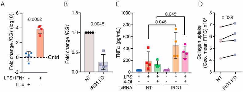
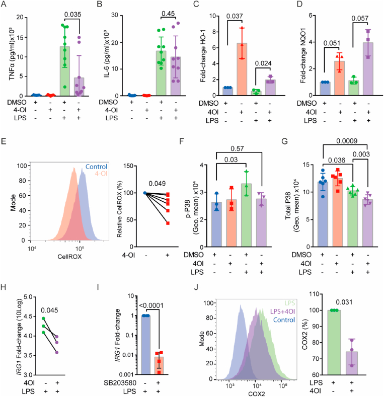
Pathological conditions associated with dysfunctional wound healing are characterized by impaired remodelling of extracellular matrix (ECM), increased macrophage infiltration, and chronic inflammation. Macrophages also play an important role in wound healing as they drive wound closure by secretion of molecules like transforming growth factor beta-1 (TGF-β). As the functions of macrophages are regulated by their metabolism, local administration of small molecules that alter this might be a novel approach for treatment of wound-healing disorders. Itaconate is a tricarboxylic acid (TCA) cycle-derived metabolite that has been associated with resolution of macrophage-mediated inflammation. However, its effects on macrophage wound healing functions are unknown. In this study, we investigated the effects of the membrane-permeable 4-octyl itaconate (4-OI) derivative on ECM scavenging by cultured human blood monocyte-derived macrophages (hMDM). We found that 4-OI reduced signalling of p38 mitogen-activated protein kinase (MAPK) induced by the canonical immune stimulus lipopolysaccharide (LPS). Likely as a consequence of this, the production of the inflammatory mediators like tumor necrosis factor (TNF)-α and cyclooxygenase (COX)-2 were also reduced. On the transcriptional level, 4-OI increased expression of the gene coding for TGF-β (TGFB1), whereas expression of the collagenase matrix metalloprotease-8 (MMP8) was reduced. Furthermore, surface levels of the anti-inflammatory marker CD36, but not CD206 and CD11c, were increased in these cells. To directly investigate the effect of 4-OI on scavenging of ECM by macrophages, we developed an assay to measure uptake of fibrous collagen. We observed that LPS promoted collagen uptake and that this was reversed by 4-OI-induced signaling of nuclear factor erythroid 2-related factor 2 (NRF2), a regulator of cellular resistance to oxidative stress and the reduced glycolytic capacity of the macrophage. These results indicate that 4-OI lowers macrophage inflammation, likely promoting a more wound-resolving phenotype.


Conflict of interest statement

Declaration of competing interest All authors declare that they have no conflicts of interest.

Figures

Image 1
Graphical abstract
Fig. 1
Fig. 1
LPS promotes collagen uptake and intracellular degradation by human blood monocyte derived macrophages (hMDM). A) Fluorescence microscopy of fibrous collagen in a cell culture dish. Collagen was labelled with FITC. Scalebar is 10 μm. B)Z-projection of live-cell confocal micrographs of macrophages seeded on fibrous FITC-collagen for 24 h. Note the large intracellular compartments (yellow arrows) containing collagen in LPS-stimulated macrophages (24 h stimulation) compared to control. Scalebar is 10 μm. C) Representative flow cytometry histogram and quantification of macrophages cultured on FITC-labelled collagen with and without LPS for 24 h, and subsequently detached with either Accutase or Trypsin (n = 5 donors, one-way ANOVA with Tukey's multiple comparison test). Data points show individual donors. D) Collagen uptake by flow cytometry of LPS-stimulated macrophages treated with the dynamin inhibitor Dynasore (n = 3, One-way ANOVA with a Dunnett's multiple comparisons test). Data points show individual donors. E)x-y projected area from confocal microscopy images of intracellular storage compartments containing FITC collagen (see Supplementary Fig. 1B). Every data point represents one cell (pooled from n = 4 donors, unpaired t-test). (For interpretation of the references to colour in this figure legend, the reader is referred to the Web version of this article.)
Fig. 2
Fig. 2
IRG1 knockdown promotes TNF-α secretion and increases collagen uptake by human blood monocyte derived macrophages (hMDM). A) mRNA levels (log10) of IRG1 in pro- (LPS and IFNγ) and anti-inflammatory (IL-4) macrophages after 24 h of polarisation normalised to untreated macrophages (Cntrl; dashed line). B) qPCR of IRG1 mRNA levels after IRG1 knockdown and 24 h of LPS stimulation, normalised to the non-targeted siRNA control (NT) (n = 4, paired t-test). C) ELISA of secreted TNF-α at 24 h after stimulation of macrophages with LPS, 4-OI or both, and with or without IRG1 knockdown (n = 4, paired t-test). D) Collagen uptake by flow cytometry of macrophages that were incubated for 24 h in FITC-collagen-coated wells with LPS after IRG1 knockdown (n = 4 donors, paired t-test).
Fig. 3
Fig. 3
4-OI increases NRF2 and decreases p38 MAPK signalling in human blood monocyte derived macrophages (hMDM) and downregulates pro-inflammatory markers. A-B) ELISA of secreted TNF-α (A) or IL-6 (B) at 24 h after stimulation of LPS, 4-OI or both (n = 9 donors, paired t-test). Data points show individual donors. C) mRNA levels of downstream NRF2 targets heme oxygenase-1 (HO1) and D) NAD(P)H quinone dehydrogenase (quinone 1) (NQO1) after stimulation of macrophages for 6 h with either LPS, 4-OI or both. DMSO: solvent control (n = 3 donors, one-way ANOVA with a Tukey's multiple comparison test). E) Representative flow cytometry histogram and quantification of cellular ROS levels by staining with CellROX after 24 h of 4-OI treatment (n = 6 donors, paired t-test). F) Phospho-flow of phosphorylated-p38 MAPK (Thr180/Tyr182) immunostaining of macrophages at 1 h after stimulation with LPS, 4-OI or both (n = 3 donors, one-way ANOVA with a Dunnette's multiple comparison test). G) Total intracellular p38 staining at 1 h after stimulation with LPS, 4-OI or both (n = 6 donors, one-way ANOVA with a Tukeys multiple comparison test).H)IRG1 levels of macrophages stimulated with LPS, with and without 4-OI, after 24 h normalised to controls without LPS (n = 3 donors, paired t-test). I) mRNA levels of IRG1 after LPS stimulation for 24 h with and without the p38 inhibitor SB203580 (n = 4 donors, paired t-test). J) Representative flow cytometry histogram and quantification of internal COX-2 levels at 24 h of LPS and 4-OI stimulation (n = 3 donors, paired t-test).
Fig. 4
Fig. 4
4-OI upregulates pro-resolution macrophage markers and reduces collagen uptake by human blood monocyte derived macrophages (hMDM). A-C) qPCR of cyclin dependent kinase inhibitor 1A (CDKN1A) (A), transforming growth factor beta-1 (TGFB1) (B) and MMP8 (C) mRNA levels after 24 h of LPS stimulation with and without 4-OI, normalised to an untreated control (dashed line) (n = 5, paired t-test). D) Collagen uptake by flow cytometry of macrophages that were incubated for 24 h in FITC-collagen-coated wells with 4-OI, LPS or both (n = 3 donors, one-way ANOVA with a Dunnette's multiple comparison test). E) FITC-labelled zymosan uptake by macrophages after 24 h of incubation with LPS, 4-OI or both (n = 6, one-way ANOVA with a Dunnett's test for multiple comparison). F) Alexa Fluor 647-labelled ovalbumin (OVA-647) uptake by macrophages after 24 h of incubation with LPS, 4-OI or both (n = 6, one-way ANOVA with a Dunnett's test for multiple comparison). G-H) Representative flow cytometry histograms and quantifications of surface levels of collagen receptors and M2-macrophage markers CD11c (G), CD206 (H), and CD36 (I) in hMDMs treated with 4-OI for 24 h (n = 6 donors, paired t-tests). Data points show individual donors.
Fig. 5
Fig. 5
4-OI blocks collagen uptake by promoting NRF2 and inhibiting p38 signalling in human blood monocyte derived macrophages (hMDM). A) Sequence alignment of mouse and human GAPDH. Red: murine cysteine 22 alkylation site. B) Glucose reduction of media in which hMDMs were cultured and treated with LPS or LPS and 4-OI (n = 3 donors, paired t-test). C) Lactate production in media in which hMDMs were cultured and treated with LPS or LPS and 4-OI (n = 3 donors, one-way ANOVA with a Tukey's multiple comparison test). D) Percentage BODIPY FL C12 uptake 24 h after incubation with LPS, 4-OI or both with flow cytometry (n = 3 donors, one-way ANOVA with a Dunnette's multiple comparison test). E) Quantification of fibrous FITC-collagen uptake by hMDMs treated with glycolysis inhibitor 2-deoxy glucose (2DG), LPS and/or 4-OI for 24 h by flow cytometry. (n = 6 donors, one-way ANOVA with a Tukey's multiple comparison test). F) Uptake of fibrous FITC-collagen by LPS-stimulated hMDMs (24 h) incubated with p38 MAPK inhibitor SB203580 by geometric mean with flow cytometry (n = 6 donors, paired t-test). G) Collagen uptake by LPS-stimulated hMDMs (24 h) with NRF2 inhibitor ML385 (n = 3 donors, paired t-test). H) Quantification of fibrous FITC-collagen uptake by macrophages treated with ML385 and/or 4-OI by flow for 24 h cytometry. DMSO: solvent control. (n = 6 donors, one-way ANOVA with a Dunnette's multiple comparison test). Data points show individual donors. (For interpretation of the references to colour in this figure legend, the reader is referred to the Web version of this article.)

References

    1. Sen C.K. Human wound and its burden: updated 2020 compendium of estimates. Adv. Wound Care. 2021;10:281–292. - PMC - PubMed
    1. Gould L., et al. Chronic wound repair and healing in older adults: current status and future research. J. Am. Geriatr. Soc. 2015;63:427. - PMC - PubMed
    1. Koh T.J., Dipietro L.A. Inflammation and wound healing: the role of the macrophage. Expet Rev. Mol. Med. 2011 doi: 10.1017/S1462399411001943. - DOI - PMC - PubMed
    1. Wynn T.A., Vannella K.M. Macrophages in tissue repair, regeneration, and fibrosis. Immunity. 2016;44:450. - PMC - PubMed
    1. J V. den B., LA O., D M. Macrophage immunometabolism: where are we (going)? Trends Immunol. 2017;38:395–406. - PubMed

Publication types

MeSH terms