

Molecular Diversity of Neuron Types in the Salamander Amygdala and Implications for Amygdalar Evolution
- PMID: 36574764
- PMCID: PMC10096051
- DOI: 10.1159/000527899
Molecular Diversity of Neuron Types in the Salamander Amygdala and Implications for Amygdalar Evolution
Abstract
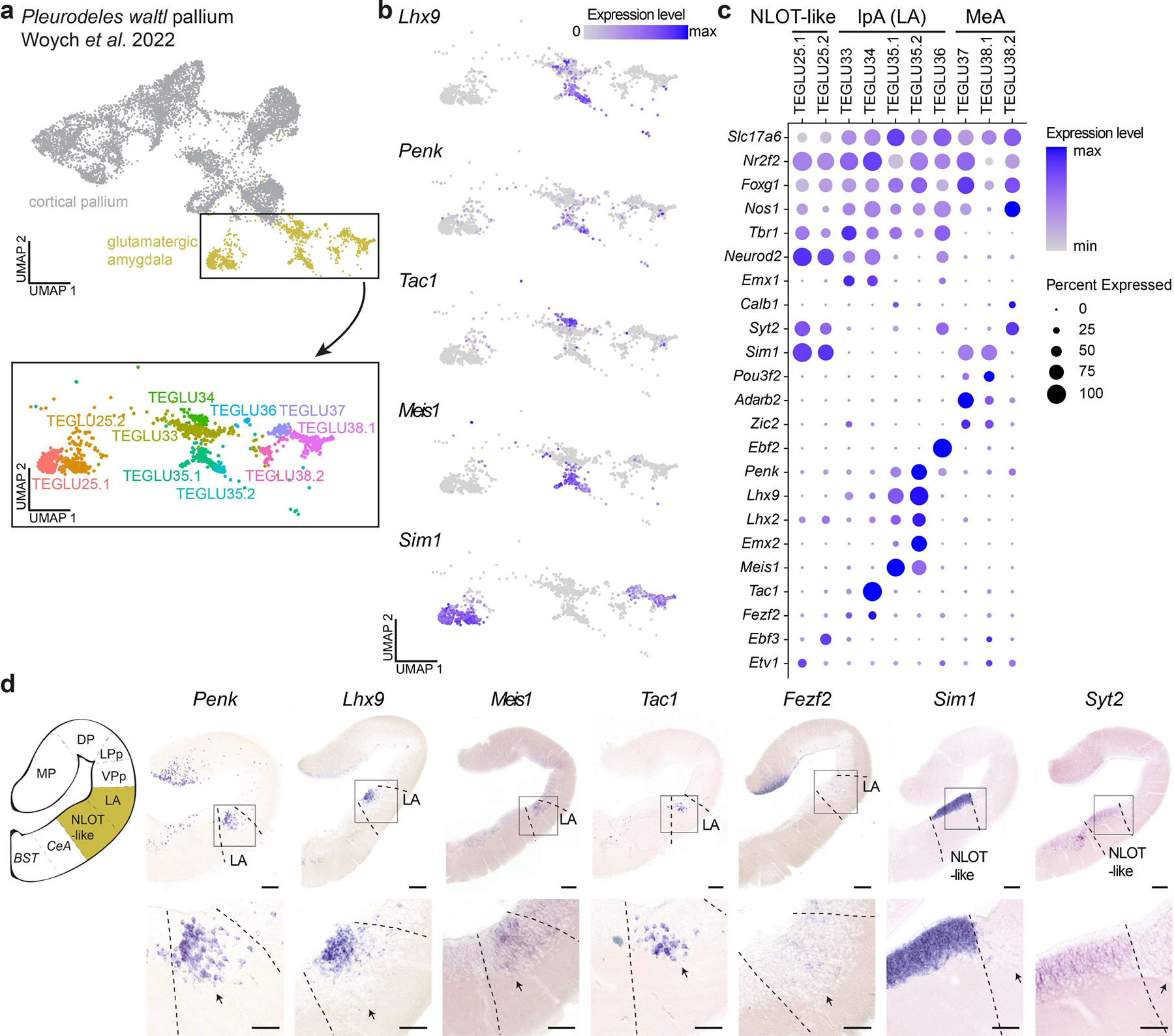
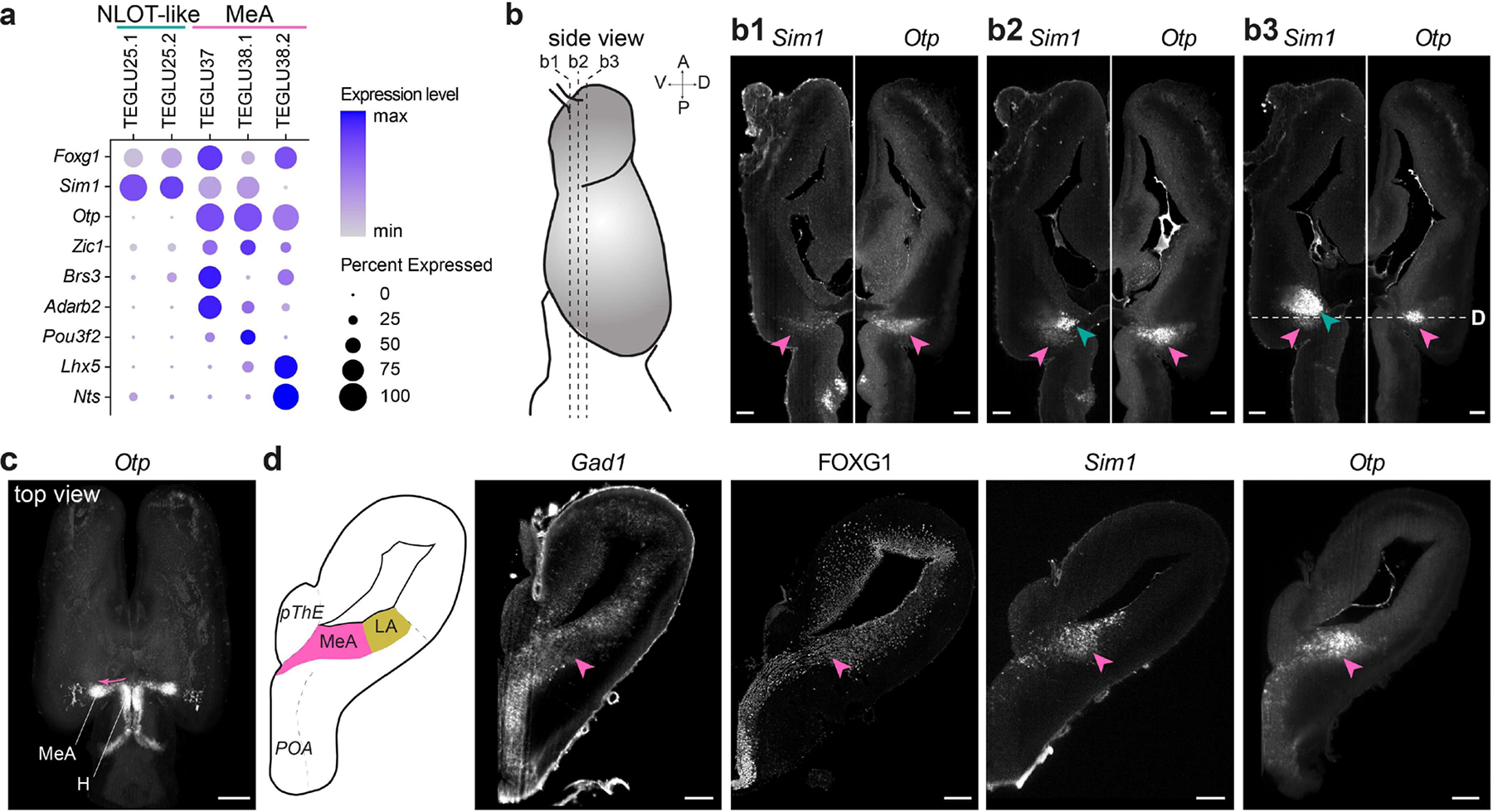
The amygdala is a complex brain structure in the vertebrate telencephalon, essential for regulating social behaviors, emotions, and (social) cognition. In contrast to the vast majority of neuron types described in the many nuclei of the mammalian amygdala, little is known about the neuronal diversity in non-mammals, making reconstruction of its evolution particularly difficult. Here, we characterize glutamatergic neuron types in the amygdala of the urodele amphibian Pleurodeles waltl. Our single-cell RNA sequencing data indicate the existence of at least ten distinct types and subtypes of glutamatergic neurons in the salamander amygdala. These neuron types are molecularly distinct from neurons in the ventral pallium (VP), suggesting that the pallial amygdala and the VP are two separate areas in the telencephalon. In situ hybridization for marker genes indicates that amygdalar glutamatergic neuron types are located in three major subdivisions: the lateral amygdala, the medial amygdala, and a newly defined area demarcated by high expression of the transcription factor Sim1. The gene expression profiles of these neuron types suggest similarities with specific neurons in the sauropsid and mammalian amygdala. In particular, we identify Sim1+ and Sim1+ Otp+ expressing neuron types, potentially homologous to the mammalian nucleus of the lateral olfactory tract (NLOT) and to hypothalamic-derived neurons of the medial amygdala, respectively. Taken together, our results reveal a surprising diversity of glutamatergic neuron types in the amygdala of salamanders, despite the anatomical simplicity of their brain. These results offer new insights on the cellular and anatomical complexity of the amygdala in tetrapod ancestors.
Keywords: Amphibian; Amygdala; Brain evolution; Neuron types.
© 2022 S. Karger AG, Basel.
Conflict of interest statement
Conflict of Interest Statement
The authors have no conflicts of interest to declare.
Figures

Similar articles
-
Distinct Subdivisions in the Transition Between Telencephalon and Hypothalamus Produce Otp and Sim1 Cells for the Extended Amygdala in Sauropsids.Front Neuroanat. 2022 May 12;16:883537. doi: 10.3389/fnana.2022.883537. eCollection 2022. Front Neuroanat. 2022. PMID: 35645737 Free PMC article.
-
Sim1-expressing cells illuminate the origin and course of migration of the nucleus of the lateral olfactory tract in the mouse amygdala.Brain Struct Funct. 2021 Mar;226(2):519-562. doi: 10.1007/s00429-020-02197-1. Epub 2021 Jan 25. Brain Struct Funct. 2021. PMID: 33492553 Free PMC article.
-
A novel telencephalon-opto-hypothalamic morphogenetic domain coexpressing Foxg1 and Otp produces most of the glutamatergic neurons of the medial extended amygdala.J Comp Neurol. 2021 Jul 1;529(10):2418-2449. doi: 10.1002/cne.25103. Epub 2021 Jan 13. J Comp Neurol. 2021. PMID: 33386618
-
Evolution and Development of Amygdala Subdivisions: Pallial, Subpallial, and Beyond.Brain Behav Evol. 2023;98(1):1-21. doi: 10.1159/000527512. Epub 2022 Oct 20. Brain Behav Evol. 2023. PMID: 36265454 Review.
-
The Everted Amygdala of Ray-Finned Fish: Zebrafish Makes a Case.Brain Behav Evol. 2022;97(6):321-335. doi: 10.1159/000525669. Epub 2022 Jun 27. Brain Behav Evol. 2022. PMID: 35760049 Review.
Cited by
-
Does a Vertebrate Morphotype of Pallial Subdivisions Really Exist?Brain Behav Evol. 2024;99(4):230-247. doi: 10.1159/000537746. Epub 2024 Jul 16. Brain Behav Evol. 2024. PMID: 38952102 Free PMC article. Review.
-
Development and function of the medial amygdala.Trends Neurosci. 2025 Jan;48(1):22-32. doi: 10.1016/j.tins.2024.11.004. Epub 2024 Dec 12. Trends Neurosci. 2025. PMID: 39672784 Review.
-
Divergence in neuronal signaling pathways despite conserved neuronal identity among Caenorhabditis species.Curr Biol. 2025 Jun 23;35(12):2927-2945.e7. doi: 10.1016/j.cub.2025.05.036. Epub 2025 May 23. Curr Biol. 2025. PMID: 40412379 Free PMC article.
References
-
- Brox A, Puelles L, Ferreiro B, Medina L: Expression of the genes GAD67 and Distal-less-4 in the forebrain of Xenopus laevis confirms a common pattern in tetrapods. J Comp Neurol 2003;461:370–393. - PubMed
Publication types
MeSH terms
Substances
Grants and funding
LinkOut - more resources
Full Text Sources
