. 2023 Jan 10;56(1):93-106.e6.
doi: 10.1016/j.immuni.2022.12.001. Epub 2022 Dec 26.

The ectonucleotidase CD39 identifies tumor-reactive CD8+ T cells predictive of immune checkpoint blockade efficacy in human lung cancer

Affiliations

The ectonucleotidase CD39 identifies tumor-reactive CD8+ T cells predictive of immune checkpoint blockade efficacy in human lung cancer

Andrew Chow et al. Immunity. .

Abstract

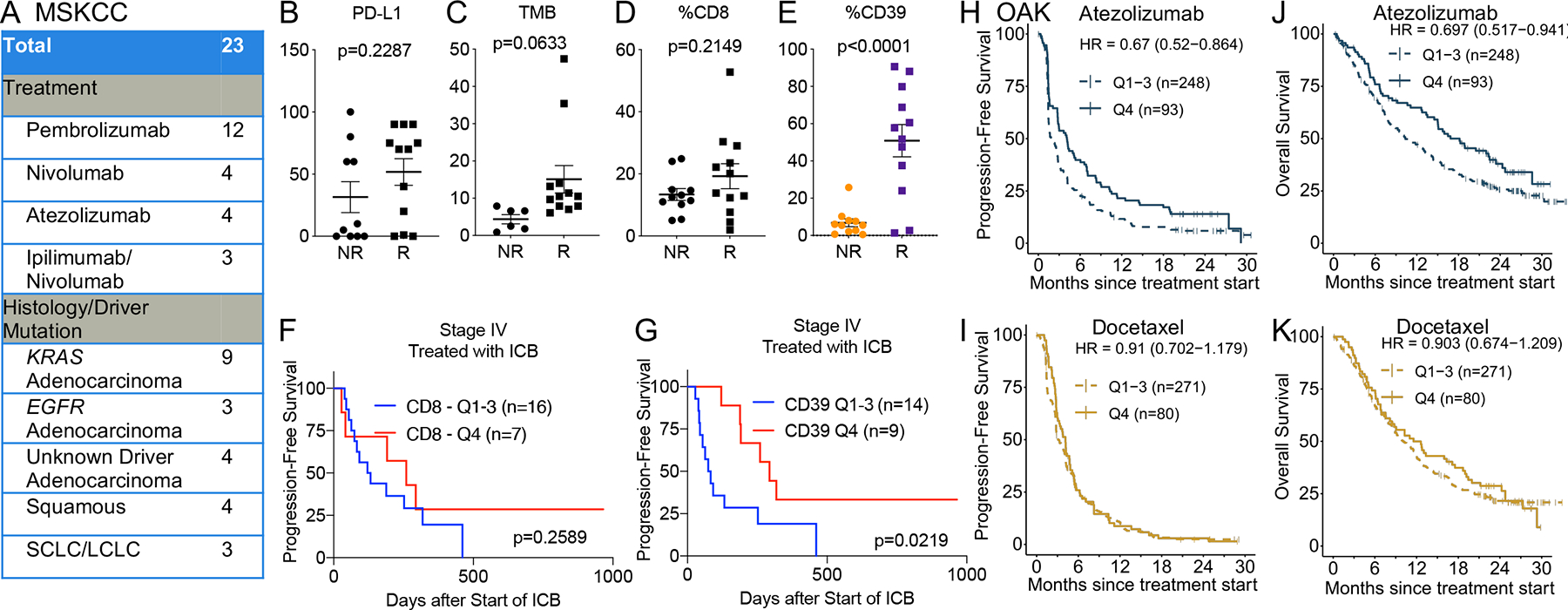
Improved identification of anti-tumor T cells is needed to advance cancer immunotherapies. CD39 expression is a promising surrogate of tumor-reactive CD8+ T cells. Here, we comprehensively profiled CD39 expression in human lung cancer. CD39 expression enriched for CD8+ T cells with features of exhaustion, tumor reactivity, and clonal expansion. Flow cytometry of 440 lung cancer biospecimens revealed weak association between CD39+ CD8+ T cells and tumoral features, such as programmed death-ligand 1 (PD-L1), tumor mutation burden, and driver mutations. Immune checkpoint blockade (ICB), but not cytotoxic chemotherapy, increased intratumoral CD39+ CD8+ T cells. Higher baseline frequency of CD39+ CD8+ T cells conferred improved clinical outcomes from ICB therapy. Furthermore, a gene signature of CD39+ CD8+ T cells predicted benefit from ICB, but not chemotherapy, in a phase III clinical trial of non-small cell lung cancer. These findings highlight CD39 as a proxy of tumor-reactive CD8+ T cells in human lung cancer.

Keywords: CD8(+) T cells; T cell receptors; immune checkpoint blockade; non-small cell lung cancer; tumor mutation burden.


Conflict of interest statement

Declaration of interests C.A.K. received research funding support from Kite/Gilead and Intima Bioscience; is on the Scientific and/or Clinical Advisory Boards of Achilles Therapeutics, Aleta BioTherapeutics, Bellicum Pharmaceuticals, Catamaran Bio, Obsidian Therapeutics, and T-knife; and has performed consulting services for Bristol Myers Squibb, PACT Pharma, and Roche/Genentech. C.A.K. is a co-inventor on patent applications related to TCRs targeting public neoantigens unrelated to the current work. M.D.H. received a research grant from BMS; personal fees from Achilles, Arcus, AstraZeneca, Blueprint, BMS, Genentech/Roche, Genzyme, Immunai, Instil Bio, Janssen, Merck, Mirati, Natera, Nektar, Pact Pharma, Regeneron, Shattuck Labs, and Syndax; and equity options from Arcus, Factorial, Immunai, and Shattuck Labs. A patent filed by MSKCC related to the use of tumor mutational burden to predict response to immunotherapy (PCT/US2015/062208) is pending and licensed by PGDx. J.D.W. is a consultant for Amgen, Apricity, Ascentage Pharma, Astellas, AstraZeneca, Bicara Therapeutics, Boehringer Ingelheim, Bristol Myers Squibb, CellCarta, Chugai, Daiichi Sankyo, Dragonfly, Georgiamune, Idera, Imvaq, Larkspur, Maverick Therapeutics, Merck, Psioxus, Recepta, Tizona, Trishula, Sellas, Surface Oncology, and Werewolf Therapeutics. J.D.W. receives grant/research support from Bristol Myers Squibb and Sephora. J.D.W. has equity in Apricity, Arsenal IO, Ascentage, Beigene, Imvaq, Linneaus, Georgiamune, Maverick, Tizona Pharmaceuticals, and Trieza. J.D.W. is a co-inventor on the following patent application: xenogeneic (canine) DNA vaccines, myeloid-derived suppressor cell (MDSC) assay, anti-PD1 antibody, anti-CTLA4 antibodies, anti-GITR antibodies and methods of use thereof, Newcastle disease viruses for cancer therapy, and prediction of responsiveness to treatment with immunomodulatory therapeutics and method of monitoring abscopal effects during such treatment. J.D.W. and T.M. are co-inventors on patent applications related to CD40 and in situ vaccination (PCT/US2016/045970). T.M. is a consultant for Immunos Therapeutics and Pfizer. T.M. is a cofounder of and equity holder in IMVAQ Therapeutics. T.M. receives research funding from Bristol Myers Squibb, Surface Oncology, Kyn Therapeutics, Infinity Pharmaceuticals, Peregrine Pharmaceuticals, Adaptive Biotechnologies, Leap Therapeutics, and Aprea Therapeutics. T.M. is an inventor on patent applications related to work on oncolytic viral therapy, alpha virus-based vaccine, neoantigen modeling, CD40, GITR, OX40, PD-1, and CTLA-4. C.M.R. has consulted regarding oncology drug development with AbbVie, Amgen, Ascentage, AstraZeneca, BMS, Celgene, Daiichi Sankyo, Genentech/Roche, Ipsen, Loxo, and PharmaMar and is on the scientific advisory boards of Elucida, Bridge, and Harpoon. B.Y.N. and X.G. are employees and stockholders of Genentech/Roche.

Figures

Figure 1.
Figure 1.. Single-cell CITE/RNA/TCR-sequencing reveals that CD39hi CD8+ T cells are enriched for features of exhaustion, tumor reactivity, and clonal proliferation in human lung cancer.
A) UMAP of sorted CD3+ T cells from four patients with lung cancer (Table S1). Clusters are annotated on left panel. Surface levels of CD4, CD8, and CD39 as assessed by CITEseq antibody-derived tags (adt) are depicted in right three panels. B) Levels of various proteins (column) across the four samples (row) as determined by CITEseq adt levels. C-G) Scaled scores for exhaustion, tumor reactivity, tumor specific, virus specific, and proliferation gene signatures (Table S4). H) Clonal proportion among CD8+ T cells of clonotypes that were categorized by mean CD39 expression. Statistical significance was determined with two-way ANOVA with Tukey’s multiple comparisons test and p value is indicated if <0.05.
Figure 2.
Figure 2.. CD39 enriches for tumor-reactive TCRs in lung cancer.
A) Schematic outlining the parallel derivation of a patient-derived xenograft for MSK 1087 and MSK 1111 and cloning and transduction of candidate CD39neg, CD39int, and CD39hi TCRs into healthy donor CD8+ T cells deleted for endogenous TCRs. The cultured PDX cells and transduced donor CD8+ T cells were co-cultured for 24 hours and 4–1BB expression was evaluated on transduced T cells. B) Flow cytometry plots of CD3 and EGFRt expression on untransduced TRAC/TRBC-edited CD8+ T (left top panel) or TRAC/TRBC-edited CD8+ T transduced with donor NY-ESO1 TCR (left bottom panel, blue box indicates transduced population). C) Flow cytometry plots of CD8+ and 4–1BB expression for NY-ESO1 TCR-transduced T cells that were cultured alone (right top panel), with H522-NYESO1 (right middle), or H522-NYESO1 with anti-MHC I (right lower). C) Flow cytometry plots of CD8+ and 4–1BB expression for MSK 1087 TCR 2-transduced T cells that were cultured alone (top panel), with MSK 1087 PDX (middle), or MSK 1087a PDX with anti-MHC I (lower). D) Bar plots of %4–1BB among EGFRt+ transduced T cells that were cultured alone (left solid bar in each series of three bars), with MSK 1087 PDX cells (middle bar with black dash), or with MSK 1087 PDX cells treated and anti-MHC I (right bar with white dash). Red bars indicate TCRs that are tumor-reactive (the %4–1BB level for the culture with PDX tumor cells is ≥5% higher than the culture with only T cells). E) Bar plots of %4–1BB among EGFRt+ transduced T cells that were cultured alone (left solid bar), with MSK 1111 PDX cells (middle bar with black dash), or with MSK 1111 PDX cells treated and anti-MHC I (right bar with white dash). Red bars indicate TCRs that are tumor-reactive. F) Tabulation of reactive TCRs after co-culture with patient-matched PDX tumor cells.
Figure 3.
Figure 3.. CD39 is durably expressed after antigen-specific stimulation.
A) Flow cytometry plots of CTV and 4–1BB levels of NY-ESO1-specific CD8+ T cells that were cultured with no tumor cells (far left plot), H522-NY-ESO1 (top row of plots), or parental H522 (bottom row of plots) for the indicated number of days. B) Flow cytometry plots of CD39 and PD-1 levels of NY-ESO1-specific CD8+ T cells that were cultured with no tumor cells (far left plot), H522-NY-ESO1 (top row of purple plots), or parental H522 (bottom row of orange plots) for the indicated number of days. C) Levels of %4–1BB+, %PD-1+, or %CD39+ among NY-ESO1-specific CD8+ T cells during co-culture with H522-NY-ESO1 (purple line) or parental H522 (orange line). D) Levels of %4–1BB+, %PD-1+, or %CD39+ among NY-ESO1-specific CD8+ T cells during co-culture with parental H522 pulsed with the NY-ESO1 altered peptide ligands SLLMWITQC (black line), SLLNWITQC (red line), SLLPWITQC (blue line), or SLLSWITQC (green line).
Figure 4.
Figure 4.. CD39 expression among intratumoral CD8+ T cells varies with lung cancer subtype.
A) Representative flow cytometry staining of CD39, Tim-3, 4–1BB, and PD-1 on DAPI CD45+ CD3+ CD8++ T cells from MSK 1105b. Left plot represents fluorescence minus one (FMO) and right plot represents the CD8+ T cells stained with the indicated antibody. B-C) Violin plots of %CD39+ (among CD8++ T cells) and %CD8+ (among CD45+) for various histological subtypes/driver mutation categories among the 440-sample cohort. Dashed lines indicate the median and 75th percentile CD39 level for entire 440-sample cohort. Red bars indicate histological subtypes/driver mutations with above median %CD39 values.
Figure 5.
Figure 5.. PD-1 axis blockade increases the frequency of CD39+ CD8+ tumor-infiltrating lymphocytes.
A-B) %CD8+ (among CD45+) or %CD39 (among CD8+ T cells) from 218 biospecimens obtained from patients with stage IV metastatic disease that did or did not receive chemotherapy or ICB therapy in the prior 3 months. Statistical significance was assessed by two-tailed student’s t test. C-D) %CD8+ (among CD45+) or %CD39 (among CD8+ T cells) from 218 biospecimens obtained from patients with stage IV metastatic disease that did or did not receive chemotherapy or ICB therapy in the prior 6 months. Statistical significance assessed by two-tailed student’s t test. E-F) %CD8+ (among CD45+) or %CD39 (among CD8+ T cells) from 208 biospecimens obtained from patients with early stage (stage I-III) NSCLC. Statistical significance was assessed by two-tailed student’s t test. *p<0.05, ***p<0.001.
Figure 6.
Figure 6.. Baseline intratumoral CD39+ CD8+ T cells portends improved outcomes from immune checkpoint blockade in lung cancer.
A) Cohort of stage IV lung cancer patients at MSKCC who received ICB monotherapy. SCLC = small cell lung cancer; LCLC = large cell lung cancer. B-E) Tumor proportion score for PD-L1, tumor mutation burden, %CD8+ (among CD45+), and % CD39 (among CD8+ T cells) among non-responders (NR) or responders (R) to ICB in the cohort described in A). Statistical significance was determined by Mantel-Cox test. F-G) Kaplan-Meier survival curve of progression-free survival for patients described in cohort A) based on stratification for top quartile (Q4) or bottom 75% level (Q1-Q3) of CD8+ T cells (F) and CD39 on CD8+ T cells (G). H-K) Progression-free and overall survival of patients with stage IV lung cancer in the phase III OAK clinical trial who were randomized to treatment with atezolizumab (H, J) or docetaxel (I, K). The patients were stratified by top quartile (Q4) or bottom 75% level (Q1–3) of a signature score for CD39+ CD8+ T cells.

References

    1. Ribas A, and Wolchok JD (2018). Cancer immunotherapy using checkpoint blockade. Science 359, 1350–1355. 10.1126/science.aar4060. - DOI - PMC - PubMed
    1. Thommen DS, Koelzer VH, Herzig P, Roller A, Trefny M, Dimeloe S, Kiialainen A, Hanhart J, Schill C, Hess C, et al. (2018). A transcriptionally and functionally distinct PD-1(+) CD8(+) T cell pool with predictive potential in non-small-cell lung cancer treated with PD-1 blockade. Nat Med 24, 994–1004. 10.1038/s41591-018-0057-z. - DOI - PMC - PubMed
    1. Hellmann MD, Ciuleanu TE, Pluzanski A, Lee JS, Otterson GA, Audigier-Valette C, Minenza E, Linardou H, Burgers S, Salman P, et al. (2018). Nivolumab plus Ipilimumab in Lung Cancer with a High Tumor Mutational Burden. N Engl J Med 378, 2093–2104. 10.1056/NEJMoa1801946. - DOI - PMC - PubMed
    1. Reck M, Rodriguez-Abreu D, Robinson AG, Hui R, Csoszi T, Fulop A, Gottfried M, Peled N, Tafreshi A, Cuffe S, et al. (2016). Pembrolizumab versus Chemotherapy for PD-L1-Positive Non-Small-Cell Lung Cancer. N Engl J Med 375, 1823–1833. 10.1056/NEJMoa1606774. - DOI - PubMed
    1. Riaz N, Havel JJ, Makarov V, Desrichard A, Urba WJ, Sims JS, Hodi FS, Martin-Algarra S, Mandal R, Sharfman WH, et al. (2017). Tumor and Microenvironment Evolution during Immunotherapy with Nivolumab. Cell 171, 934–949 e916. 10.1016/j.cell.2017.09.028. - DOI - PMC - PubMed

Publication types

MeSH terms

Substances