. 2023 Jan 4;15(1):evac181.
doi: 10.1093/gbe/evac181.

Global Gene Expression Analysis Reveals Complex Cuticle Organization of the Tribolium Compound Eye

Affiliations

Global Gene Expression Analysis Reveals Complex Cuticle Organization of the Tribolium Compound Eye

Qing Chen et al. Genome Biol Evol. .

Abstract

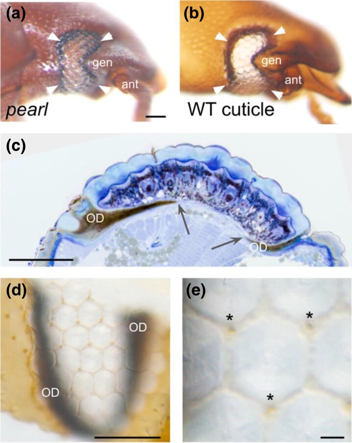
The red flour beetle Tribolium castaneum is a resource-rich model for genomic and developmental studies. To extend previous studies on Tribolium eye development, we produced transcriptomes for normal-eyed and eye-depleted heads of pupae and adults to identify differentially transcript-enriched (DE) genes in the visual system. Unexpectedly, cuticle-related genes were the largest functional class in the pupal compound eye DE gene population, indicating differential enrichment in three distinct cuticle components: clear lens facet cuticle, highly melanized cuticle of the ocular diaphragm, which surrounds the Tribolium compound eye for internal fortification, and newly identified facet margins of the tanned cuticle, possibly enhancing external fortification. Phylogenetic, linkage, and high-throughput gene knockdown data suggest that most cuticle proteins (CPs) expressed in the Tribolium compound eye stem from the deployment of ancient CP genes. Consistent with this, TcasCPR15, which we identified as the major lens CP gene in Tribolium, is a beetle-specific but pleiotropic paralog of the ancient CPR RR-2 CP gene family. The less abundant yet most likely even more lens-specific TcasCP63 is a member of a sprawling family of noncanonical CP genes, documenting a role of local gene family expansions in the emergence of the Tribolium compound eye CP repertoire. Comparisons with Drosophila and the mosquito Anopheles gambiae reveal a steady turnover of lens-enriched CP genes during insect evolution.

Keywords: Tribolium; cuticle; evodevo; eye; gene family evolution; lens proteins.


Figures

Fig. 1.
Fig. 1.
External morphologies of WT and eye-depleted adult and pupal heads in Tribolium. (a–d) Lateral views with anterior to the right. Ant, antenna. (a) Adult WT adult head. Dorsal and ventral borders of the compound eye are indicated by arrowheads. Scale bar: 100 µm. (b) Eye-depleted Ey/Es mutant adult head. Arrowheads indicate approximate margins of the area filled by the compound eye in WT Tribolium. Same scale as in a. (c) Pupal WT Tribolium head. Arrowheads indicate the dorsal margins of developing compound eye. Hatched line indicates border between the head and the thorax. Dark brown dots in the differentiating retinal field represent early emerging photoreceptor clusters. Scale bar: 100 µm. (d) Pupal head of eye-depleted eyaRNAi animal. Arrowheads indicate the approximate dorsal area filled by the developing compound eye in WT animals. Same scale as in c.
Fig. 2.
Fig. 2.
Enriched cuticle gene expression in the developing Tribolium compound eye. (a) Bee swarm box plot of averaged binary log (log2) fold differences in cuticle gene transcript levels in eyaRNAi and TEDRNAi pupal heads compared with WT pupal heads. Blue overlay indicates range of high-confidence compound eye DE genes based on transcript reduction in eye-depleted pupal heads compared with WT pupal heads. Orange overlay indicates range of cuticle DE genes that are significantly transcript enriched in WT compared with eye-depleted pupal heads. Whiskers extend from 5th to 95th percentile of data point distribution. NC, noncanonical cuticle genes; CP, non-CPR subfamily cuticle protein genes; RR-1, CPR RR-1 subfamily CP genes; RR-2, CPR RR-2 subfamily CP genes. (b) Five microliters of semiquantitative RT-PCR amplicons of TcasCPR15, TcasCP63, TcasCPR68, and TcasCPR77 from adult stage cDNA preparations from WT head, WT trunk, sq head, and Ey/Es head tissue separated in 1% agarose gel. RT-PCR amplicons of the photoreceptor-specific benchmark genes LW-opsin and Chp are shown for comparison. Armadillo2 (Arm2) amplicons visualize relative cDNA input. (c–f) Lateral view of 65% pupal head labeled for TcasCPR15 (c), TcasCP63 (d), TcasCPR77 (e), and TcasCPR68 (f) expression as detected by colorimetric whole-mount in situ hybridization. The scale bar in c corresponds to 50 µm for d–f. (g–j) High magnification view of distal layer of single ommatidial unit labeled for TcasCPR15 (g), TcasCP63 (h), TcasCPR77 (i), and TcasCPR68 (j) expression as detected by colorimetric whole-mount in situ hybridization. (k–o) Differential interference contrast microscopy high magnification view of distal layer of single ommatidial units in LacZ negative control (k) versus TcasCPR15 (l), TcasCP63 (m), TcasCPR77 (n), and TcasCPR68 (o) knockdown specimens. Asterisks denote ommatidial bristles. Arrows point at the central lens cuticle core, which was not detectable in the TcasCP15 knockdown specimens (l). The scale bar in panel k corresponds to 10 µm for k–o.
Fig. 3.
Fig. 3.
Age distributions of compound eye transcript-enriched cuticle genes. Bar graph summarizing time windows of origins for noncanonical, CPR RR-1 (RR-1), CPR RR-2 (RR-2), and other CP genes deployed in the Tribolium compound eye. Gene age estimates derived from the OrthoDB (O) (Waterhouse et al. 2013) and Ensembl Metazoa (E) (Kinsella et al. 2011) databases are shown for comparison.
Fig. 4.
Fig. 4.
Genomic linkage and approximate phylogenetic relationships of compound eye-enriched CPR genes. (a) Maximum likelihood gene tree of the CPR-RR2 gene family members from T. castaneum (Tcas), A. gambiae (Agam), and D. melanogaster (Dmel). Topology unscaled and midpoint rooted. Internal branches with bootstrap support below 60 lack branch support value numbers. Dipteran homologs are indicated by blue branches. Homolog gene cluster associations are visualized by differential background colors specified by the color at bottom right. Tribolium homologs are indicated by brown branches and protein identification numbers. High confidence Tribolium compound eye DE genes are indicated by terminal blue dots. Tentative Tribolium compound eye DE genes are indicated by light blue dots with dark blue margins. The major lens cuticle gene TcasCPR15 is highlighted by a blue margin and extended blue dot. Body wall cuticle Tribolium DE genes are indicated by brown dots. Searchable tree accessible at: https://itol.embl.de/tree/141217516110951620682560. See figure 1 in supplementary 2, Supplementary Material online for CPR RR-1 tree and gene cluster visualization. (b) Physical linkage of high-confidence and tentative compound eye DE paralogs (gene numbers given in parentheses) in CPR and noncanonical CP gene families. Closely linked paralogs are defined as separated by interspersed genes from other gene families. (c) Comparison of average and maximal cluster sizes of high-confidence and tentative compound eye DE paralogs in CPR and noncanonical gene families. Numbers in parantheses represent standard deviations.
Fig. 5.
Fig. 5.
Gene criticality related to cuticle gene enrichment classes. Bars show number of genes with no, mild, strong, and embryonic cuticle depletion phenotypes in the iBeetle-Base high-throughput RNAi screen (Schmitt-Engel et al. 2015) parsed by DE classes. Eye DE: high-confidence and tentative compound eye DE genes. BW DE: high-confidence body wall cuticle DE genes.
Fig. 6.
Fig. 6.
Shared versus body compartment-specific cuticle gene enrichment in the Tribolium forewing, hindwing, and compound eye. Numbers represent numbers of cuticle genes that are uniquely enriched in the developing compound eye or regular body wall cuticle (regular head) or coenriched in either of the former and the developing fore- or hindwings. See spreadsheet page P8 of supplementary 1, Supplementary material online for gene identities.
Fig. 7.
Fig. 7.
Clear, tanned, and melanized cuticle elements in the Tribolium compound eye. (a–c) Anterior to the right. (a) The lateral head of live pearl mutant Tribolium which lacks eye pigment to the effect that the internalized, melanized black cuticle of the OD that furnishes the compound eye margin (arrowheads) is visible. Scale bar: 100 µm. (b) The lateral head cuticle of soft tissue cleared WT head revealing that the fully preserved OD is part of the cuticular head exoskeleton. (c) Histological section through the WT Tribolium compound eye, demonstrating that the OD is a massive internal protrusion at the compound eye margin that reaches underneath the retinal floor. Arrows point at internal edges of the OD. Scale bar: 100 µm. (d) Horizontal overview of the ventral compound eye cuticle revealing a grit of hexagonal tanned cuticle margins around each facet. Scale bar: 100 µm. (e) High magnification view of a single facet and its frame of tanned cuticle. Asterisks indicate interommatidial bristles. ant, antenna; gen, gena. Scale bar: 10 µm.
Fig. 8.
Fig. 8.
Structural model of differential transcript enrichment in eyed versus eye-depleted Tribolium heads. Simplified schematic of major cuticle elements in eyed (WT) versus eye-depleted head. Genes hypothesized to be enriched in the compound eye due to differential transcriptional activation (TcasCPR15, TcasCP63, and Th) are indicated by vertical arrows pointing up. CP gene products hypothesized to be enriched in the compound eye due to structural enrichment in the optic diaphragm are indicated by bullet points. Smaller font sizes used for these gene names in the eye-depleted condition reflect the smaller structural representation compared with WT condition. Hatched grey horizontal bars in the lateral cuticle of the eye-depleted head indicate the approximate extent of the missing compound eye area.

Similar articles

Cited by

References

    1. Anders S, Pyl PT, Huber W. 2015. HTSeq—a python framework to work with high-throughput sequencing data. Bioinformatics 31:166–169. - PMC - PubMed
    1. Andersen SO, Chase AM, Willis JH. 1973. The amino-acid composition of cuticles from Tenebrio molitor with special reference to the action of juvenile hormone. Insect Biochem. 3:171–180.
    1. Ansari S, et al. . 2018. Double abdomen in a short-germ insect: zygotic control of axis formation revealed in the beetle Tribolium castaneum. Proc Natl Acad Sci U S A. 115:1819–1824. - PMC - PubMed
    1. Arakane Y, et al. . 2005. The PLoS Genet. chitin synthase genes PLoS Genet. and PLoS Genet. are specialized for synthesis of epidermal cuticle and midgut peritrophic matrix. PLoS Genet. 14:453–463. - PubMed
    1. Arakane Y, et al. . 2012. Formation of rigid, non-flight forewings (elytra) of a beetle requires two major cuticular proteins. PLoS Genet. 8:e1002682. - PMC - PubMed

Substances