. 2022 Dec 27;8(1):138.
doi: 10.1038/s41421-022-00484-1.

High-precision mapping reveals rare N6-deoxyadenosine methylation in the mammalian genome

Affiliations

High-precision mapping reveals rare N6-deoxyadenosine methylation in the mammalian genome

Li-Qian Chen et al. Cell Discov. .

Abstract

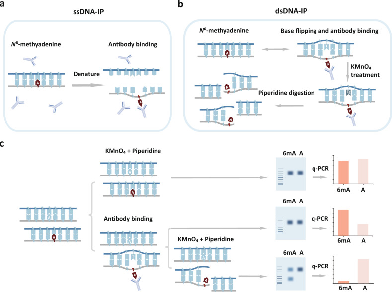
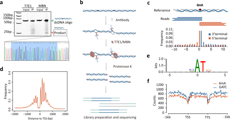
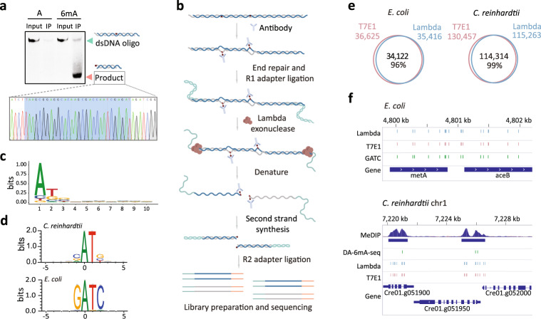
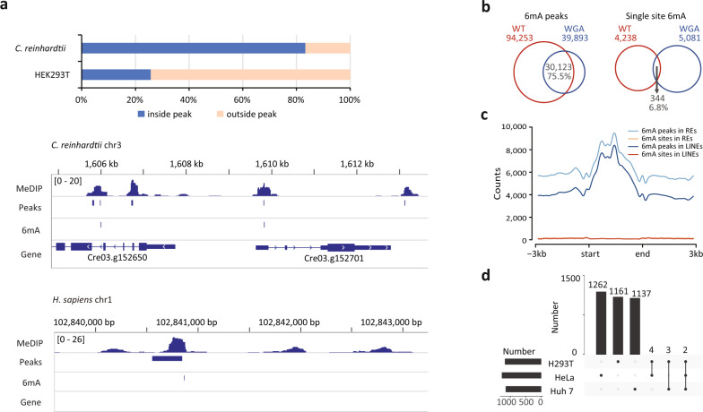
N6-deoxyadenosine methylation (6mA) is the most widespread type of DNA modification in prokaryotes and is also abundantly distributed in some unicellular eukaryotes. However, 6mA levels are remarkably low in mammals. The lack of a precise and comprehensive mapping method has hindered more advanced investigations of 6mA. Here, we report a new method MM-seq (modification-induced mismatch sequencing) for genome-wide 6mA mapping based on a novel detection principle. We found that modified DNA bases are prone to form a local open region that allows capture by antibody, for example, via a DNA breathing or base-flipping mechanism. Specified endonuclease or exonuclease can recognize the antibody-stabilized mismatch-like structure and mark the exact modified sites for sequencing readout. Using this method, we examined the genomic positions of 6mA in bacteria (E. coli), green algae (C. reinhardtii), and mammalian cells (HEK239T, Huh7, and HeLa cells). In contrast to bacteria and green algae, human cells possess a very limited number of 6mA sites which are sporadically distributed across the genome of different cell types. After knocking out the RNA m6A methyltransferase METTL3 in mouse ES cells, 6mA becomes mostly diminished. Our results imply that rare 6mA in the mammalian genome is introduced by RNA m6A machinery via a non-targeted mechanism.


Conflict of interest statement

The authors declare no competing interests.

Figures

Fig. 1
Fig. 1. KMnO4 footprinting assay to detect the 6mA-induced mismatch-like structure.
a The dsDNA is denatured prior to antibody binding during IP in the canonical MeDIP procedure. b Antibody captures transiently flipped N6-methyadenine base and releases the opposite thymine. The unpaired thymine can be oxidized by KMnO4 and subsequently digested by piperidine. c The products of KMnO4 footprinting assay can be quantified by q-PCR to indicate the presence of 6mA. Substrates with 6mA are prone to form a mismatch-like structure with the antibody binding and cleaved by piperidine.
Fig. 2
Fig. 2. Development of endonuclease facilitated MM-seq method for 6mA mapping in C. reinhardtii and E. coli.
a T7E1 and MBN endonuclease can recognize and digest 6mA sites in dsDNA oligos after immunoprecipitation. Sanger sequencing traces of the cleavage products are shown below. b A schematic diagram of MM-seq using T7E1 and MBN endonucleases. c Cleavage signals of reads mapped to the known 6mA sites in a genome-wide scale. d Accumulative plot of the distances between the 6mA sites and the transcription start sites (TSS). e Sequence logo showing VATB motif sequences of the 6mA sites detected in C. reinhardtii. f The distribution of the 6mA sites identified in E. coli displays a similar pattern to the genome-wide GATC motif. TSS transcription start site, TTS transcription termination site.
Fig. 3
Fig. 3. Exonuclease facilitated MM-seq and its application in multiple organisms.
a A fragment resulting from Lambda exonuclease digestion of the synthetic oligo with a 6mA site. Sanger sequencing traces for the cleavage products is shown below. b A schematic diagram showing optimized MM-seq using the Lambda exonuclease. c Nucleotides composition at the 5’ terminal of NGS sequence reads. d Consensus sequences of the genomic context to the 6mA sites identified in C. reinhardtii and E. coli. e High number of overlapping 6mA sites detected between T7E1 endonuclease and Lambda exonuclease in C. reinhardtii and E. coli. f IGV illustration of 6mA sites identified in this study compared to published results from MeDIP-seq and DA-6mA-seq.
Fig. 4
Fig. 4. Application of MM-seq in mammalian cell lines.
a The percentage of 6mA sites identified by MM-seq that fall within the 6mA peaks in C. reinhardtii and human HEK293T cells. The single-base 6mA sites and peaks are illustrated by IGV. b Venn diagrams showing 6mA peaks that were detected in HEK293T cells highly overlapped with those detected in WGA samples. c Accumulative distribution of the 6mA peaks and single-base 6mA sites at repetitive elements (REs) and LINEs. d Bar graph showing the total number of total 6mA sites that appear in each cell line and the total number of 6mA sites that are common between the cell lines as indicated by the dotted lines. Horizontal bars represent the total number of 6mA sites in each sample, and vertical bars with numbers represent the specific or common sites in one or more samples. The dotted lines indicate the site classification (specific or common).

References

    1. Weinhold B. Epigenetics: the science of change. Environ. Health Perspect. 2006;114:a160–a167. doi: 10.1289/ehp.114-a160. - DOI - PMC - PubMed
    1. Smith ZD, Meissner A. DNA methylation: roles in mammalian development. Nat. Rev. Genet. 2013;14:204–220. doi: 10.1038/nrg3354. - DOI - PubMed
    1. Jones PA. Functions of DNA methylation: islands, start sites, gene bodies and beyond. Nat. Rev. Genet. 2012;13:484–492. doi: 10.1038/nrg3230. - DOI - PubMed
    1. Fu Y, et al. N6-methyldeoxyadenosine marks active transcription start sites in Chlamydomonas. Cell. 2015;161:879–892. doi: 10.1016/j.cell.2015.04.010. - DOI - PMC - PubMed
    1. Wion D, Casadesús J. N6-methyl-adenine: an epigenetic signal for DNA–protein interactions. Nat. Rev. Microbiol. 2006;4:183–192. doi: 10.1038/nrmicro1350. - DOI - PMC - PubMed