

Laser speckle contrast imaging for blood flow monitoring in predicting outcomes after cerebral ischemia-reperfusion injury in mice
- PMID: 36575381
- PMCID: PMC9795726
- DOI: 10.1186/s12868-022-00769-x
Laser speckle contrast imaging for blood flow monitoring in predicting outcomes after cerebral ischemia-reperfusion injury in mice
Abstract
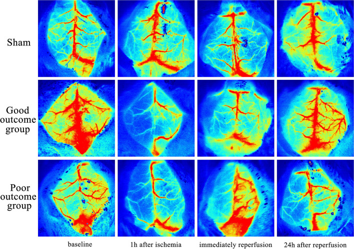
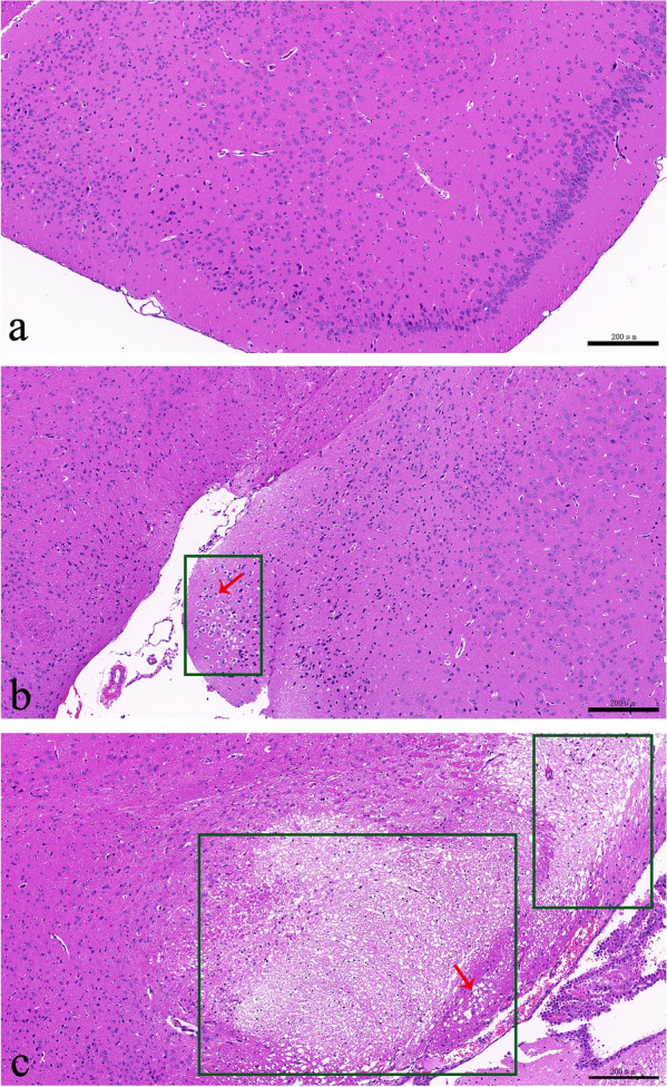
Background: In the treatment of ischemic cerebral stroke (ICS), most conventional treatments, including carotid endarterectomy and carotid artery stenting, may cause cerebral ischemia-reperfusion injury (CIRI). For treated ICS patients, changes in cerebral blood flow are directly related to brain function. At present, computed tomography perfusion, dynamic susceptibility contrast-enhanced perfusion weighted imaging and magnetic resonance arterial spin labeling perfusion imaging are used to monitor cerebral blood flow, but they still have some limitations. Our study aimed to monitor the changes in cerebral cortical blood flow by laser speckle contrast imaging (LSCI) in CIRI model mice and to propose a new method for predicting outcomes after CIRI. C57BL/6 N mice were used to establish a mouse CIRI model based on a modified thread-occlusion method and divided into a good outcome group and a poor outcome group according to survival within 7 days. The cerebral cortical blood flow of the area supplied by the left middle cerebral artery was monitored by LSCI at baseline (before modeling), 1 h after ischemia, immediately after reperfusion and 24 h after reperfusion. Then, the brains of the mice were removed immediately and stained with hematoxylin and eosin to observe the pathological changes in brain neurons.
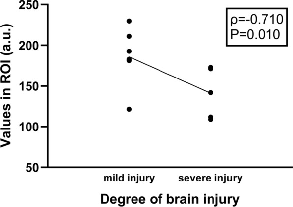
Results: The cerebral cortical blood flow in the poor outcome group was obviously reduced compared with that less in the good outcome group at 24 h after reperfusion (180.8 ± 20.9 vs. 113.9 ± 6.4, p = 0.001), and at 24 h after reperfusion, the cerebral cortical blood flow was negatively correlated with the severity of brain tissue injury (p = - 0.710, p = 0.010).
Conclusions: LSCI can monitor the changes in cerebral cortical blood flow during CIRI in mice and could be used as a feasible method for predicting outcomes after CIRI in mice.
Keywords: Brain tissue injury; Cerebral blood flow; Cerebral ischemia-reperfusion injury; Laser speckle contrast imaging; Outcome prediction.
© 2022. The Author(s).
Conflict of interest statement
All authors claim that there are no competing interests.
Figures
References
Publication types
MeSH terms
Grants and funding
LinkOut - more resources
Full Text Sources
