Review
. 2022 Dec 27;41(1):360.
doi: 10.1186/s13046-022-02555-3.

Cellular senescence in cancer: clinical detection and prognostic implications

Affiliations
Review

Cellular senescence in cancer: clinical detection and prognostic implications

Andreas Domen et al. J Exp Clin Cancer Res. .

Abstract

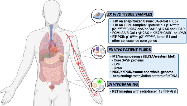
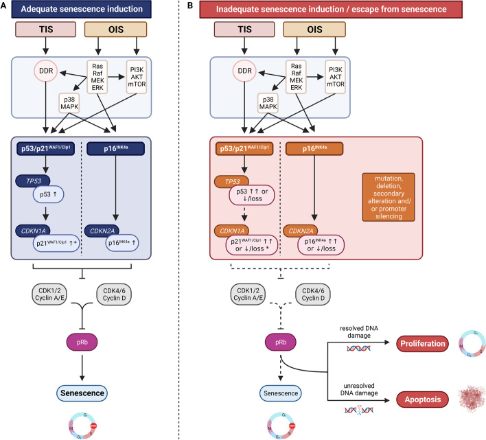
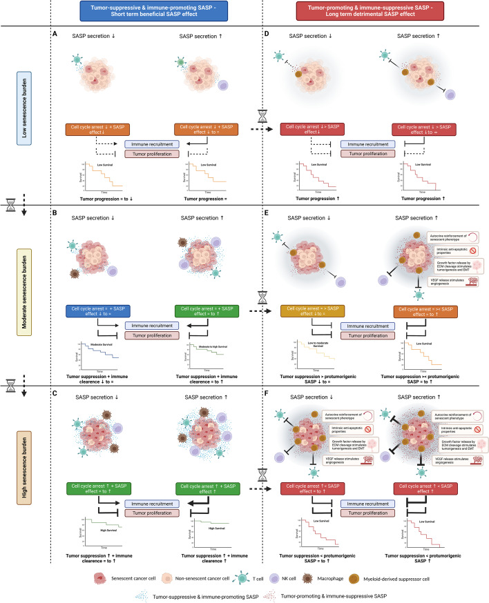
Cellular senescence is a state of stable cell-cycle arrest with secretory features in response to cellular stress. Historically, it has been considered as an endogenous evolutionary homeostatic mechanism to eliminate damaged cells, including damaged cells which are at risk of malignant transformation, thereby protecting against cancer. However, accumulation of senescent cells can cause long-term detrimental effects, mainly through the senescence-associated secretory phenotype, and paradoxically contribute to age-related diseases including cancer. Besides its role as tumor suppressor, cellular senescence is increasingly being recognized as an in vivo response in cancer patients to various anticancer therapies. Its role in cancer is ambiguous and even controversial, and senescence has recently been promoted as an emerging hallmark of cancer because of its hallmark-promoting capabilities. In addition, the prognostic implications of cellular senescence have been underappreciated due to the challenging detection and sparse in and ex vivo evidence of cellular senescence in cancer patients, which is only now catching up. In this review, we highlight the approaches and current challenges of in and ex vivo detection of cellular senescence in cancer patients, and we discuss the prognostic implications of cellular senescence based on in and ex vivo evidence in cancer patients.

Keywords: Cancer; Detection; Oncogene-induced senescence; Prognosis; SASP; Senescence; Therapy-induced senescence.


Conflict of interest statement

The authors declare that they have no competing interests.

Figures

Fig. 1
Fig. 1
Overview of approaches of ex and in vivo detection of cellular senescence with corresponding senescence markers in cancer patients. IHC, immunohistochemistry; SA-β-Gal, senescence-associated beta-galactosidase; FFPE, formalin-fixed paraffin-embedded; SAHF, senescence-associated heterochromatin foci; γH2AX, phosphorylated H2AX; FCM, flow-cytometry; HGMB1, high mobility group box 1; RT-PCR; reverse transcription–polymerase chain reaction; MS, mass spectrometry; SASP, senescence-associated secretory phenotype; EVs, extracellular vesicles; NGS, next-generation sequencing; ddPCR, digital droplet polymerase chain reaction; cfDNA, cell-free DNA; PET-imaging, positron emission tomography-imaging
Fig. 2
Fig. 2
Molecular pathways of OIS and TIS. A Adequate senescence induction via participation of the DDR and the Ras-Raf-MEK-ERK, PI3K/AKT/mTOR and p38/MAPK signaling pathways resulting in functionally activated cell cycle inhibitor pathways (solid arrows) and upregulation of tumor suppressor proteins p53, p21WAF1/Cip1 and p16INK4a. Functional p21WAF1/Cip1 and/or p16INK4a induce a stable cell cycle arrest by inhibition of CDK (i.e., CDK1, CDK2, CDK4 and CDK6)—cyclin (i.e., cyclin A, E and D) complexes, thereby preventing phosphorylation of the retinoblastoma protein (solid inhibitor lines), which blocks S-phase entry and induces senescence (solid arrow). B Inadequate senescence induction or escape from senescence due to (acquired) mutations, deletions, secondary alterations and/or promoter silencing affecting cellular control genes TP53 (encoding p53), CDKN1A (encoding p21WAF1/Cip1) CDKN2A (encoding p16INK4a), resulting in absent or dysfunctional cell cycle inhibitor pathway activation (dotted arrow) and absent or dysfunctional tumor suppressor proteins to induce or maintain senescence (dotted inhibitor lines and arrow). Depending on whether the DNA damage is repaired, the cell may resume proliferation or go into apoptosis. * p21WAF1/Cip1 can also be activated by pathways that are independent of p53 [125]. OIS, oncogene-induced senescence; TIS, therapy-induced senescence; DDR, DNA damage response; CDK, cyclin-dependent kinase; pRb, retinoblastoma protein
Fig. 3
Fig. 3
Model for differential prognostic outcomes of cellular senescence (OIS and TIS) in cancer patients. Tumor-suppressive and immune-promoting SASP—Short term beneficial SASP effect. In case of a net tumor-suppressive and immune-promoting SASP or short term presence of SASP, immune recruitment will result in immune clearance of senescent cancer cells as well as non-senescent cancer cells, thereby reinforcing cellular senescence to provide adequate tumor suppression. However, in case of a (A) low senescence burden, the effects of the SASP are expected to be less profound as the overall SASP levels secreted by the low number of tumoral senescent cells are lower compared to SASP levels in case of a moderate or high senescence burden. Therefore, the senescence-associated cell cycle arrest as well as the SASP levels are expected to be insufficient to provide an adequate tumor suppression. In case of a (B) moderate senescence burden, the senescence-associated cell cycle arrest can increasingly be reinforced in case of low and high SASP secretion, respectively, to provide adequate tumor suppression. In case of a (C) high senescence burden, the senescence-associated cell-cycle arrest can be reinforced by the tumor-suppressive and immune-promoting SASP in case of high as well as low SASP secretion due to the large number of tumoral senescent cells. As such, in case of a net tumor-suppressive and immune-promoting SASP, a high senescence burden result in improved outcome. Tumor-promoting and immune-suppressive SASP—Long term detrimental SASP effect. In case of a net tumor-promoting and immune-suppressive SASP or long term presence of SASP, the senescence-associated cell cycle arrest can be opposed by the SASP by molding an immune-suppressive and protumorigenic TME and stimulating immune evasion. However, in case of a (D) low senescence burden, the effects of the SASP are expected to be less profound as the overall SASP levels secreted by the low number of tumoral senescent cells are lower compared to SASP levels in case of a moderate or high senescence burden. Therefore, the senescence-associated cell cycle arrest in case of a low senescence burden is not opposed by the SASP, however, the senescence-associated cell cycle arrest is insufficient to prevent tumor proliferation. In case of a (E) moderate senescence burden, the senescence-associated cell cycle arrest can be opposed by the SASP in case of high SASP secretion, whereas in case of low SASP secretion the senescence-associated cell cycle arrest overrules the lower SASP levels, resulting in differential tumor-promoting and tumor-suppressive effects, respectively. In case of a (F) high senescence burden, the senescence-associated cell cycle arrest can be opposed and overruled by the protumorigenic effects of the SASP in case of high as well as low SASP secretion, as the overall SASP levels produced by the large number of tumoral senescent cells are elevated, even in case of low SASP secretion. As such, in case of a net tumor-promoting and immune-suppressive SASP, a high senescence burden can paradoxically result in worse outcome. ECM, extracellular matrix; EMT, epithelial-mesenchymal transition; VEGF, vascular endothelial growth factor; NK cell, natural killer cell; SASP, senescence-associated secretory phenotype; ↑, high; = , moderate; ↓, low; > , greater-than; < , less-than; ⌛, time

Similar articles

Cited by

References

    1. Gorgoulis V, Adams PD, Alimonti A, Bennett DC, Bischof O, Bishop C, et al. Cellular Senescence: Defining a Path Forward. Cell. 2019;179(4):813–827. doi: 10.1016/j.cell.2019.10.005. - DOI - PubMed
    1. Muñoz-Espín D, Cañamero M, Maraver A, Gómez-López G, Contreras J, Murillo-Cuesta S, et al. Programmed cell senescence during mammalian embryonic development. Cell. 2013;155(5):1104–1118. doi: 10.1016/j.cell.2013.10.019. - DOI - PubMed
    1. Storer M, Mas A, Robert-Moreno A, Pecoraro M, Ortells MC, Di Giacomo V, et al. Senescence is a developmental mechanism that contributes to embryonic growth and patterning. Cell. 2013;155(5):1119–1130. doi: 10.1016/j.cell.2013.10.041. - DOI - PubMed
    1. Demaria M, Ohtani N, Youssef SA, Rodier F, Toussaint W, Mitchell JR, et al. An essential role for senescent cells in optimal wound healing through secretion of PDGF-AA. Dev Cell. 2014;31(6):722–733. doi: 10.1016/j.devcel.2014.11.012. - DOI - PMC - PubMed
    1. Jun JI, Lau LF. The matricellular protein CCN1 induces fibroblast senescence and restricts fibrosis in cutaneous wound healing. Nat Cell Biol. 2010;12(7):676–685. doi: 10.1038/ncb2070. - DOI - PMC - PubMed